
Methods and apparatus for electromagnetic source imaging using deep neural networks

Pending Publication Date: 2021-11-11
CARNEGIE MELLON UNIV
View PDF0 Cites 3 Cited by
  • Summary
  • Abstract
  • Description
  • Claims
  • Application Information

AI Technical Summary

Benefits of technology

The invention is a neural network that can be used for various applications including imaging and localizing epilepsy sources. It can outperform conventional ESI methods and is effective in analyzing different types of signals. This means that the invention can provide better performance regardless of the signals it is collecting.

Problems solved by technology

However, scalp topographical maps can only provide a rough estimation of the underlying sources due to the volume conduction effect.
However, it is challenging to localize and image the brain electrical activity precisely and reliably with only a few hundred sensors, because of the ill-posed nature of the inverse problem, as different source distributions can produce similar potentials / fields at the scalp.
These methods require parameter tuning to balance between fitting the recorded data and satisfying the regularization constraints when solving every instance of a given problem (i.e., for every given EEG / MEG measurement), which could be a time-consuming process.

Method used


Image

  • Methods and apparatus for electromagnetic source imaging using deep neural networks
  • Methods and apparatus for electromagnetic source imaging using deep neural networks
  • Methods and apparatus for electromagnetic source imaging using deep neural networks

Examples


Embodiment Construction

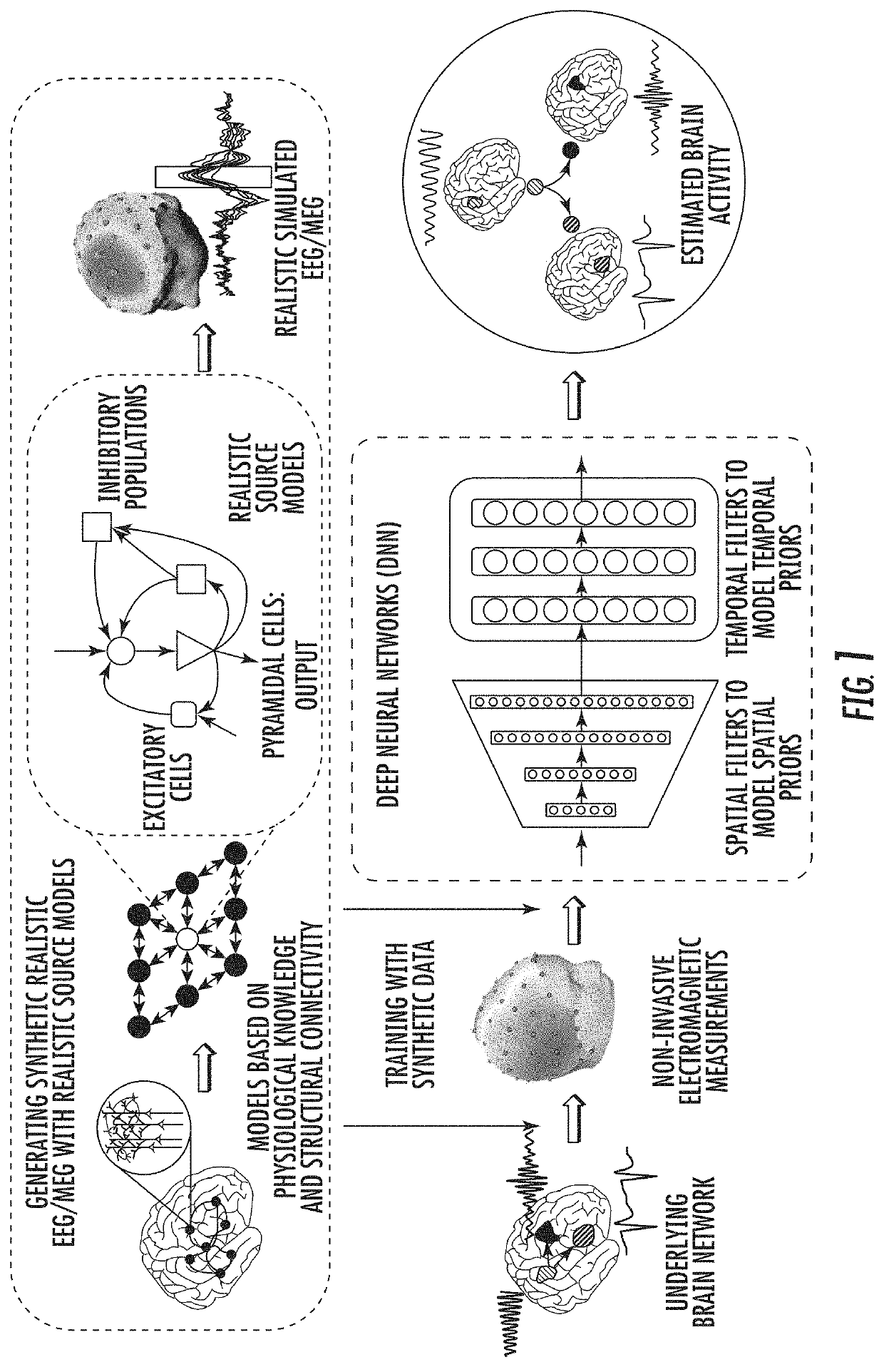
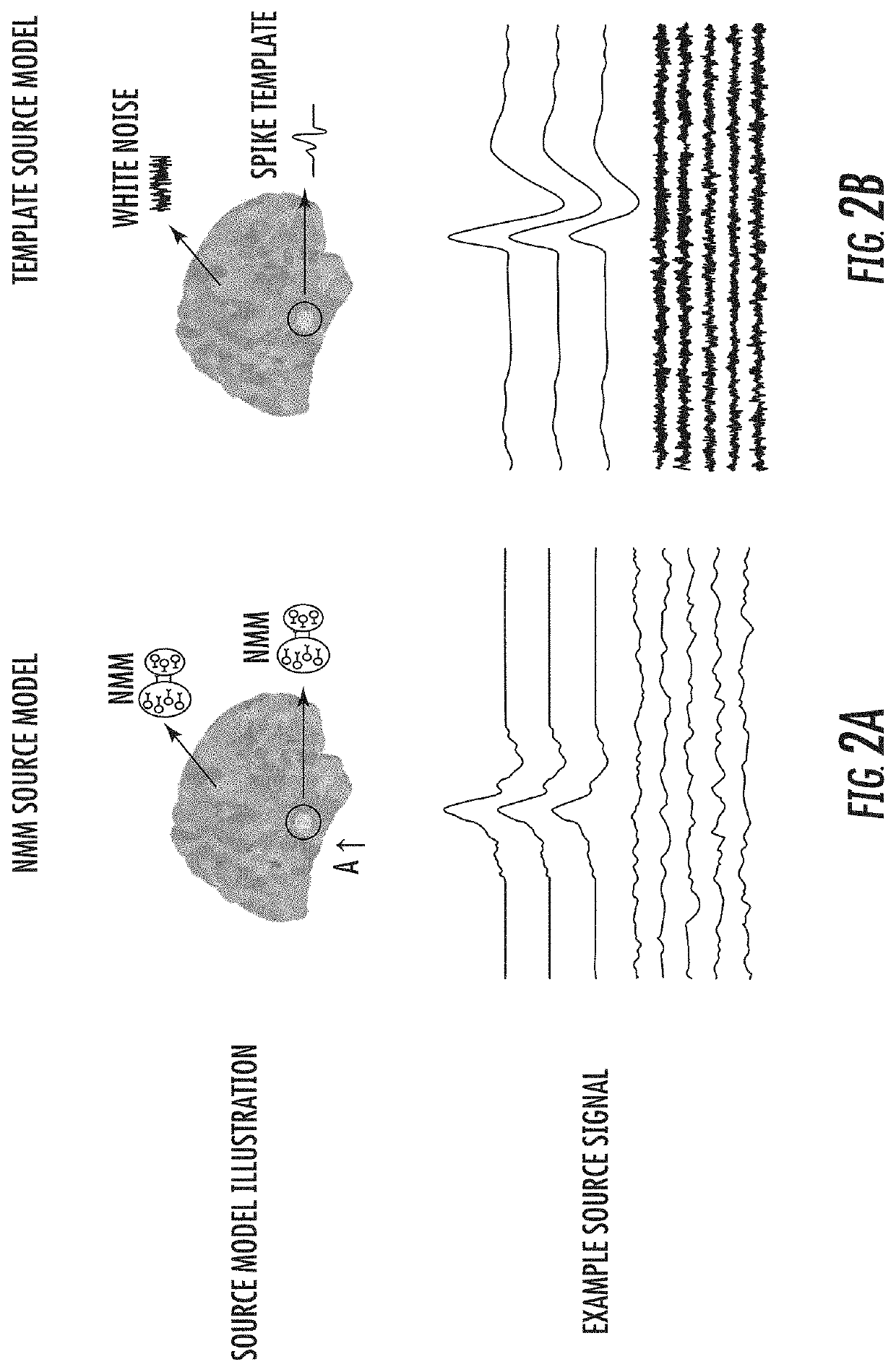
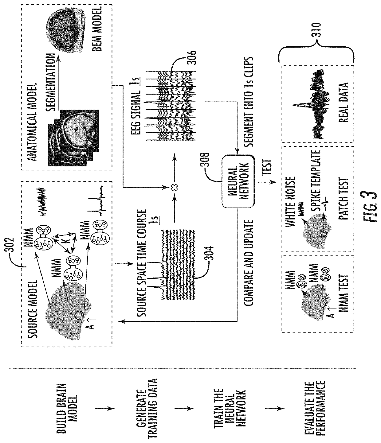
[0031]While the present invention can be applied to EEG, MEG and even intracranial EEG data, for simplicity, the invention will be explained in the context of its application to EEG data. Examples of applying the invention on multiple types of scalp data, i.e. EEG / MEG, will be shown. The invention addresses the fundamental problems for training a deep neural network to image and localize brain electrical activity. The invention first addresses the challenge of generating enough training data that resembles real EEG data. Because an actual mapping of source signals to an EEG trace would require invasive probing of the brain of a subject, a suitable quantity of data for training a deep neural network is unavailable. Additionally, the invention addresses the robustness of a model trained on these source models in comparison to variations in the input data. Finally, the invention addresses how well a model trained in accordance with the present invention performs, given real EEG or MEG ...


PUM


Abstract

Disclosed herein are methods and apparatus for the imaging of brain electrical activity from electromagnetic measurements, using deep learning neural networks where a simulation process is designed to model realistic brain activation and electromagnetic signals to train generalizable neural networks and a residual convolutional neural network and / or a recurrent neural network is trained using the simulated data, capable of estimating source distributions from electromagnetic measurements, and their temporal dynamics over time, for pathological signals in diseased brains, such as interictal activity and ictal signals, and physiological brain signals such as evoked brain responses and spontaneous brain activity.

Description

RELATED APPLICATIONS[0001]This application claims the benefit of U.S. Provisional Patent Application No. 63 / 022,876, filed May 11, 2020, the contents of which are incorporated herein in their entirety.GOVERNMENT INTEREST[0002]This invention was made with government support under grants from the National Institutes of Health, Nos. EB021027, NS096761, EB029354, MH114233 and AT009263. The government has certain rights in this invention.BACKGROUND[0003]Electroencephalography (EEG) and magnetoencephalography (MEG) are noninvasive techniques that measure synchronized activities of neuronal ensembles in the brain. Scalp EEG / MEG topography provides spatial information about the underlying brain electrical activity. However, scalp topographical maps can only provide a rough estimation of the underlying sources due to the volume conduction effect. Electrophysiological source imaging (ESI) was developed to significantly enhance the spatial resolution of noninvasive EEG / MEG. ESI is the process ...

Claims


Application Information

Patent Timeline
IPC IPC(8): A61B34/10A61B5/245A61B5/369G06F30/23G06N3/08
CPCA61B34/10A61B5/245A61B2034/105G06F30/23G06N3/08A61B5/369A61B5/246A61B5/378A61B5/383A61B5/7267A61B5/725G06N3/044G06N3/045A61B5/372A61B5/37
Inventor HE, BINSUN, RUISOHRABPOUR, ABBAS
Owner CARNEGIE MELLON UNIV
Learn More