
Method and Assembly for Securing an Implantable Medical Device on a Delivery System

a technology for implantable medical devices and delivery systems, applied in the field of delivery of implantable medical devices, can solve the problems of affecting the insertion process, and carries a so as to minimize the risk of piercing the balloon and prevent the end of the balloon from flaring

Active Publication Date: 2022-04-07
VENUS MEDTECH (HANGZHOU) INC
View PDF0 Cites 2 Cited by
  • Summary
  • Abstract
  • Description
  • Claims
  • Application Information

AI Technical Summary

Benefits of technology

The present invention provides a way to securely attach an implantable medical device to a delivery system and deploy it at a treatment location while minimizing movement during crimping and deployment. This is achieved by using rings at the ends of the device that hold a locking wire, which prevents flaring of the device and piercing of the balloon.

Problems solved by technology

While the current crimping and deployment of devices tend to be effective for most procedures, there can be some issues with the balloon expandable medical devices in certain situations.
First, the crimped medical devices are exposed to the surrounding environment (e.g., the introducing sheath, blood vessels, etc.), which may cause the medical device to experience movement during insertion.
When crossing the aortic arch, the exposed medical device tends to flare at its distal end on the outer surface, which can be a hazard to the artery.
Second, the shoulders used in securing the medical device on the balloon can hold the medical device in place but carries a risk of piercing the balloon, as the medical device tends to move and presses against the shoulders.

Method used


Image

  • Method and Assembly for Securing an Implantable Medical Device on a Delivery System
  • Method and Assembly for Securing an Implantable Medical Device on a Delivery System
  • Method and Assembly for Securing an Implantable Medical Device on a Delivery System

Examples


Embodiment Construction

[0028]The following detailed description is of the best presently contemplated modes of carrying out the invention. This description is not to be taken in a limiting sense, but is made merely for the purpose of illustrating general principles of embodiments of the invention. The scope of the invention is best defined by the appended claims.

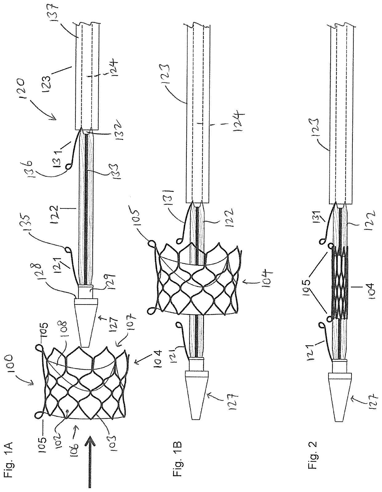
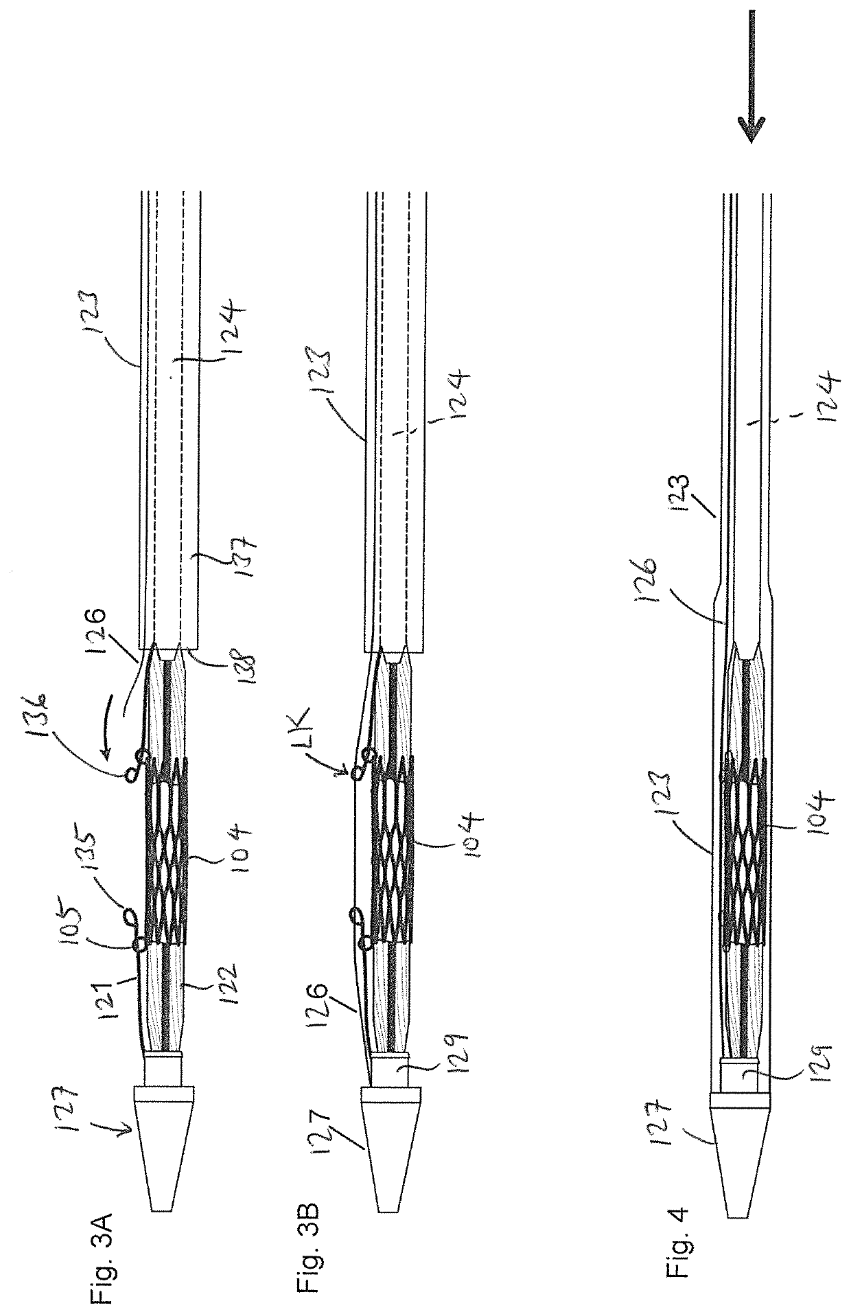
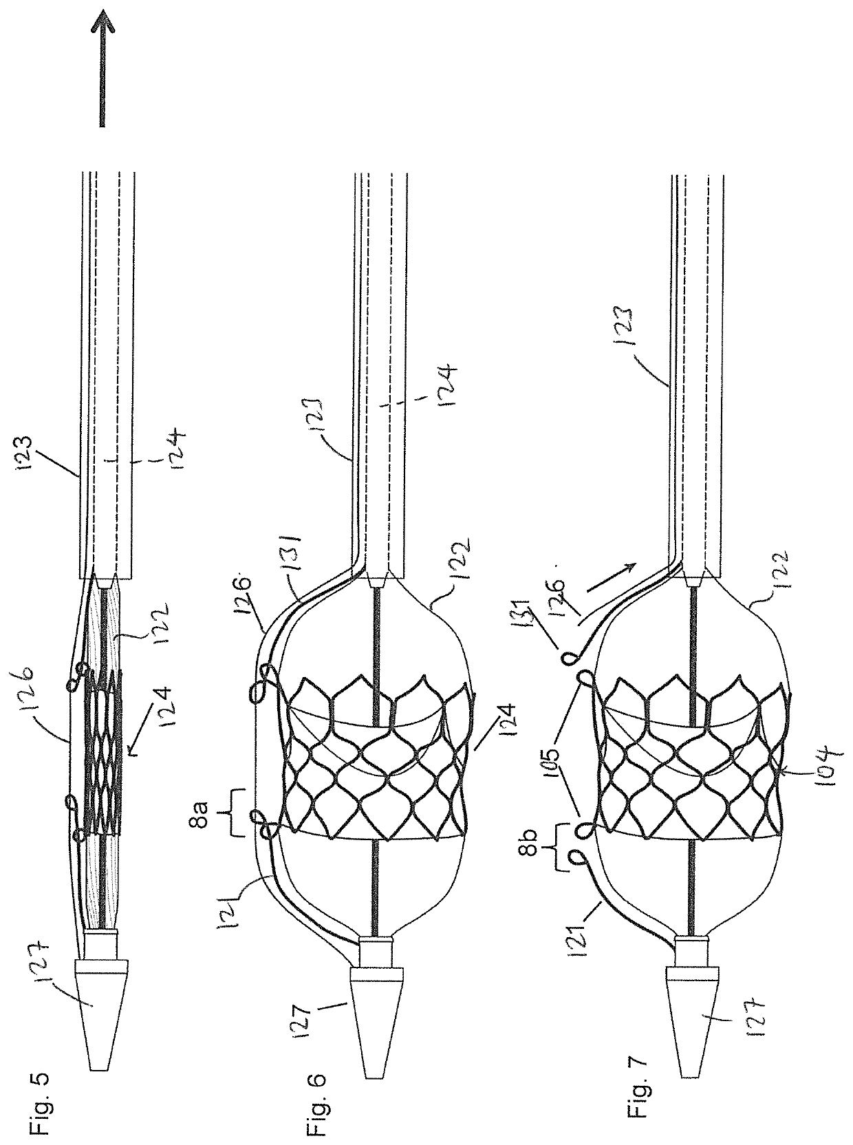
[0029]FIG. 1A is an exploded side view illustrating a medical device 100 and a balloon catheter 120 according to one embodiment of the present invention. The medical device 100 can be any medical device that is intended to be implanted into the human body, such as a stent, transcatheter heart valve, stent-graft assembly, closure device, and plug, among others. The drawings in the present invention are illustrated in connection with a transcatheter heart valve assembly 104 which can an expandable stent, or a stent-frame for use with a transcatheter heart valve. The stent heart valve assembly 104 can have a generally cylindrical stent body that has ...


PUM


Abstract

A method and an assembly for securing a crimped medical device over a deflated balloon of a balloon catheter is provided. The medical device is positioned in its expanded state over the deflated balloon of the balloon catheter, and is then crimped over the deflated balloon. First and second eyelets of first and second strings, respectively, are then threaded through first and second rings, respectively, that are provided on the medical device. Next, a locking wire is advanced through a lumen defined between the sheath and the catheter body to exit the distal end of the sheath, and then advanced through the first and second eyelets and into the distal tip of the balloon catheter. The sheath is then advanced over the crimped medical device to the distal tip to completely cover the crimped medical device.

Description

BACKGROUND OF THE INVENTION1. Field of the Invention[0001]The present invention relates to the delivery of implantable devices into the human body, and in particular, to a method and an assembly for securing an implantable medical device to a delivery system for delivery to a treatment location, and for deployment of the implant at the treatment location.2. Description of the Prior Art[0002]Non-invasive deployment of certain medical devices has become the routine method for treating certain medical conditions. For example, stents and prosthetic heart valves are now delivered to treatment locations inside blood vessels and inside the heart via a transcatheter procedure that avoids invasive surgery.[0003]To facilitate delivery to desired treatment locations, for certain procedures, the medical device (e.g., stents and prosthetic heart valves) is crimped on a deflated balloon catheter, and then threaded through an introducer or a sheath which is inserted into the vasculature to the tar...

Claims


Application Information

Patent Timeline
Patent Type & Authority Applications(United States)
IPC IPC(8): A61F2/958A61F2/95A61F2/966
CPCA61F2/958A61F2/9522A61F2002/9665A61F2002/9511A61F2002/9583A61F2/9662A61F2/2433A61F2/2436A61F2/9517A61F2/2418A61F2250/0018A61F2250/0039
Inventor PHAM, TRINH VANZENG, ASHLEY RACHEL
Owner VENUS MEDTECH (HANGZHOU) INC
Learn More