
An Auxiliary Diagnostic Model and an Image Processing Method for Detecting Acute Ischemic Stroke

a diagnostic model and image processing technology, applied in the field of medical image processing, can solve the problems of poor sensitivity to the early small infarction, heavy stress on the observer, and huge consumption for patients, and achieve the effect of rapid diagnosis of strok

Pending Publication Date: 2022-05-12
WEST CHINA HOSPITAL SICHUAN UNIV +1
View PDF0 Cites 1 Cited by
  • Summary
  • Abstract
  • Description
  • Claims
  • Application Information

AI Technical Summary

Benefits of technology

The present invention provides an auxiliary diagnostic model and an image processing method for detecting acute ischemic stroke. The model uses a generative adversarial network (GAN) to learn the mapping relationships from NECT to FLAIR images, and converts raw CT to synthetic MRI with higher sensitivity. This improves the efficiency of emergency scanning in acute ischemic stroke, reaching both sensitivity and immediacy that are poor in CT interpretation and limited in MRI examination. The technical effects of this invention include improved efficiency of emergency screening for stroke and overcoming clinical predicaments that limit the current technology's sensitivity and acquisition time of magnetic resonance images.

Problems solved by technology

Acute ischemic stroke is the most common type of cerebrovascular diseases, and a significant contributor to the global disease burden, bringing a heavy stress and huge consumption to the patients, their families, and the society.
However, it suffers from observer reliability and poor sensitivity to the early small infarction.
This easily leads to delayed image interpretation and misdiagnosis, thus affecting timely intervention in the stroke patients.
Magnetic resonance imaging (MRI) holds advantages in detecting small and early cerebral ischemic changes, wherein T2-weighted fluid-attenuation inversion recovery (FLAIR) images demonstrate hyperintensities within 3 to 6 hours after onset of symptoms, but with lower availability, higher expense, and slower image acquisition.
This limits MRI to be used in the real emergency settings and degrades it as a supplementary examination for a minority of harsh indications.
Briefly, NECT is rapid but insensitive, whereas MRI is sensitive but time-consuming in early imaging assessment of acute ischemic stroke.

Method used


Image

  • An Auxiliary Diagnostic Model and an Image Processing Method for Detecting Acute Ischemic Stroke
  • An Auxiliary Diagnostic Model and an Image Processing Method for Detecting Acute Ischemic Stroke
  • An Auxiliary Diagnostic Model and an Image Processing Method for Detecting Acute Ischemic Stroke

Examples


embodiment 1

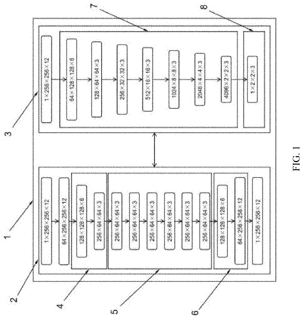
[0035] An auxiliary diagnostic model for detecting acute ischemic stroke, as shown in FIG. 1, comprising the generative adversarial network model 1, and the generative adversarial network model 1 comprises the first three-dimensional convolutional neural network and the second three-dimensional convolutional neural network, the first three-dimensional convolutional neural network is the generator G2 that is used to complete 3D image-to-image conversion, and the second three-dimensional convolutional neural network is the discriminator D3 that is used to distinguish the authenticity of the input images. The generator G2 comprises two first three-dimensional convolutional layers 4 for downsampling, residual blocks 5 and three-dimensional transposed convolutional layers 6 for upsampling. The discriminator D3 comprises second three-dimensional convolutional layers 7 and output layers 8.

[0036]In the embodiment, the auxiliary diagnostic model for detecting stroke is a generative adversari...

embodiment 2

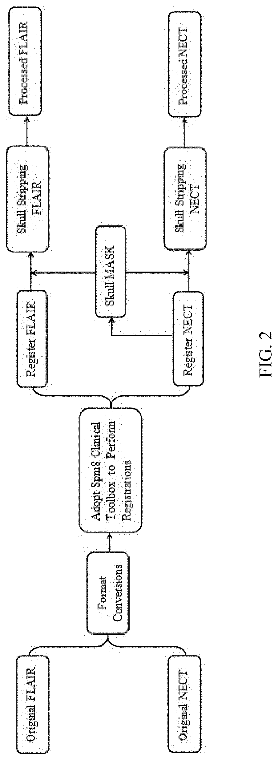
[0043] an image processing method for detecting acute ischemic stroke, as shown from FIG. 2 to FIG. 5, comprising the following steps:

[0044]S1, data normalization, collect NECT images of stroke patients and FLAIR images corresponding to NECT images from the hospital, then make data processing of the collected NECT images and FLAIR images, then make data normalization of the collected NECT images and FLAIR images;

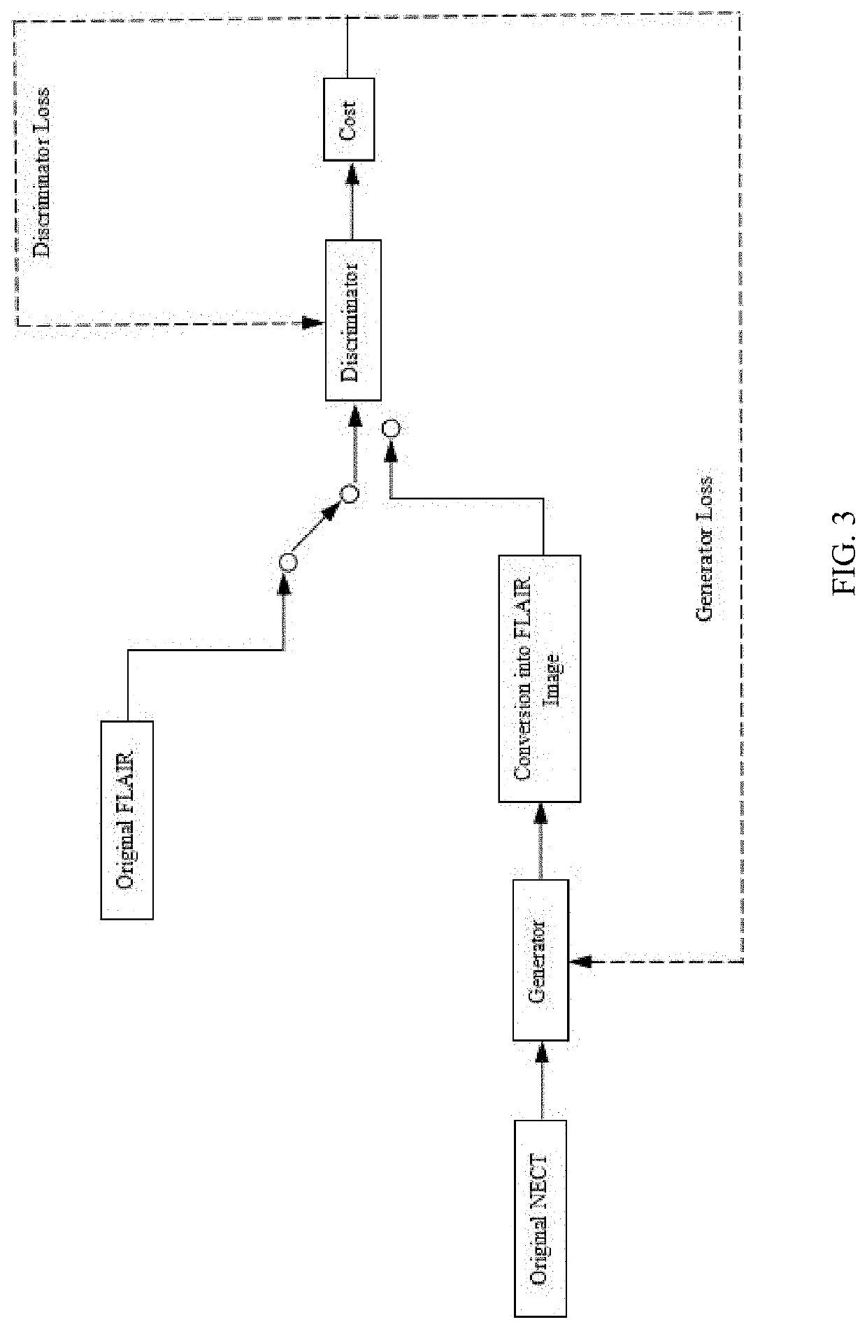
[0045]S2, model creation, create the generator G2 to complete 3D image-to-image conversion and the discriminator D3 to distinguish the authenticity of the input images, and create the generative adversarial network model, the generator G2 and the discriminator D3 are two different three-dimensional convolutional neural networks;

[0046]S3, model training, define the complete training loss of the generative adversarial network model created in step S2 as

G*=arg⁢minG⁢maxD⁢LGAN⁡(G,D)+λ⁢⁢LL⁢⁢1⁡(G),

and train the generative adversarial network model 1, in which, a gradient penalty te...


PUM


Abstract

This invention discloses an auxiliary diagnostic model and an image processing method for detecting acute ischemic stroke. This refers to the technical field of medical image processing. The technical essentials are described as follow: the presented deep-learning model is based on generative adversarial networks (GANs), comprising a generator (G) and a discriminator (D). G is the first three-dimensional convolutional neural network, used to synthesize realistic images from raw data, while D is the second three-dimensional convolutional neural network, used to classify images as real or fake (synthetic). The presented GAN model can learn the mapping relationship from non-enhanced computed tomography (NECT) images to T2-weighted fluid-attenuation inversion recovery (FLAIR) magnetic resonance imaging (MRI), then converting the raw CT to synthetic FLAIR with high sensitivity. This improves the efficiency of emergency scanning in acute ischemic stroke, reaching sensitivity that is poor in CT interpretation and immediacy that is limited in MRI examination.

Description

TECHNICAL FIELD[0001]This invention refers to the technical field of medical image processing, and discloses an auxiliary diagnostic model and an image processing method for detecting acute ischemic stroke.BACKGROUND[0002]Acute ischemic stroke is the most common type of cerebrovascular diseases, and a significant contributor to the global disease burden, bringing a heavy stress and huge consumption to the patients, their families, and the society. Brain assessment of patients with acute ischemic stroke requires both immediacy and sensitivity. In clinical routine, non-enhanced computed tomography (NECT) is the first-line examination in emergency practice. However, it suffers from observer reliability and poor sensitivity to the early small infarction. This easily leads to delayed image interpretation and misdiagnosis, thus affecting timely intervention in the stroke patients.[0003]Magnetic resonance imaging (MRI) holds advantages in detecting small and early cerebral ischemic changes...

Claims


Application Information

Patent Timeline
IPC IPC(8): G06V10/82G06T3/40G06N3/04
CPCG06V10/82G06T3/4046G06T2207/20081G06T2207/20084G06N3/04G16H50/20G16H50/50G16H30/20G06N3/08G06N3/045G06T11/00G16H30/40G06V2201/03G06N3/047
Inventor HU, NALV, SUGU, SHIZHANG, TIANWEI
Owner WEST CHINA HOSPITAL SICHUAN UNIV
Learn More