
Implantable constructs and uses thereof

a technology of implantable constructs and constructs, which is applied in the field of implantable constructs, can solve the problems of fibrosis (walling off), subsequent implant failure, and modest patient response to these approaches, and achieve the effect of improving the efficacy of a therapeutic agent, modulating activation and/or programming

Pending Publication Date: 2022-10-06
RICE UNIV
View PDF0 Cites 0 Cited by
  • Summary
  • Abstract
  • Description
  • Claims
  • Application Information

AI Technical Summary

Benefits of technology

This patent describes implantable constructs that can be used to treat diseases such as cancer. These constructs contain antigenic and therapeutic agents that are protected from the host immune response, allowing for controlled and sustained release of the agents. The implantable constructs can also modulate the response of host cells without inducing their expansion. This technology has the potential to improve efficacy and minimize side effects.

Problems solved by technology

However, in many instances, the percentage of patients responsive to these approaches remain modest (Park et al., Sci. Transl. Med. 10(433) 2018).
A fundamental barrier to successful device-based therapies is the lack of biocompatible transplantation devices.
Implanted biomaterials lead to foreign body responses (FBRs), which are cascades of inflammatory events and wound-healing processes that lead to fibrosis (walling off) and subsequent implant failure.
At present, techniques for achieving the dual goals of FBR and controlled delivery of therapeutic agents are lacking.

Method used


Image

  • Implantable constructs and uses thereof
  • Implantable constructs and uses thereof
  • Implantable constructs and uses thereof

Examples


example 1

neering and Cell Culture

[0100]Reagents. Cell culture media and associated reagents, such as transfection media, were purchased through Fisher Scientific. Lipofection reagents (Lipofectamine 3000®) and selection media (puromycin) were purchased from Invitrogen. Expression vectors and helper plasmids were developed and subsequently purchased from VectorBuilder. CellTiter-Glo® was purchased from Promega. TrypanBlue® and Live Dead® stains were purchased from Fisher Scientific. Alginate compounds were purchased from PRONOVA. The encapsulation apparatus used contains syringe pumps from Harvard Apparatus and co-axial nozzles from Ramé-Hart. All animals were purchased from Jackson Labs. Alginate compounds were purchased through PRONOVA.

[0101]Cell Culture. ID8 / MOSEC cells were obtained commercially from EMD Millipore, Sigma. PAN02 or PANC02 mouse cells were obtained from The Division of Cancer Treatment and Diagnosis (DCTD), National Cancer Institute. Human ARPE-19 cells were obtained from A...

example 2

ation of Production by ELISA

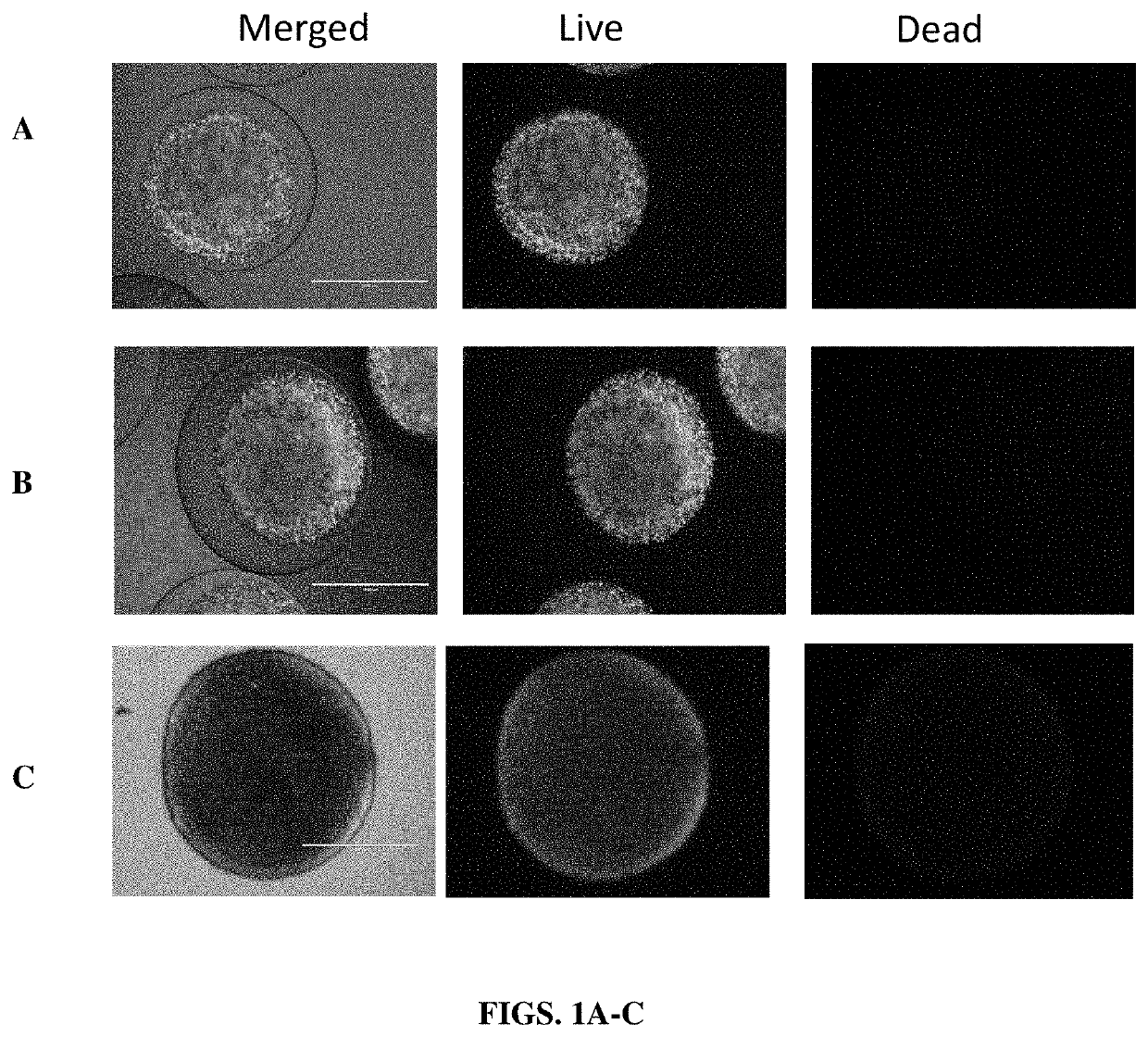
[0106]Cells from each group were grown up into a T-150 flask in DMEM media with 10% FBS, 1% Penstrep, and 0.5 μl / 500 ml puromycin. At confluency, culture media was aspirated and 10 ml TrypLE was added. Cells were incubated for 3 minutes at 37° C. in a 5% CO2 humidified atmosphere. Once the cell layer was dispersed, 10 ml's complete media was added to stop the reaction. The cell suspension was transferred to a 50 ml conical tube and centrifuged at 250G for 5 minutes. The supernatant was aspirated, and cells were re-suspended in 1 ml complete media. Cell concentration was counted using a Countess™ hemocytometer. The volume required to achieve a concentration of 10,000 cells in 200 μl was calculated based on the concentration of live cells in the sample. Of the remaining cell suspension volume, 1 million cells were resuspended in 1 ml alginate to be synthesized into core shell capsules. All groups were incubated for 24 hours at 37° C. in a 5% CO2 humidified ...

example 3

oliferation Assay

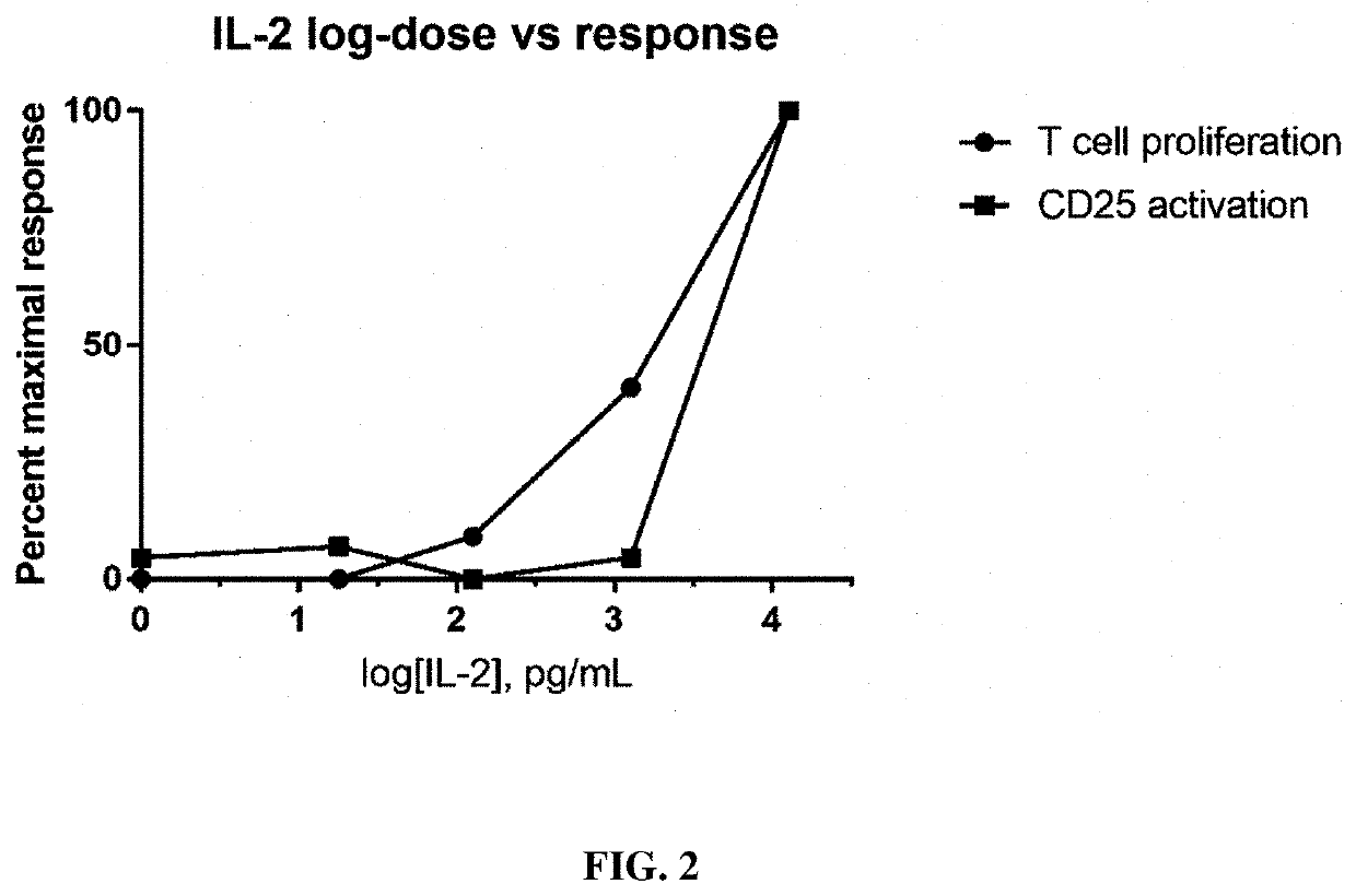
[0107]A EasySep T Cell Isolation Kit (StemCell Technologies) and associated reagents and devices were used to isolate T cell from C57BL6 mouse spleens (Jackson Labs). Cells were plated in 96 well plates at 10,000 cells / well in 100 ul RPMI 1640. Cells were supplemented with 0.1, 1, 5 or 10 ng / mL of cell produced native IL2 or 1 or 10 ng / mL of recombinant IL-2 (Miltenyi). 24 or 72 hours after incubation at 37 C, cell proliferation (and viability) was measured using CellTiter-Glo Luminescent Cell Viability Assay (Promega).


PUM

PropertyMeasurementUnit
timeaaaaaaaaaa
average molecular weightaaaaaaaaaa

Abstract

The present disclosure relates to implantable constructs designed to deliver antigenic therapeutic reagents to a subject while providing protection from host immune responses. In certain aspects, the constructs are designed to degrade over time or upon a particular signal, thereby providing control of the length of time the therapeutic agent is delivered to the subject.

Description

PRIORITY CLAIM[0001]This application is a national phase application under 35 U.S.C. § 371 of International Application No. PCT / US2020 / 045471, filed Aug. 7, 2020, which claims benefit or priority to U.S. Provisional Application Ser. Nos. 62 / 884,397 and 63 / 012,693 filed Aug. 8, 2019 and Apr. 20, 2020, respectively, the entire contents of each application being hereby incorporated by reference.STATEMENT REGARDING FEDERALLY FUNDED RESEARCH[0002]This invention was made with government support under Grant No. R01DK120459, awarded by the National Institutes of Health. The government has certain rights in the invention.BACKGROUNDI. Field[0003]The present disclosure relates to the fields of biology, medicine, bioengineering and medicals devices. More particular, it relates to the development and use of implantable constructs designed to deliver antigenic therapeutic reagents to a subject and protect them from immune responses generated by the host. In particular, the constructs are designed...

Claims


Application Information

Patent Timeline
Patent Type & Authority Applications(United States)
IPC IPC(8): A61K9/00A61K45/06
CPCA61K9/0024A61K45/06A61K38/00A61K35/00A61K9/5036C07K14/54C07K14/5418C07K14/5434C07K14/5428C07K14/5443C07K14/55C12N2510/00C12N5/0012C12N2533/74A61P35/00A61K38/19A61K38/2013A61K38/2046
Inventor VEISEH, OMIDNASH, AMANDAJARVIS-RUOCCO, MARIA ISABELMUKHERJEE, SUDIPDOERFERT, MICHAEL DAVIDAGHLARA-FOTOVAT, SAMIRAZHANG, DAVID YU
Owner RICE UNIV
Learn More