Method for creation of drug delivery and/or stimulation pockets in myocardium

a technology of myocardium and drug delivery, applied in the field of myocardium drug delivery and/or stimulation pockets, can solve the problems of increasing peri- and post-operative bleeding, little success, and difficult to maintain a sufficient concentration of such compounds within the channel

Inactive Publication Date: 2000-05-30
ECLIPSE SURGICAL TECH
View PDF5 Cites 62 Cited by
  • Summary
  • Abstract
  • Description
  • Claims
  • Application Information

AI Technical Summary

Benefits of technology

Broadly, an advantage of the present invention is to provide an apparatus and method for creating pockets within mu

Problems solved by technology

The surgical method through incision into the chest wall does require perforation of the epicardium to create channels through the myocardium and endocardium which may result in increased peri- and post-operative bleeding.
With little success, attempts have been made by practitioners to maintain the patency of the lumen of the channels through administration of appropriate pharmacologically active compounds.
Maintaining a sufficient concentration of such compounds within the channels is ver

Method used


Image

  • Method for creation of drug delivery and/or stimulation pockets in myocardium
  • Method for creation of drug delivery and/or stimulation pockets in myocardium
  • Method for creation of drug delivery and/or stimulation pockets in myocardium

Examples


Embodiment Construction

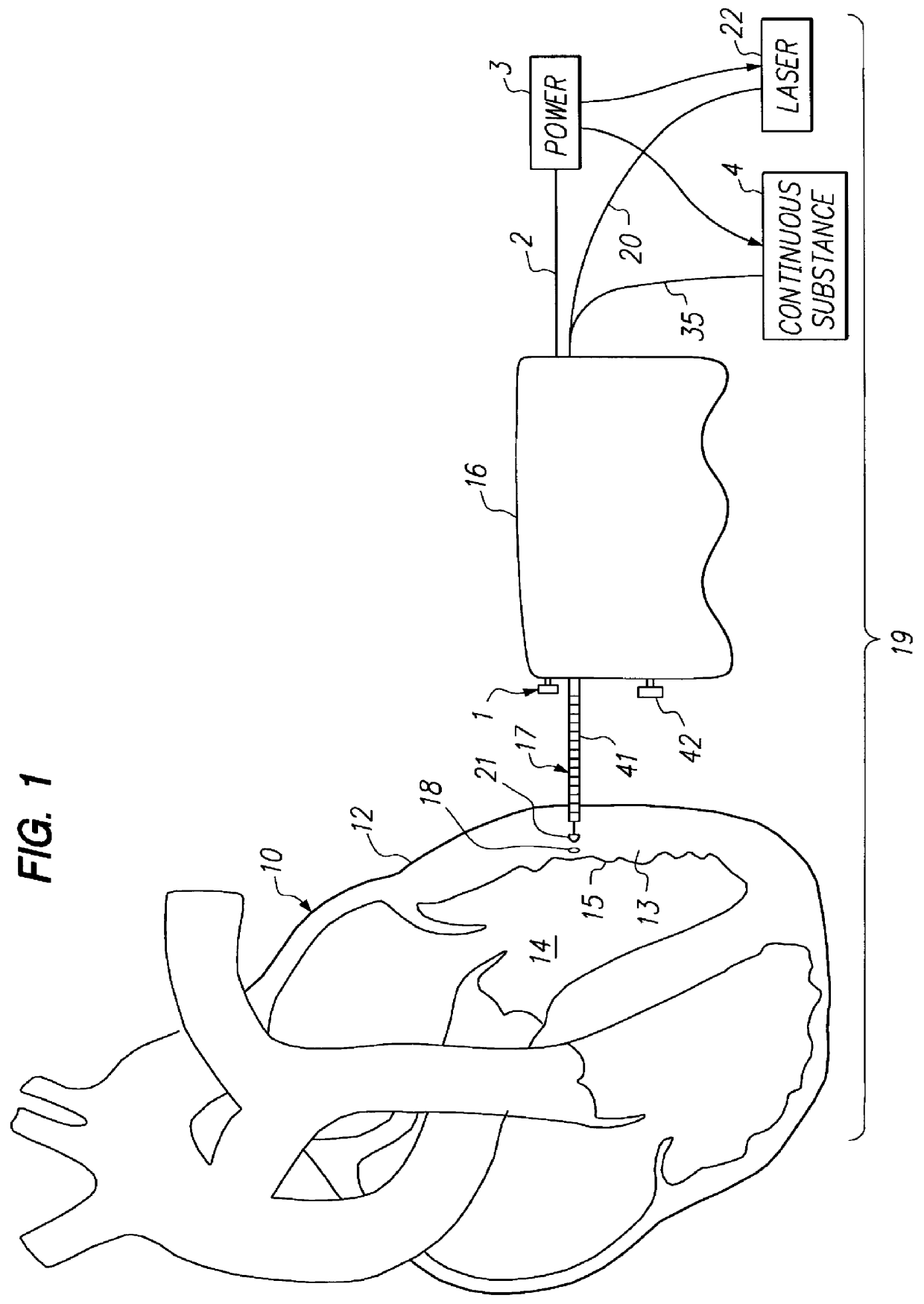
With reference to the drawing, FIG. 1 diagrammatically depicts a human heart with the epicardium 12 of the left ventricle 14 exposed where a stimulation / drug pocket formation procedure according to the invention is to be performed. Preliminary to the procedure, the surgeon makes an incision in the patient's chest to expose the outer wall (epicardium) of the heart's left ventricle. In a human heart, the wall of the left ventricle is comprised of an outer layer, the epicardium 12, the main muscle thickness, the myocardium 13, and the inner layer or endocardium 15. The epicardium is comprised of a smooth moist serous membrane which is somewhat tougher than the other tissue layers of the heart muscle.

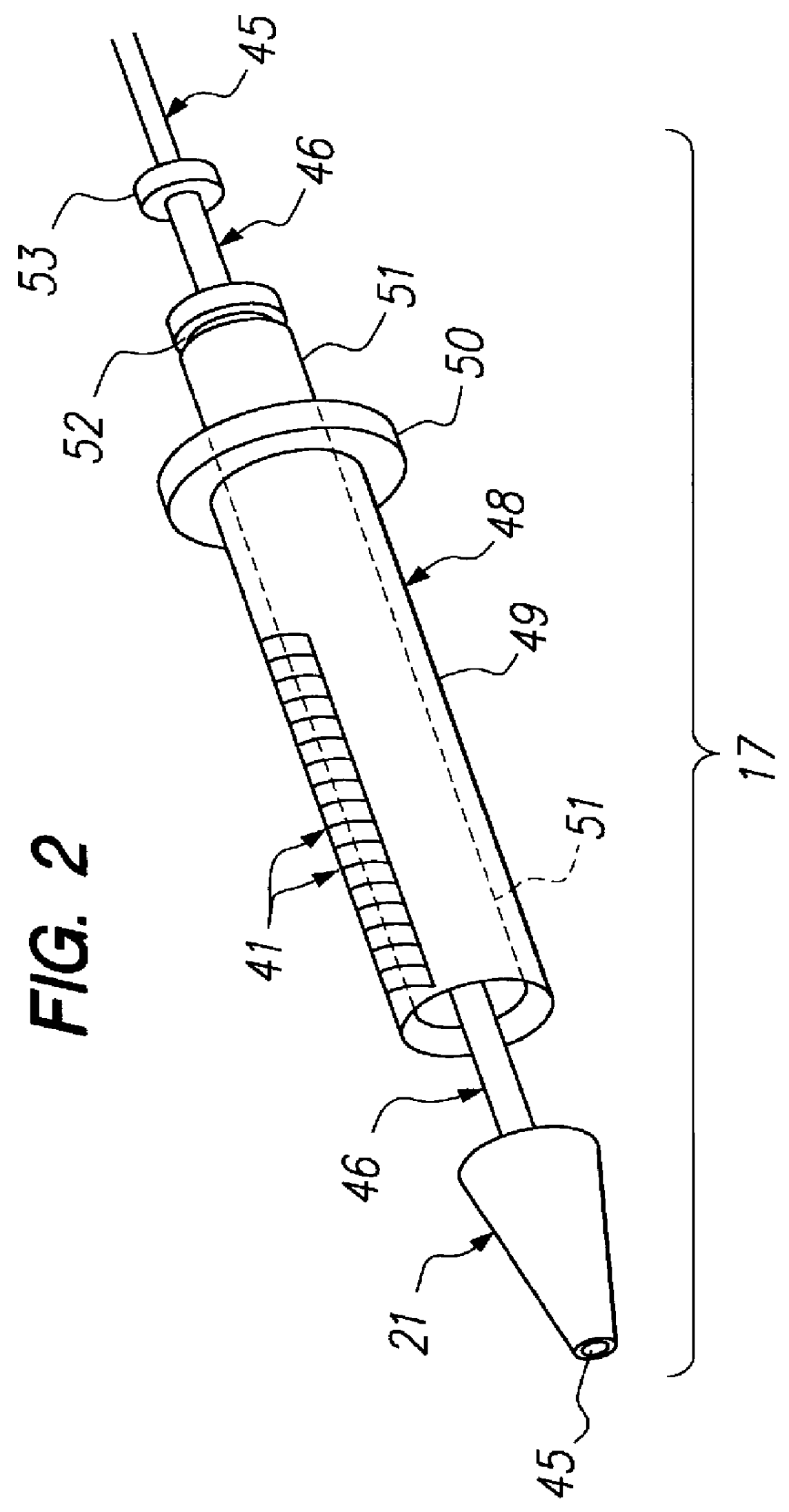
In accordance with the method of the present invention, the surgeon uses a hand-held control device 16 attached to an excising assembly 17 which is manipulated and operated to form a series of drug filled stimulation pockets 18 in the myocardium of the patient's heart at selected spaced apa...


PUM


Abstract

An apparatus and method for creating drug-filled pockets within muscle tissue, such as myocardium of the heart for increasing angiogenesis. More particularly, the apparatus has an excising assembly with a dilator tip for penetrating and advancing through the surface and body of a muscle or organ, such as the heart. Preferably, the dilator tip has a low level laser optical fiber emission to ease the passage of the excising assembly and provide thermal damage which also stimulates angiogenesis. More preferably, the dilator tip also disperses a pharmacologically active substance as the apparatus is passed through the tissue and/or creates pockets. The excising assembly is connected to a hand-held control device from which the operator pushes a switch to activate a punching mechanism within the excising assembly. The punching mechanism cuts a discrete piece of muscle tissue and traps it within the excising assembly leaving a pocket in the remaining muscle tissue. The excising assembly may also optionally release a bolus of the pharmacologically active substance into the pocket so created. Most preferably, there is a timing mechanism to measure the contraction of the heart, and the timing mechanism is synchronized with the operator's switch on the hand-held device to ensure that the punching occurs at maximum contraction of systole. A measurement guide determines how much excised tissue is trapped in the reservoir of the excising assembly. At a threshold level of filling, the surgeon will remove the excising assembly from the hand-held control device and open the punching mechanism for release of tissue. More preferably, the threshold level of filling will automatically turn off the switch to the punching mechanism to indicate to the surgeon the need to empty the excising assembly of tissue.

Description

This invention relates to the field of microsurgery in creating pocket spaces within muscle tissue, and more particularly to creating intramyocardial pockets for the purposes of drug delivery and / or stimulation of angiogenesis of the myocardium of the heart.Various surgical techniques have been developed to counteract ischemic conditions of the heart, including coronary bypass grafts, angioplasty and for patients who are not suitable candidates for these procedures, or in conjunction with these procedures, transmyocardial revascularization (TMR). In TMR generally, the surgeon creates many narrow channels of approximately one millimeter width that span from an opening at the endocardial surface of a ventricle of the heart, preferably the left ventricle, into the myocardium and then terminating before the epicardial surface. The surgeon generally uses laser to create the channels by either accessing the endocardium through a percutaneous route or the epicardium through an incision int...

Claims


Application Information

Patent Timeline
IPC IPC(8): A61B18/04A61B18/08A61B17/22A61B17/00
CPCA61B18/08A61B2018/00392A61B2017/22082A61B2017/00247
Inventor MUELLER, RICHARD L.
Owner ECLIPSE SURGICAL TECH
Why Eureka
Try Eureka