Analyte detection device and process

Inactive Publication Date: 2001-02-20
BAYER CORP
View PDF19 Cites 20 Cited by
  • Summary
  • Abstract
  • Description
  • Claims
  • Application Information

AI Technical Summary

Problems solved by technology

The need for a device using small sample volumes is particularly relevant to the detection of...

Method used


Image

  • Analyte detection device and process
  • Analyte detection device and process
  • Analyte detection device and process

Examples


example 1

Construction of Analyte Detection Device

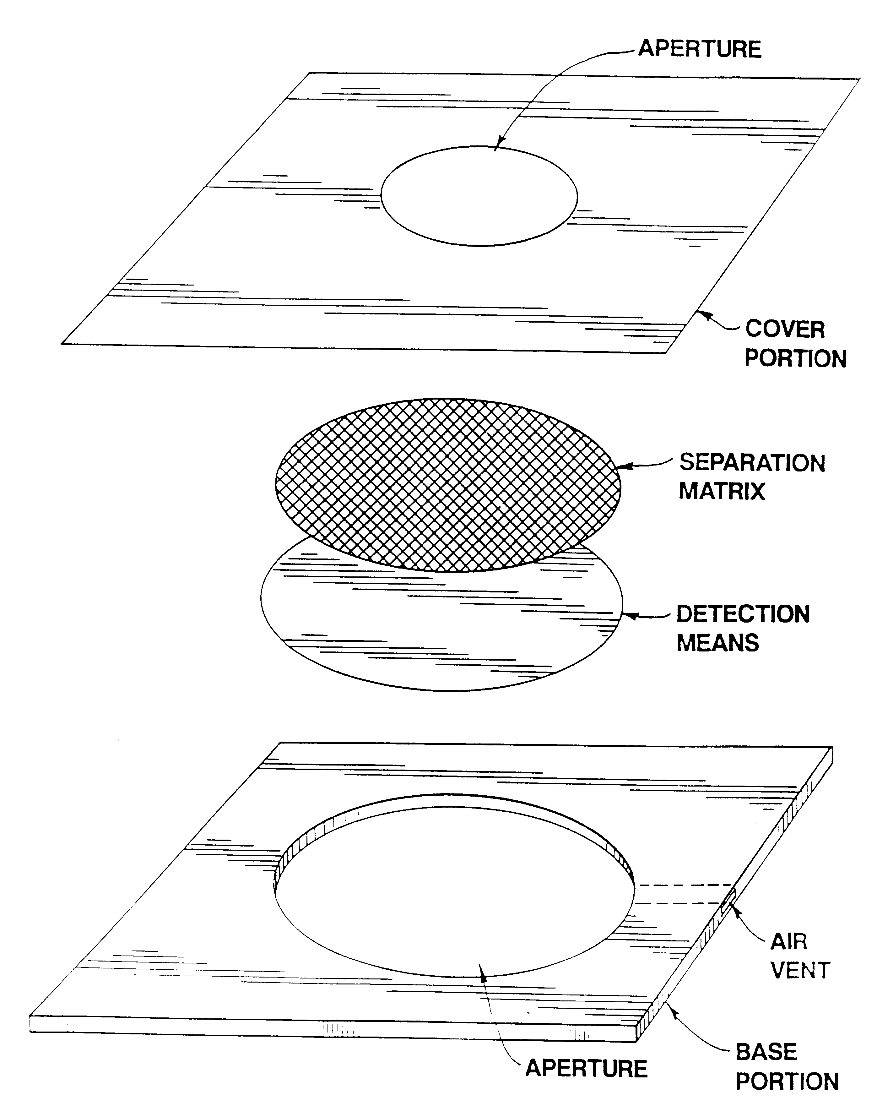
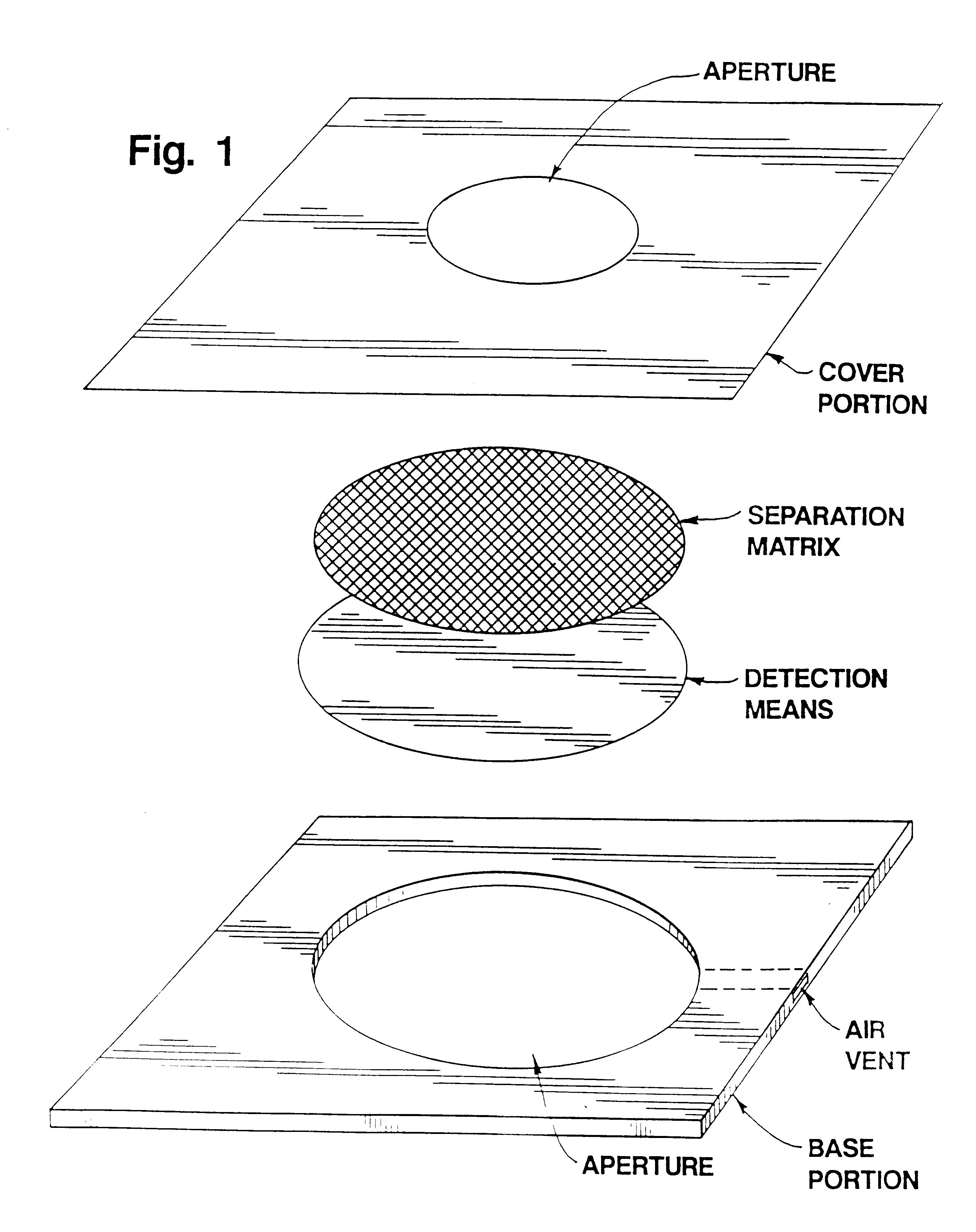
Multiple layers of thin plastic sheets and adhesive tape were used to construct the detection device. The overall thickness of the device was varied by using varying numbers of layers. Such various layers can be adhered to each other using a suitable adhesive. An exemplary and preferred adhesive is double-sided adhesive tape purchased from 3M. The layers of the device were constructed of high density polystyrene sheets (0.006" thick) purchased from American Can.

After adhering a desired number of plastic sheets together, holes were punched through the adhered layers to make a cavity for the separation and detection layers. A clear bottom layer (polycarbonate) was added, the detection and separation layers inserted and a top layer with application site was added to enclose the layers within the device. Polyethylene terephthalate can be used for the top and bottom layers. A schematic diagram of a detection device of the present invention is shown...

example 2

Effect of HEPES Buffer

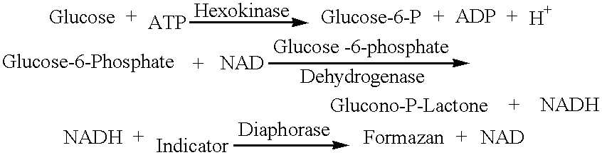
A device was prepared as described in EXAMPLE 1 with the addition that a glass fiber filter, used as the separation layer, contained HEPES buffer, potato lectin and Cremophor EL. The glass fiber filter was impregnated with those substances by soaking the cut filters in an aqueous solution of 150 mM HEPES, pH 7.5, 57,600 Units / L potato lectin and 1.3 percent (w / w) Cremophor EL. After soaking, the fibers were removed from the solution and dried in an oven at 50.degree. C. for about 15 minutes.

A synthetic membrane fabricated of nylon (Biodyne A, 0.45 micron pore size, PALL Biosupport, Long, Island, N.Y.) was impregnated with ATP, NAD, hexokinase, glucose-6-phosphate (G-6-P) dehydrogenase, diaphorase and NBT (a tetrazolium salt, Sigma Chem. Co.) and employed as the detection layer. Impregnation was accomplished by soaking the synthetic membrane in an aqueous solution having the following final concentrations:

5.5% (w / w) Tetrazolium indicator (e.g. NBT)

1% (w / w) NAD

4....

example 3

Other Analytes

A detection device for other analytes can be prepared using the procedures set forth above. The following impregnation solutions are exemplary of solutions that can be used to impregnate detection matrices for use with the detection of cholesterol, triglyceride and amylase.


PUM


Abstract

The present invention provides a device and a process for detecting an analyte in a biological fluid. The device comprises a separating matrix for separating analyte from the fluid and means for detecting the analyte, where the separating matrix contains HEPES buffer, preferably in an amount between 70 and 150 millimolar. The process comprises applying the sample to the device having a separating matrix and then detecting analyte in the sample using the detection means. The presence of HEPES in the separation layer shortened the detection time.

Description

The present invention relates to a device and a process for detecting an analytes in a biological fluid. A device comprises a separating matrix for separating an analyte from a sample fluid, where the separating matrix contains HEPES buffer. A process comprises applying a sample fluid to a device having a separating matrix and then detecting an analyte in the sample using detection means in a detection layer adjacent to the separating matrix.DESCRIPTION OF THE BACKGROUND ARTThere is a need in the end point detection art for devices and methods that allow for the rapid, accurate determination of analyte (e.g., glucose, triglyceride, cholesterol, etc.) concentration in very small sample volumes of biological fluid. The need for a device using small sample volumes is particularly relevant to the detection of blood glucose because of the difficulty and inconvenience of obtaining blood samples from subjects.Currently available such devices include ACCUCHEK EASY TEST STRIPS.TM. and ACCUTR...

Claims


Application Information

Patent Timeline
IPC IPC(8): C12Q1/54G01N33/52G01N31/22G01N33/66
CPCC12Q1/54G01N33/525Y10T436/25375Y10T436/144444
Inventor CHU, AMY H.WILCOX, MICHAEL J.
Owner BAYER CORP
Why Eureka
Try Eureka