
Method for detecting neuronal degeneration and anionic fluorescein homologue stains therefor

a technology of anionic fluorescein and neuronal degeneration, which is applied in the field of postmortem detection and localization of degenerated neurons in brain slices, can solve the problems of prone to false positives, shrunken and hyperchromatic cells, and difficult to demonstrate this phenomenon, etc., and achieves simple and reliable use, easy to interpret, and high sensitive

Inactive Publication Date: 2001-05-08
SCHMUED LAURENCE C
View PDF4 Cites 16 Cited by
  • Summary
  • Abstract
  • Description
  • Claims
  • Application Information

AI Technical Summary

Benefits of technology

The principal object of the present invention is to provide an improved histochemical tracer which is simple and reliable to use as well as highly sensitive and easy to interpret.
A further object of the present invention is to provide an improved histochemical tracer of the foregoing character which is simple to use and produces reliable results.
The present invention is embodied in an anionic fluorochrome capable of selectively staining degenerating neurons in brain slices, the method of making the anionic fluorochrome, and the method of using the same for detecting neuronal degeneration. The histochemical application of this fluorochrome results in a simple, sensitive and reliable method for staining degenerating neurons and their processes. The technique will detect neuronal degeneration resulting from exposure to a variety of neurotoxic insults. The fluorochrome can be combined with other fluorescent methodologies including immunofluorescence, fluorescent axonal tract tracing, and fluorescent Nissl counterstaining. Compared to conventional methodologies, the fluorochrome of the present invention is a more sensitive and definitive marker of neuronal degeneration than conventional H&E or Nissl type stains, while being comparably sensitive yet considerably simpler and more reliable than suppressed silver techniques.

Problems solved by technology

The demonstration of this phenomenon, however, is often difficult from both a technical and an interpretational point of view.
Unfortunately, such changes are not necessarily indicative of neuronal degeneration and may be due to processing artifacts or non-lethal alterations in cellular morphology.
Processing artifacts can result in both shrunken and hyperchromatic cells.
Not only are such techniques prone to false positives, but it is also possible to miss degenerating neurons since all cells stain with these dyes while only relatively subtle morphological differences exist between normal and degenerating neurons.
These interpretational difficulties make the analysis of conventionally stained material time consuming.
The main drawbacks associated with suppressed silver techniques lie in their labor intensive and capricious nature.

Method used


Image

  • Method for detecting neuronal degeneration and anionic fluorescein homologue stains therefor
  • Method for detecting neuronal degeneration and anionic fluorescein homologue stains therefor
  • Method for detecting neuronal degeneration and anionic fluorescein homologue stains therefor

Examples


Embodiment Construction

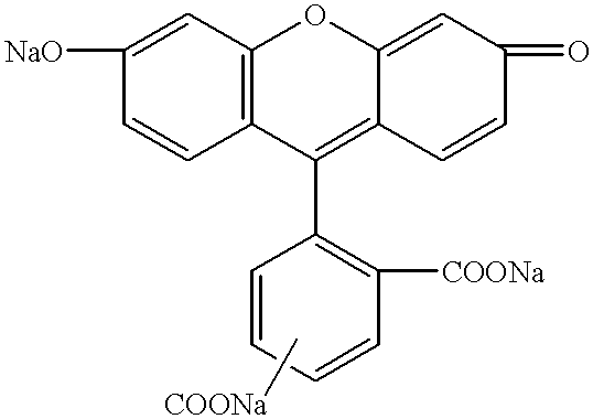
The two anionic fluorescein based fluorescent stains embodying the present invention are 1) the water soluble, tribasic salt of a mixture of the fluorescein isomers 5-carboxyfluorescein and 6-carboxyfluorescein and 2) the water soluble tetrabasic salt of 5,6 dicarboxyfluorescein. The two preferred fluorescent stains of the present invention are the trisodium fluorescein salt, trisodium 5 / 6-carboxyfluorescein, which has the following structure: ##STR1##

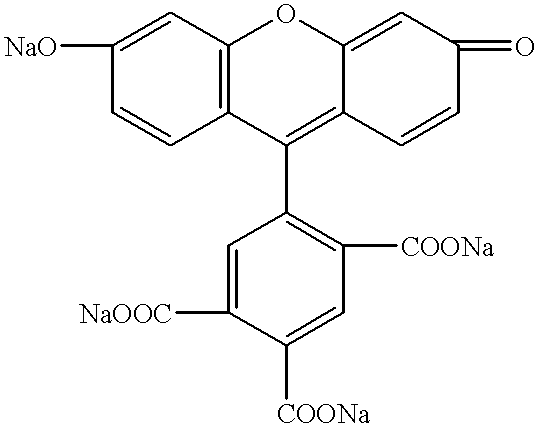
And the tetrasodium 5,6-tricarboxyfluorescein which has the following structure: ##STR2##

The abovetrisodium 5,6-carboxyfluorescein and tetrasodium 5,6-carboxyfluorescein both provide a simple, sensitive, and reliable new method for demonstrating and localizing neuronal degeneration.

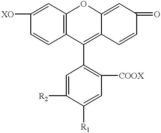
The respective tribasic and tetrabasic salts of 5 / 6-carboxyfluorescein and 5,6-dicarboxyfluorescein embodying the present invention may be shown by the following general formula: ##STR3##

wherein R.sub.1 and R.sub.2 =--COOX, or --H (provided that both do not=...


PUM


Abstract

An anionic fluorescein homologue capable of selectively staining degenerating neurons in brain slices, the method of making the anionic fluorescein homologue, and the method of using the same for detecting neuronal degeneration. Two anionic fluorescein based fluorescent stains are the water soluble tribasic salt of a mixture of the fluorescein isomers 5-carboxyfluorescein and 6-carboxyfluorescein, and the water soluble tetrabasic salt of 5,6 dicarboxyfluorescein.

Description

1. Field of the InventionThe present invention relates to a method for the post-mortem detection and localization of degenerating neurons in brain slices, and more particularly to anionic fluorescent type homologue therefor.2. Prior ArtMany neuroscientists including toxicologists, pathologists, pharmacologists, anatomists and histochemists share an interest in detecting neuronal degeneration. The demonstration of this phenomenon, however, is often difficult from both a technical and an interpretational point of view. Conventional techniques such as H&E (hematoxylin and eosin), or Nissl type stains (crystal violet, thionin, etc.) are technically simple and can be used to infer degeneration based on changes such as neuronal shrinkage, vacuolation, and hyperchromatism. Chassan, J. L., "Nervous System" in W. A. D. Anderson and J. M. Kissane (Eds.), Pathology, Mosby, St. Louis, 1977, pp. 2074-2148; Garcia, J. H. and Kamijiyo, Y., "Cerebral Infraction: Evolution of Histopathological Chang...

Claims


Application Information

Patent Timeline
IPC IPC(8): C07D311/80C07D311/00C09B11/08C09B11/00G01N1/30G01N33/68
CPCC07D311/80C09B11/08G01N1/30G01N33/6896G01N2001/302
Inventor SCHMUED, LAURENCE C.
Owner SCHMUED LAURENCE C
Learn More