Methods for detecting chromosomal aberrations using chromosome-specific paint probes
a technology of chromosomes and paint probes, applied in the field of methods for detecting chromosomal aberrations using chromosome-specific paint probes, can solve the problems of low resolution of methods, small rearrangements, including some deletions and duplications, and inability to detect small changes
- Summary
- Abstract
- Description
- Claims
- Application Information
AI Technical Summary
Benefits of technology
Problems solved by technology
Method used
Image
Examples
example i
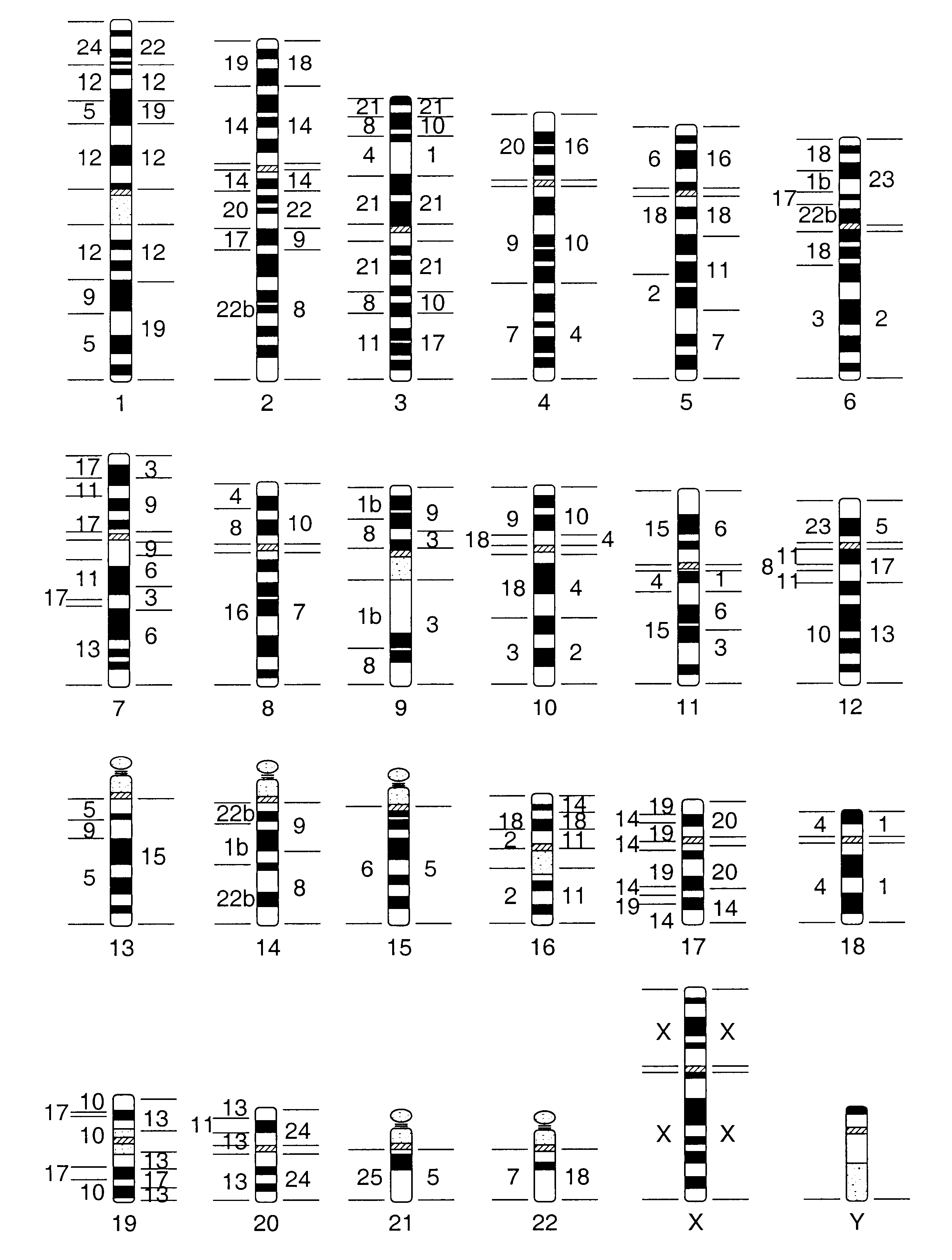
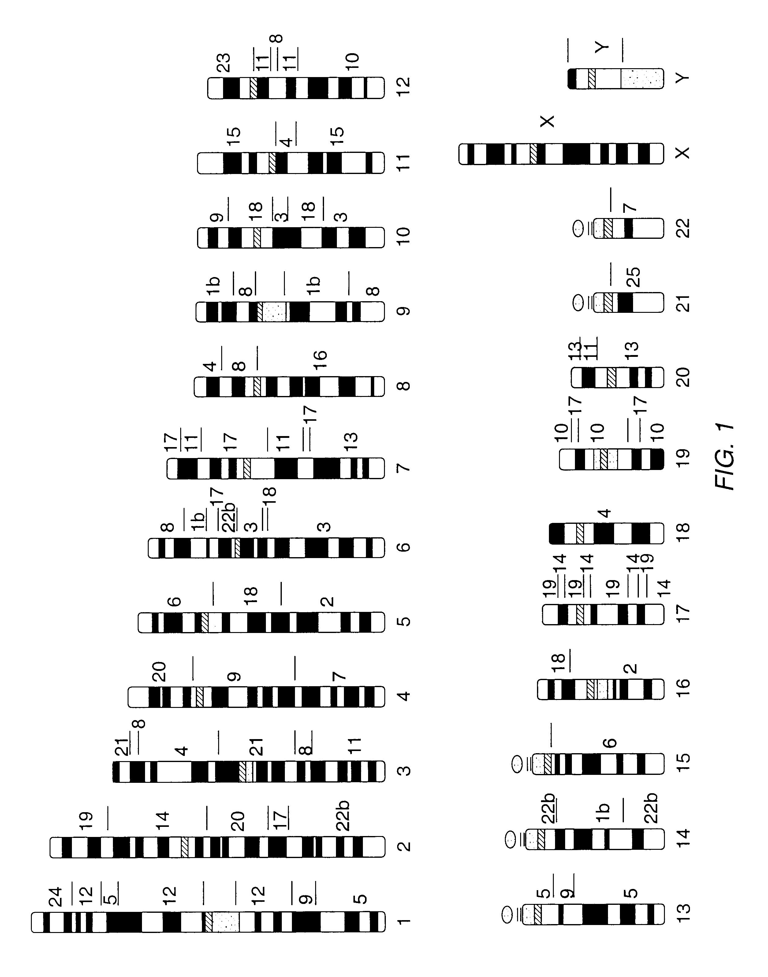
Metaphase spreads from peripheral blood lymphocytes and lymphoblastoid cell lines were prepared and blood samples were obtained from a normal human male and from clinical cases showing chromosome abnormalities. A lymphoblastoid cell line was obtained from a male Concolor gibbon (Hylobates concolor, 2n=52). Flow sorting of concolor gibbon (HCO) chromosomes was performed on a dual laser cell sorter (FACStar Plus, Becton Dickinson ImmunoCytometry Systems). Bivariate flow cytometry yielded 25 peaks which were identified by comparison of their hybridization patterns to homologies revealed by human chromosome specific paints on HCO chromosomes. This revealed that single chromosome-specific paints could be obtained from 17 chromosomes (Table 1). Four peaks contained two chromosomes (HCO X+5, HCO 9+11, HCO 12+15, and HCO 6+7). Chromosome numbering followed that of Koehler et al., 1995, Genomics 30:287-92. Three hundred to five hundred chromosomes of each type were amplified by a preliminary...
example ii
a) Preparation and Characterization of Chromosome-Specific Probes
Using FACS and subsequent DOP-PCR, chromosome-specific probes from Hylobates concolor and H. syndactylus chromosomes were prepared. Briefly, gibbon chromosomes were sorted using a dual laser flow sorting system as described for other species (Rabbitts et al., 1995, Nature Genet. 9:369-75; Ferguson-Smith, 1997, Europ. J. Hum. Genet. 5:253-65) Chromosome specific probes were generated by degenerate oligonucleotide primed PCR (DOP-PCR) directly from the flow sorted chromosomes using PCR primers and amplification conditions as described (Telenius et al., 1992, Genes, Chromosomes Cancer 4:257-63).
To identify the content of each peak in the flow karyotype, hapten labeled probes derived from the individual peaks were hybridized to the DAPI banded chromosomes of the particular species. FIG. 2 shows the flow karyotype of the two species. Many peaks contained single chromosomes only, however, especially in H. syndactylus, a numb...
example iii
This example describes the use of an H. concolor CSC-banding probe in human karyotyping. CSC-banding probe is prepared as described in Example I, supra, and labeled according to the scheme of Table 2. The probe is suspended, typically at a concentration of about 10-30 nanograms per microliter of each chromosome-specific probe, in formamide hybridization buffer (50% formamide, 10% dextran sulfate, 2.times.SSC, 40 mM NaPO4 (pH 7), 1.times.Denhardt's Solution (Ausubel et al., 1997, CURRENT PROTOCOLS IN MOLECULAR BIOLOGY, Greene Publishing and Wiley-Interscience, New York), 120 ng / .mu.l salmon sperm DNA).
Metaphase preparations are prepared from a human lymphocyte cell culture using standard methods. The cell suspension is gently mixed and a drop applied to the slide by dropping from approximately one-inch above the slide. A drop of freshly prepared 4.degree. C fixative methanol:glacial acetic acid (3:1) is added and the slide is allowed to air dry. Following examination of the metaphase...
PUM
| Property | Measurement | Unit |
|---|---|---|
| concentration | aaaaa | aaaaa |
| volume | aaaaa | aaaaa |
| temperature | aaaaa | aaaaa |
Abstract
Description
Claims
Application Information
 - R&D
- Intellectual Property
- Life Sciences
- Materials
- Tech Scout
- Unparalleled Data Quality
- Higher Quality Content
- 60% Fewer Hallucinations
Browse by: Latest US Patents, China's latest patents, Technical Efficacy Thesaurus, Application Domain, Technology Topic, Popular Technical Reports.
© 2025 PatSnap. All rights reserved.Legal|Privacy policy|Modern Slavery Act Transparency Statement|Sitemap|About US| Contact US: help@patsnap.com


