

Method of estimating skeletal status
a skeletal status and status technology, applied in the field of estimating skeletal status, can solve the problems of significant affecting fracture risk, disproportinately large loss of biomechanical competence, overlap in bmd measurement,
- Summary
- Abstract
- Description
- Claims
- Application Information
AI Technical Summary
Benefits of technology
Problems solved by technology
Method used
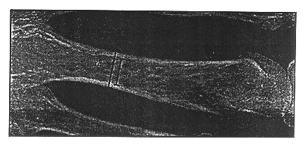
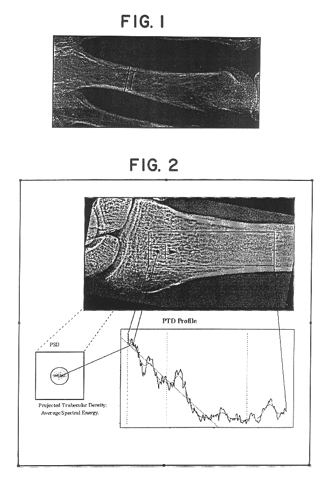
Image

Examples
example 2
Another Preferred Embodiment
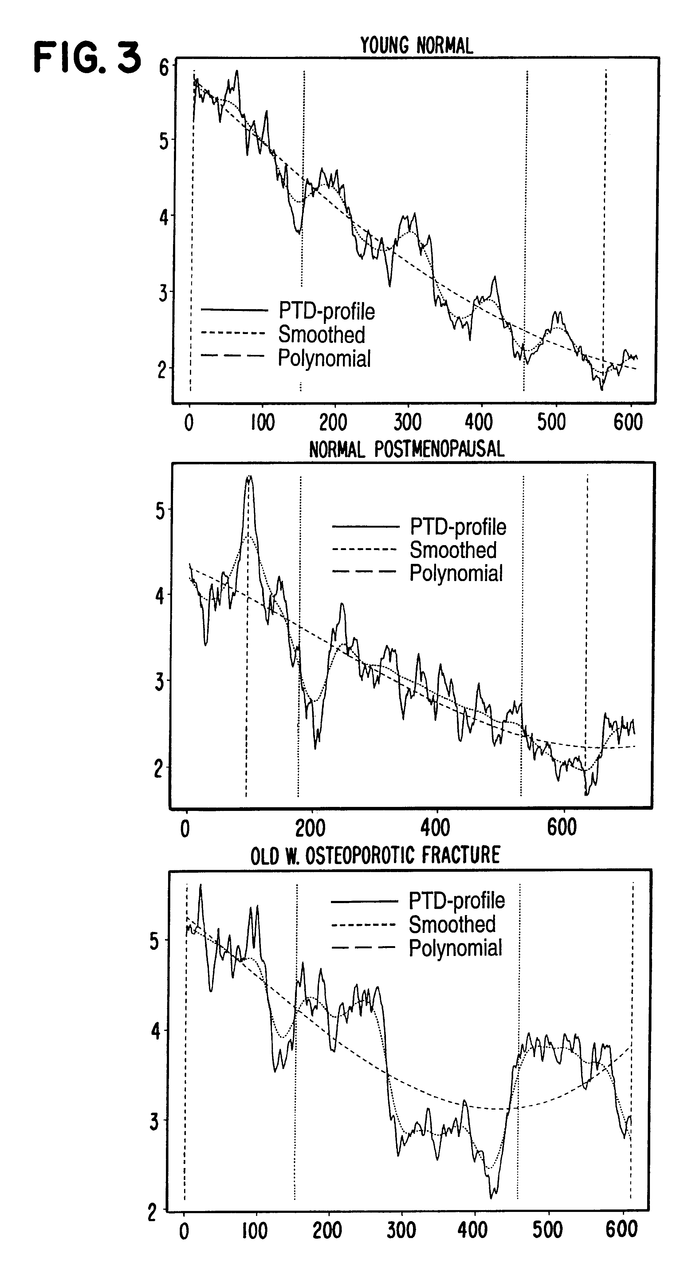
Another method of deriving features from the graph defined by the PTD values may be to calculate the morphological opening or closing of the graph or of a smoothed version thereof and to subsequently derive the standard deviation between those two graphs. From this standard deviation, information relating to the skeletal status may be derived.
Using the following notation
* PTD:
Original PTD-profile
* smooth(x,wsiz):
A function that smooths the profile, x, with a moving average of size wsiz
* open(x, struc, ntim):
A function that performs a morphological opening of the profile x using a structural element of the size struc. The opening is performed as ntim erosions followed by ntim dilations, using the structural element specified.
* close(x, struc, ntim):
A function that performs a morphological closing of the profile x using a structural element of the size struc. The opening is performed as ntim dilations followed by ntim erosions, using the structural element ...
example 3
A Third Preferred Embodiment
In this example, an embodiment of a background correction is described wherein information relating to the absorption of cortical bone is obtained for the area wherein the PTD profile is determined. This information may be used for subtracting information relating to the absorption of cortical bone from the image data and, thus, for generating information relating to the absorption of the trabecullar bone in the same area.
In FIG. 5, a hand radiograph is illustrated wherein the positions of determining the CCT (four metacarpal bones and the distal radius) are illustrated. From these assessed thicknesses and the absorption caused thereby, as determined from the image data, a relation of cortical thickness and caused absorption may be derived. The absorption caused by the determined thickness of cortical bone is preferably determined at the part of the bone where it is the narrowest and at center of the bone at the position in question.
In FIG. 6, the distal ...
PUM
 Abstract
Description
Claims
Application Information