Method and apparatus for safety delivering medicants to a region of tissue using imaging, therapy and temperature monitoring ultrasonic system

a technology of ultrasonic system and medical device, which is applied in the direction of ultrasonic/sonic/infrasonic image/data processing, ultrasonic/sonic/infrasonic diagnostics, therapy, etc., can solve the problems of limited use of surface treatment or invasive procedures, poor control of energy deposition, and high absorption of medicants in tissu

Inactive Publication Date: 2003-09-23
GUIDED THERAPY SYSTEMS LLC
View PDF39 Cites 312 Cited by
  • Summary
  • Abstract
  • Description
  • Claims
  • Application Information

AI Technical Summary

Problems solved by technology

In contrast with ultrasound, RF energy is characterized by long wavelengths in tissue, with limited to poor control of energy deposition, and high absorption.
On the other hand, the optical energy which is typically emitted from lasers can be precisely controlled in beamwidth, but the opacity and high absorption in tissue also limits its use to surface treatment or invasive procedures.
Furthermore, laser and RF energy are emitted from ionizing radiation sources which are typically associated with some risk, unlike acoustic transducers which are typically used for generating ultrasound.
However, in contrast with the diagnostic uses, the therapeutic uses of ultrasound such as hyperthermia and non-invasive surgery have seen relatively little progress due to several technological barriers.
The primary impediment has been a lack of the ability to monitor temperature in the treatment region during the therapeutic treatment process.
However, the thermocouple probes are highly invasive because they have to be inserted into the region-of-interest.
In addition, use of the thermocouple probes has necessarily led to very poor spatial resolution since only a small number of probes could be safely embedded in the region-of-interest.
Furthermore, the embedded thermocouple probes are likely to disturb the acoustic propagation in the tissue and typically cause excessive heating at the probe interface during the therapeutic treatment process.
This results in an undesirably modified temperature distribution as well as erroneous measurements.
Another factor which has curtailed progress in the therapeutic uses of ultrasound has been the design of the conventional acoustic transducers.
However, the conventional acoustic transducers which are typically employed for therapeutic purposes are acoustically large, often single-element devices having narrow bandwidth in the frequency domain.
Although they are designed to efficiently transmit acoustic energy to the target tissue, the conventional acoustic transducers are typically unsuited for imaging of the treatment region and / or monitoring the temperature profile therein.
This precludes development and implementation of these vital functions for performing a desirable precise therapeutic treatment process.
The system disclosed therein, however, does not include temperature monitoring of the target tissue (treatment region).
Consequently, an actual real-time monitoring of the temperature may be difficult in the disclosed method and apparatus.
Moreover, although the method and apparatus can detect changes in the temperature of the target, an absolute value of the target temperature may not be obtained therefrom.
In addition, any movement of the target may introduce changes in the differential image, which may cause erroneous results.
While some prior art temperature monitoring techniques exist, see, for example, U.S. Pat. No. 4,807,633 issued to Fry on Feb. 28, 1989, such techniques are complex and have limited applicability.
That is, use of such techniques essentially preclude use of the system for purposes of imaging, unless one were to use multiple transducers.
In that regard, while two or more physically separated transducers can be used to accomplish imaging and therapy, typically with one configured for imaging and the other for therapy, such a-system is susceptible to the generation of imprecise data and is overly complex and expensive.
For example, U.S. Pat. No. 5,149,319 issued to Linger discloses a method for heat treating biological tissues which includes administering a therapeutically effective amount of a hyperthermic potentiator to the tissue and then applying ultrasound to heat the tissue to a temperature of at least about 43 degrees C. Although the potentiators disclosed in this method result in making the hyperthermic ultrasound a more selective and more effective therapeutic method for treating tissue, there is no inclusion of a monitoring means to safeguard against potential overheating and its resulting tissue damage.
Further, it should be noted that the hyperthermic potentiators are used to increase the acoustic heterogeneity and generate cavitation nuclei in tumors and tissues, and that this cavitation effect can be harmful to the tissue.
This method also fails to provide means for monitoring the temperature of tissue in order to prevent tissue damage that can occur from overheating.
Although these references disclose the use of ultrasound therapy with drug containing liposomes to effect the treatment of tissue, these references do not disclose the use of imaging and temperature monitoring functions to facilitate treatment planning and feedback to ensure the safe treatment of the tissue.
Further, these references do not disclose heat treatment of the liposomes themselves, nor the use of heat sensitive liposomes.

Method used


Image

  • Method and apparatus for safety delivering medicants to a region of tissue using imaging, therapy and temperature monitoring ultrasonic system
  • Method and apparatus for safety delivering medicants to a region of tissue using imaging, therapy and temperature monitoring ultrasonic system
  • Method and apparatus for safety delivering medicants to a region of tissue using imaging, therapy and temperature monitoring ultrasonic system

Examples


Embodiment Construction

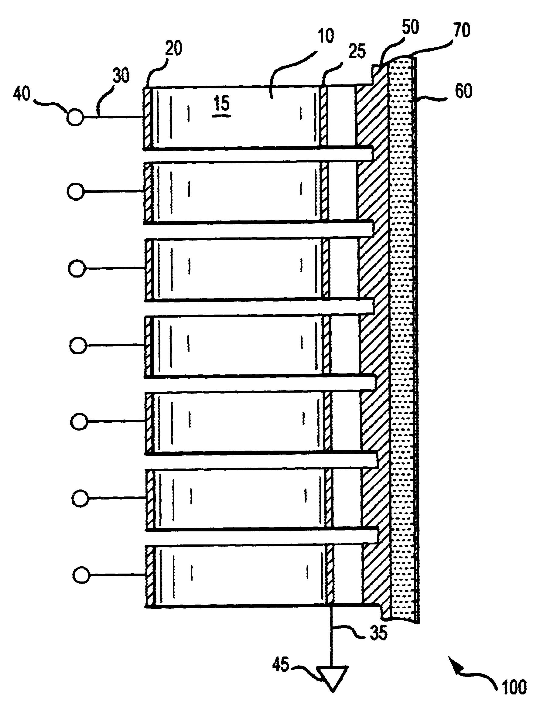
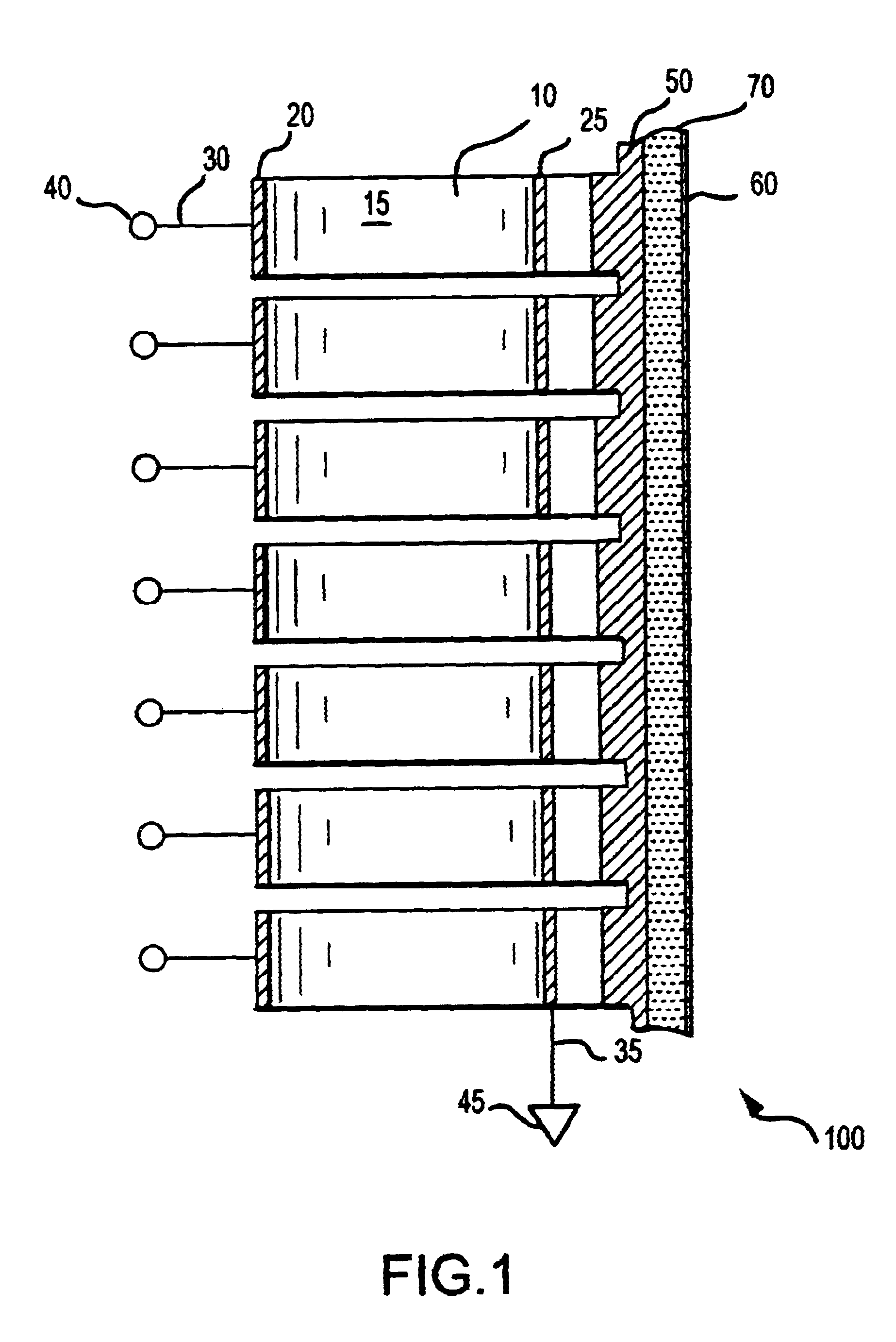
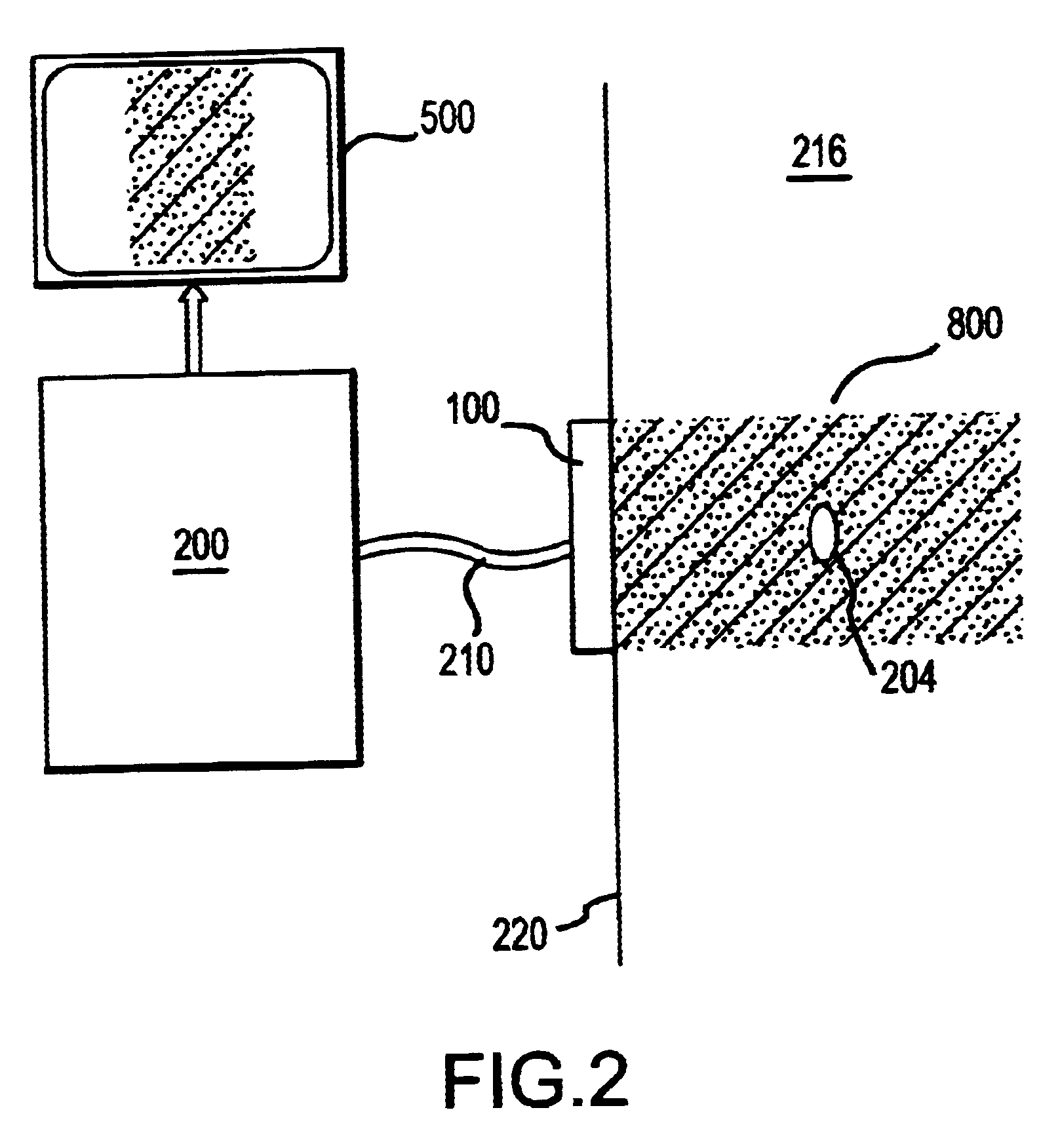
A system for achieving successful ultrasonic therapy procedures in accordance with the present invention includes four major subsystems or components. Specifically, they are an acoustic transducer assembly, an imaging subsystem, a therapy subsystem (also referred to as a "therapeutic heating subsystem"), and a temperature monitoring subsystem, which are illustrated in FIGS. 1 through 4, respectively. Although not shown in the drawing figures, the system further includes components typically associated with a therapy system, such as any required power sources, memory requirements, system control electronics, and the like.

With reference to FIG. 1, the acoustic transducer assembly 100 included in the system of the present invention will be described in detail below. As shown in the cross-sectional view of FIG. 1, the acoustic transducer assembly 100 includes a piezoelectric ceramic plate 10. The air-backed side of the ceramic plate 10 may be partially diced to have a plurality of curve...


PUM


Abstract

A method and apparatus for controlling the safe delivery of thermosensitive liposomes containing medicant to a targeted tissue region using ultrasound. Thermosensitive liposomes containing medicants are delivered to a region of interest, the region of interest is located using ultrasound imaging, ultrasound therapy is applied to heat the region of interest, and the temperature of the region is monitored to determine whether a designated threshold temperature has been reached which allows for the release of medicants from the liposomes. If the threshold temperature is reached, and the liposomes are melted, the treatment stops. If the threshold temperature has not been reached, the application of ultrasound therapy and ultrasound imaging are alternated until the threshold temperature is reached. The ultrasound imaging, temperature monitoring and ultrasound therapy are preferably performed with a single transducer.

Description

1. Field of the InventionThis invention generally relates to a non-invasive therapeutic ultrasonic system, and more particularly, to a system which is capable of acoustically imaging and heating a certain region to be treated ("the treatment region") in target tissue for therapeutic purposes as well as acoustically monitoring the temperature profile of the treatment region. Further, the present invention relates to a method and apparatus for safely treating a region of body tissue using a single transducer and control unit to monitor and image a temperature profile of the tissue, and using the same transducer and control unit to melt a heat-activated, liposome encapsulated medicant disposed within the tissue.2. Description of the Related ArtThe absorption of energy in tissue, for example, in the human body produces an increase in temperature, which can be exploited for therapeutic purposes. The irradiation of ultrasound to the target tissue such as in the human body, which has been ...

Claims


Application Information

Patent Timeline
Patent Type & Authority Patents(United States)
IPC IPC(8): A61B8/08A61N7/02A61N7/00A61B17/22
CPCA61B5/01A61B8/0833A61B8/5223A61N7/02A61B2017/22028G16H50/30
Inventor SLAYTON, MICHAEL H.BARTHE, PETER G.LISHKO, VALERYI
Owner GUIDED THERAPY SYSTEMS LLC
Why Eureka
Try Eureka