
Glue-on tissue mount

a tissue mount and glue technology, applied in the field of surgery, can solve the problems of difficult to cut, trim, split or divide smaller than thumbnail size pieces of biologic tissue when detached, irregular shape of small tissue pieces, and difficulty in holding, distorting or stretching the shape, etc., to improve the visualization of tissue

Inactive Publication Date: 2003-11-25
WONG IRA G
View PDF5 Cites 5 Cited by
  • Summary
  • Abstract
  • Description
  • Claims
  • Application Information

AI Technical Summary

Benefits of technology

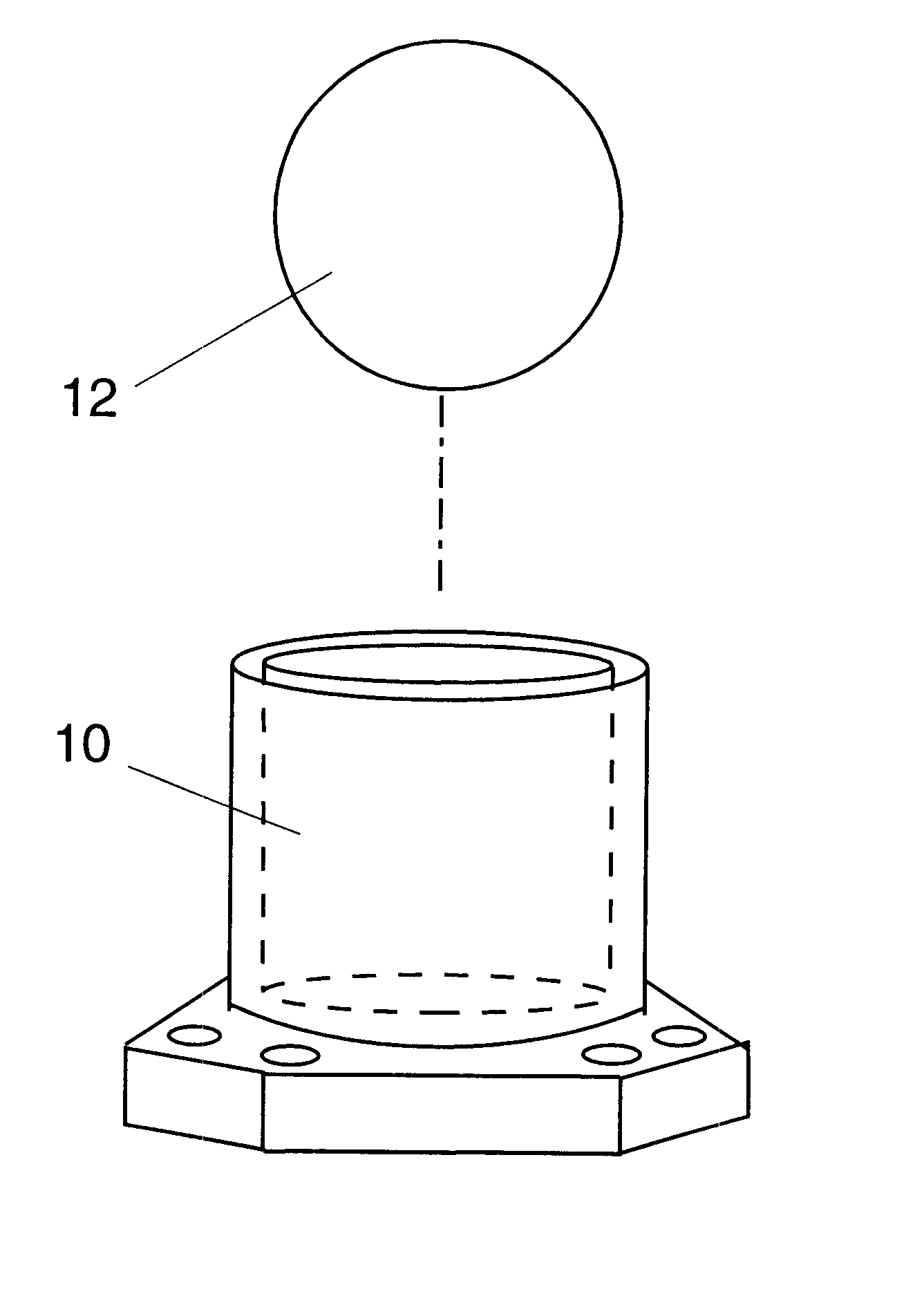
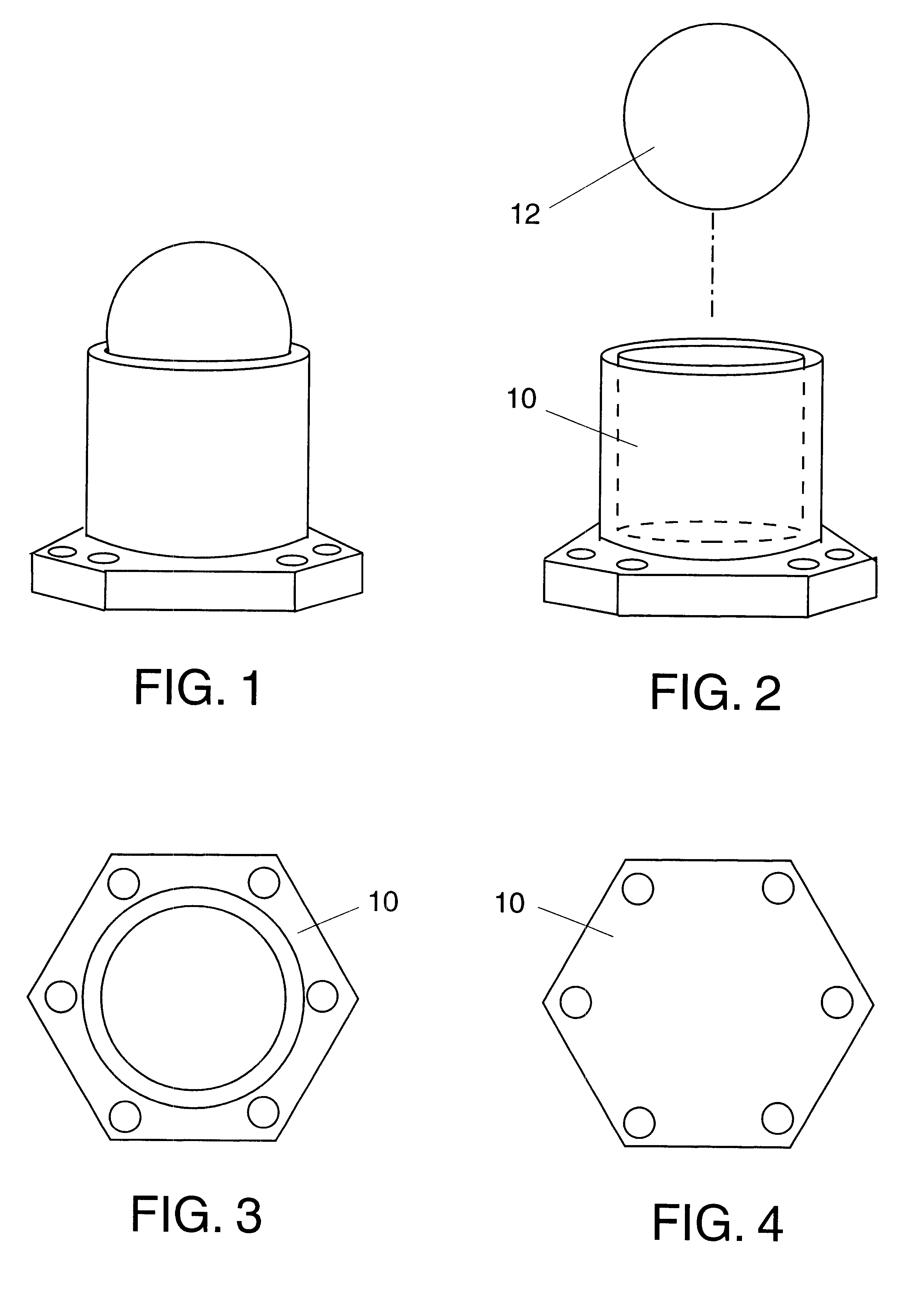
The base 10 can be grasped with fingers or stand freely on a flat surface under an operating microscope. The base 10 can be rotated or tilted on one its hexagonal edges to improve visualization of the tissue. The hexagonal-shaped footing of the base 10 prevents rocking when the apparatus is tilted for observation or manipulation.

Problems solved by technology

Cutting, trimming, splitting or dividing smaller than thumbnail size pieces of biologic tissue is difficult when detached.
A small piece of tissue may be irregularly shaped and difficult to hold.
Just holding tissue with a pair of forceps may distort or stretch the shape and affect the accuracy and precision of a cut.
Holding and shaping a small piece of skin is awkward.
Forceps holding tissue may obstruct or distort the cutting or trimming.
Holding a small piece of tissue under an operating microscope is awkward.
As the surgeon dissects and pulls against the forceps the tissue distorts and stretches, an undesirable effect.
Without the support of the rest of the eye, donor cornea tissue is difficult to hold.
The needles interfere with the cutting.
Both procedures are awkward, time-consuming and potentially damaging to the donor tissue.
The ball does not provide a stable base for cutting.

Method used


Image

  • Glue-on tissue mount
  • Glue-on tissue mount
  • Glue-on tissue mount

Examples


Embodiment Construction

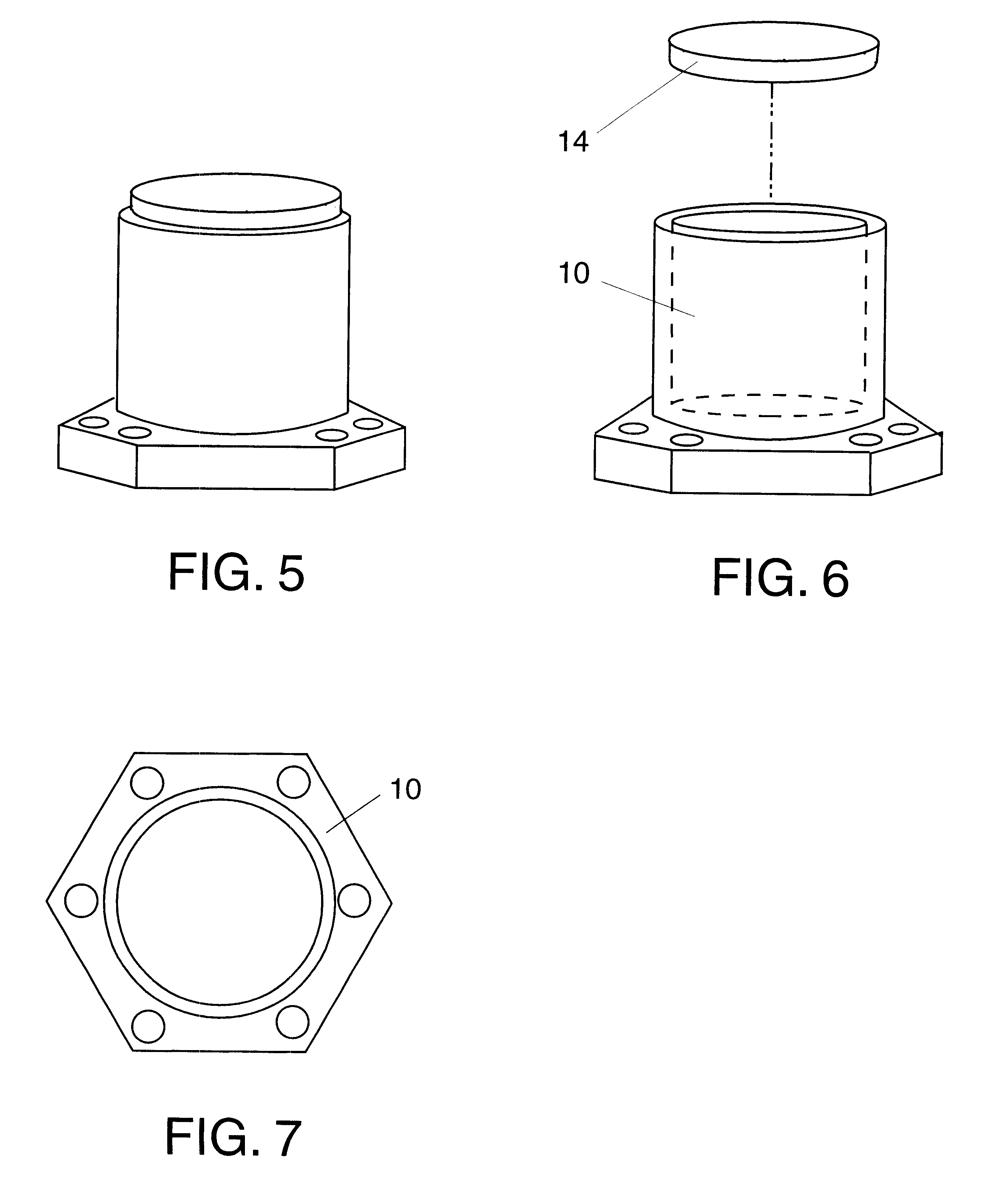
FIGS. 6, 7, 8

Depending on the size and shape of tissue, gluing a tissue onto a flat surface may be preferred to a spherical surface. For tissue like skin, a flat mounting surface is preferred. For donor cornea tissue a spherical mounting surface is preferred.

FIG. 5 shows the mounting block with a flat cutting surface and FIG. 6 shows an exploded view. The flat cutting surface is an acrylic disk 14 attached to the base 10 as illustrated in FIG. 5. FIG. 7 is a top view. The bottom view is a duplicative view of the first embodiment, FIG. 4.

The operation of the flat surface mount is the same as the spherical mount. A small amount of cyanoacrylate glue is applied to the tissue and the tissue is set onto the flat acrylic surface. A minute later the tissue is ready to be manipulated, cut, trimmed, divided or split.

CONCLUSION, RAMIFICATIONS AND SCOPE OF INVENTION

Thus the reader will see that the glue-on tissue mount provides a simple, quick method to perform precise and accurate cutting, tr...


PUM

PropertyMeasurementUnit
sizeaaaaaaaaaa
operating microscopeaaaaaaaaaa
areaaaaaaaaaaa

Abstract

The glue-on tissue mount is a device to hold small and / or irregular-shaped biologic tissue so tissue can be manipulated, cut, trimmed, split or divide for transplant and reconstructive surgery. Biologic tissue is quickly mounted onto an acrylic mount with fast-bonding cyanoacrylate glue. On the mount tissue can be held firmly. The tissue can be cut on the mount like on a cutting board or dissected into layers. The mount can rest on flat surfaces and be used with an operating microscope or held between fingers. The glue-on tissue mount has a spherical acrylic mounting surface 12 or flat acrylic mounting surface 14 upon which the tissue is glued. The size and shape of the tissue determines if a spherical or flat mount is used. The mounting surface is attached to a cylindrical base 10 that is easily held. If the glue adhesion breaks the tissue can be anchored with sutures tied to the eyelets in the hexagonal footing of the base 10. The glue-on tissue mount can be used for cutting a variety of biologic tissue or biologic material.

Description

FEDERALLY SPONSORED RESEARCHNot ApplicableSEQUENCE LISTING OR PROGRAMNot ApplicableBACKGROUND--FIELD OF INVENTIONThis invention relates to surgery, specifically a medical device to improve the method of cutting, trimming, splitting or dividing biologic tissue.BACKGROUND--DISCUSSION OF PRIOR ARTCutting, trimming, splitting or dividing smaller than thumbnail size pieces of biologic tissue is difficult when detached. A small piece of tissue may be irregularly shaped and difficult to hold. Biologic soft tissue is malleable. Just holding tissue with a pair of forceps may distort or stretch the shape and affect the accuracy and precision of a cut.In transplant and reconstructive surgery tissue is removed, altered and replaced. When a tissue is removed it may be cut, trimmed, split or divided before it is attached.For example, in grafting skin, the skin graft is trimmed to fits its future site. Holding and shaping a small piece of skin is awkward. Forceps holding tissue may obstruct or dis...

Claims


Application Information

Patent Timeline
Patent Type & Authority Patents(United States)
IPC IPC(8): A61B19/00A61F2/14A61B17/00G01N1/28G01N1/36
CPCA61B17/00491A61B19/00A61F2/14G01N2001/2873G01N1/36A61B2017/00969A61F2/142A61B90/00
Inventor WONG, IRA G.
Owner WONG IRA G
Learn More