
Measurement of cardiac output and blood volume by non-invasive detection of indicator dilution

a non-invasive detection and cardiac output technology, applied in the field of cardiac output and blood volume measurement by non-invasive detection of indicator dilution, can solve the problem of minimal invasiveness, achieve the effect of improving system sensitivity or accuracy, reducing signal to noise, and improving system sensitivity

Inactive Publication Date: 2004-06-29
ALFRED E MANN INST FOR BIOMEDICAL ENG AT THE UNIV OF SOUTHERN CALIFORNIA
View PDF15 Cites 68 Cited by
  • Summary
  • Abstract
  • Description
  • Claims
  • Application Information

AI Technical Summary

Benefits of technology

Such minimally invasive procedures are advantageous over other methods of evaluating the cardiovascular system. First, complications and patient discomfort caused by the procedures are reduced. Second, such practical and minimally invasive procedures are within the technical ability of most doctors and nursing staff, thus, specialized training is not required. Third, this minimally invasive methods may be performed at a patient's bedside or on an out-patient basis. Finally, methods may be used on a broader patient population, including patients whose low risk factors may not justify the use of central arterial measurements of cardiovascular parameters.
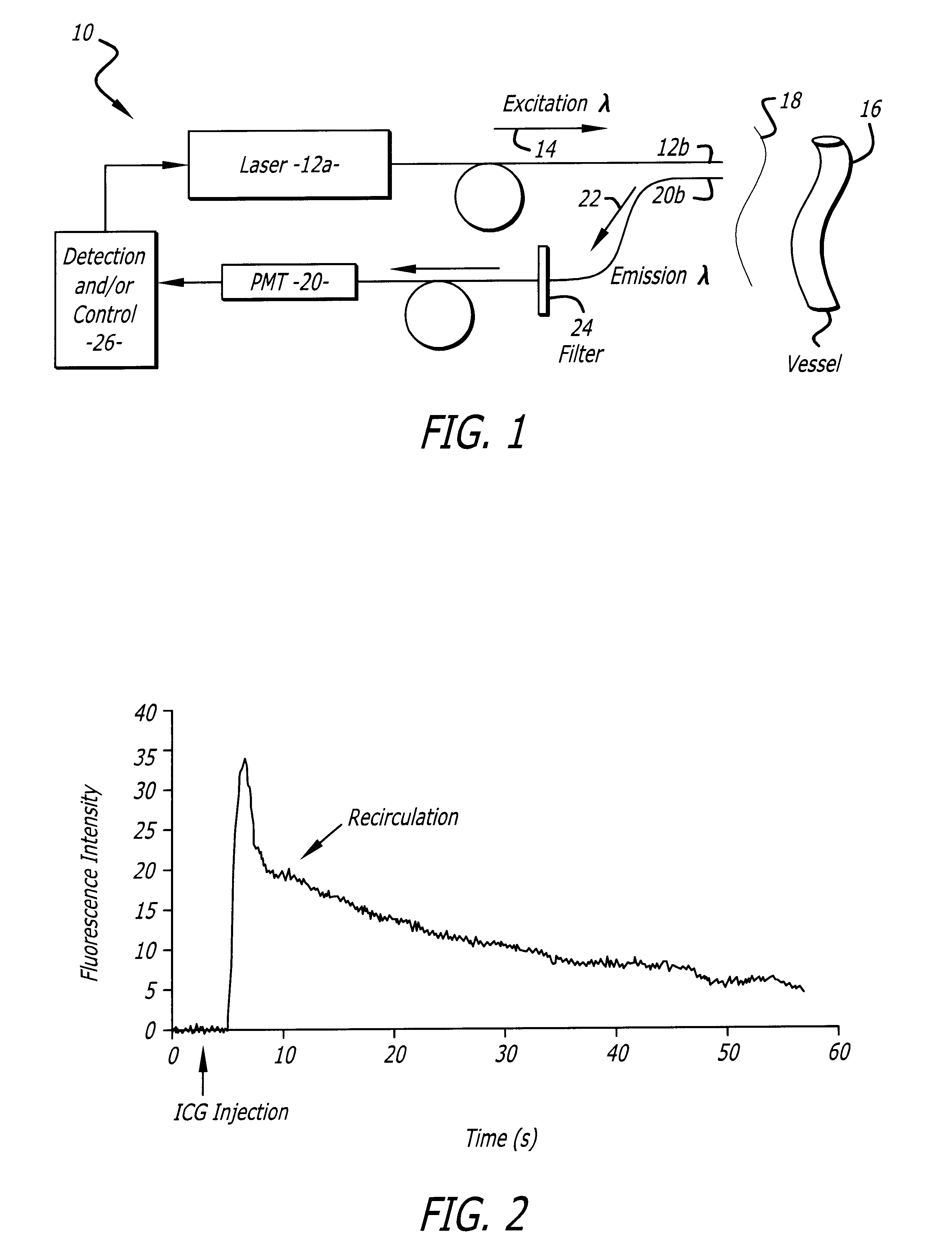
In some embodiments a photomultiplier tube is utilized as or operably connected with another photodetector to enhance the sensitivity of the system. Finally, in some embodiments, additional features, such as filters, may be utilized to minimize the background of the emission signals detected. For example, a filter may be selected which corresponds to the peak wavelength range or around the peak wavelength range of the indicator emission.

Problems solved by technology

Third, this minimally invasive methods may be performed at a patient's bedside or on an out-patient basis.
Finally, methods may be used on a broader patient population, including patients whose low risk factors may not justify the use of central arterial measurements of cardiovascular parameters.

Method used


Image

  • Measurement of cardiac output and blood volume by non-invasive detection of indicator dilution
  • Measurement of cardiac output and blood volume by non-invasive detection of indicator dilution
  • Measurement of cardiac output and blood volume by non-invasive detection of indicator dilution

Examples


example 2

A. A Sample Method and System for Measuring Cardiac Output and Blood Volume.

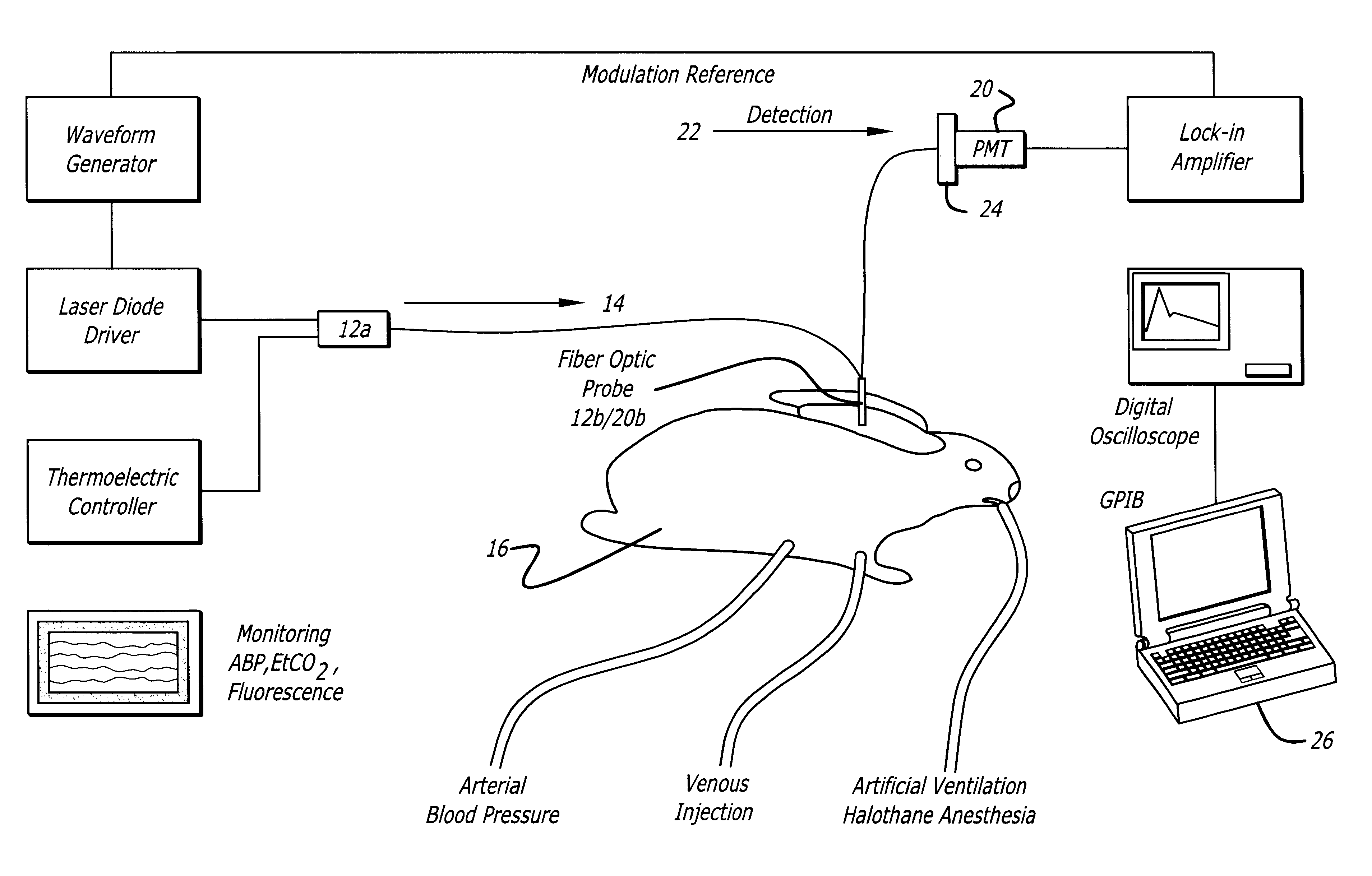
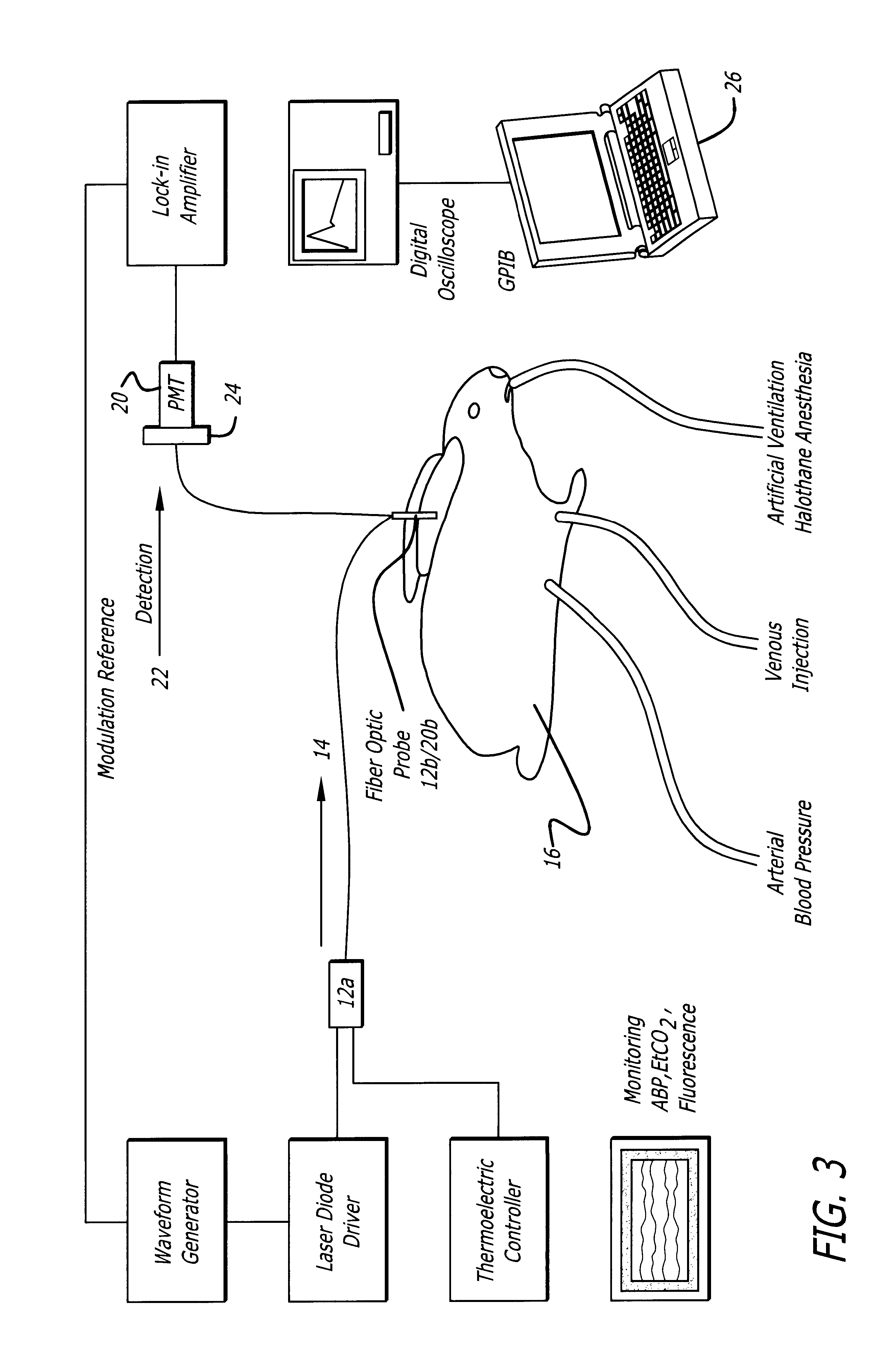
Experiments have been performed in New Zealand White rabbits (2.8-3.5 Kg) anesthetized with halothane and artificially ventilated with an oxygen-enriched gas mixture (FiO2.about.0.4) to achieve a Sa2above 99% and an end-tidal CO2between 28 and 32 mm Hg (FIG. 4). The left femoral artery was cannulated for measurement of the arterial blood pressure throughout the procedure. A small catheter was positioned in the left brachial vein to inject the indicator, ICG. Body temperature was maintained with a heat lamp.

Excitation of the ICG fluorescence was achieved with a 780 nm laser (LD head: Microlaser systems SRT-F780s.sup.-12) whose output was sinusoidally modulated at 2.8 KHz by modulation of the diode current at the level of the laser diode driver diode (LD Driver: Microlaser Systems CP 200) and operably connected to a thermoelectric controller (Microlaser Systems: CT15W). The near-infrared light output was forwa...


PUM


Abstract

A system for evaluating the cardiovascular system parameters using indicator dilution and non-invasive or minimally invasive detection methods is disclosed. Intravascular indicators are stimulated, and emission patterns detected for computation of cardiac output, cardiac index, blood volume and other indicators of cardiovascular health.

Description

1. Field of the InventionThis invention pertains to the detection of parameters of cardiovascular system of a subject.2. General Background and State of the ArtCardiac output is a central part of the hemodynamic assessment in patients having heart disease, acute hemodynamic compromise or undergoing cardiac surgery, for example. Cardiac output is a measure of the heart's effectiveness at circulating blood throughout the circulatory system. Specifically, cardiac output (measured in L / min) is the volume of blood expelled by the heart per beat (stroke volume) multiplied by the heart rate. An abnormal cardiac output is at least one indicator of cardiovascular disease.The current standard method for measuring cardiac output is the thermodilution technique (Darovic, G. O. Hemodynamic monitoring: invasive and noninvasive clinical application. 2nd Ed. W. B. Saunders, 1995). Generally, the technique involves injecting a thermal indicator (cold or heat) into the right side of the heart and det...

Claims


Application Information

Patent Timeline
Patent Type & Authority Patents(United States)
IPC IPC(8): A61B5/026A61B5/029A61B5/0275
CPCA61B5/029A61B5/0261A61B5/0275
Inventor RUBINSTEIN, EDUARDO H.SCREMIN, OSCAR V.HOLSCHNEIDER, DANIEL P.MAAREK, JEAN-MICHEL I.
Owner ALFRED E MANN INST FOR BIOMEDICAL ENG AT THE UNIV OF SOUTHERN CALIFORNIA
Learn More