Apparatus for treating a tumor or the like and articles incorporating the apparatus for treatment of the tumor

a tumor and apparatus technology, applied in the field of tumor apparatus for treatment, can solve the problems of limiting the therapeutic effect of such treatments, reducing the ability to distinguish between tumor cells and normal cells, and not being able to achieve meaningful stimulatory effects

Inactive Publication Date: 2005-03-15
NOVOCURE GMBH
View PDF10 Cites 207 Cited by
  • Summary
  • Abstract
  • Description
  • Claims
  • Application Information

AI Technical Summary

Benefits of technology

A major use of the present apparatus is in the treatment of tumors by selective destruction of tumor cells with substantially no affect on normal tissue cells, and thus, the exemplary apparatus is described below in the context of selective destruction of tumor cells. It should be appreciated however, that for purpose of the following description, the term “cell” may also refer to a single-celled organism (eubacteria, bacteria, yeast, protozoa), multi-celled organisms (fungi, algae, mold), and plants as or parts thereof that are not normally classified as “cells”. The exemplary apparatus enables selective destruction of cells undergoing division in a way that is more effective and more accurate (e.g., more adaptable to be aimed at specific targets) than existing methods. Further, the present apparatus causes minimal damage, if any, to normal tissue and, thus, reduces or eliminates many side-effects associated with existing selective destruction methods, such as radiation therapy and chemotherapy. The selective destruction of dividing cells using the present apparatus does not depend on the sensitivity of the cells to chemical agents or radiation. Instead, the selective destruction of dividing cells is based on distinguishable geometrical characteristics of cells undergoing division, in comparison to non-dividing cells, regardless of the cell geometry of the type of cells being treated.
The movement of the cellular organelles towards the bridge disrupts the cell structure and results in increased pressure in the vicinity of the connecting bridge membrane. This pressure of the organelles on the bridge membrane is expected to break the bridge membrane and, thus, it is expected that the dividing cell will “explode” in response to this pressure. The ability to break the membrane and disrupt other cell structures can be enhanced by applying a pulsating alternating electric field that has a frequency from about 50 KHz to about 500 KHz. When this type of electric field is applied to the tissue, the forces exerted on the intercellular organelles have a “hammering” effect, whereby force pulses (or beats) are applied to the organelles numerous times per second, enhancing the movement of organelles of different sizes and masses towards the bridge (or neck) portion from both of the sub-cells, thereby increasing the probability of breaking the cell membrane at the bridge portion. The forces exerted on the intracellular organelles also affect the organelles themselves and may collapse or break the organelles.

Problems solved by technology

Thus, unfortunately, the sensitivity of tumor cells is not sufficiently higher than that many types of normal tissues.
This diminishes the ability to distinguish between tumor cells and normal cells, and therefore, existing cancer treatments typically cause significant damage to normal tissues, thus limiting the therapeutic effectiveness of such treatments.
Furthermore, the inevitable damage to other tissue renders treatments very traumatic to the patients and, often, patients are unable to recover from a seemingly successful treatment.
Also, certain types of tumors are not sensitive at all to existing methods of treatment.
The applied electrical current destroys substantially all cells in the vicinity of the target tissue.
Thus, this type of electrical method does not discriminate between different types of cells within the target tissue and results in the destruction of both tumor cells and normal cells.
These currents can be calculated by Ohm's law and can have dangerous stimulatory effects on the heart and CNS and can result in potentially harmful ion concentration changes.
Also, if the currents are strong enough, they can cause excessive heating in the tissues.
This limitation is due to the fact that excitation induced by electric stimulation is normally mediated by membrane potential changes, the rate of which is limited by the RC properties (time constants on the order of 1 ms) of the membrane.
Regardless of the frequency, when such current inducing fields are applied, they are associated with harmful side effects caused by currents.
For example, one negative effect is the changes in ionic concentration in the various “compartments” within the system, and the harmful products of the electrolysis taking place at the electrodes, or the medium in which the tissues are imbedded.
An increase in [Ca++] is harmful to most cells and if sufficiently high will lead to the destruction of the cells.
In view of the above observations, long term current application to living organisms or tissues can result in significant damage.
Another major problem that is associated with such electric fields, is due to the electrolysis process that takes place at the electrode surfaces.
These can cause significant damage to organic molecules, especially macromolecules and thus damage the living cells and tissues.
Therefore, the dominant field effect is that due to dielectric losses and heating.
One disadvantage of this method is that the ability to discriminate is highly dependent upon cell type, for example, the size difference between normal cells and tumor cells is significant only in certain types of cells.
Another drawback of this method is that the voltages which are applied can damage some of the normal cells and may not damage all of the tumor cells because the differences in size and membrane dielectric properties are largely statistical and the actual cell geometries and dielectric properties can vary significantly.

Method used


Image

  • Apparatus for treating a tumor or the like and articles incorporating the apparatus for treatment of the tumor
  • Apparatus for treating a tumor or the like and articles incorporating the apparatus for treatment of the tumor
  • Apparatus for treating a tumor or the like and articles incorporating the apparatus for treatment of the tumor

Examples


example

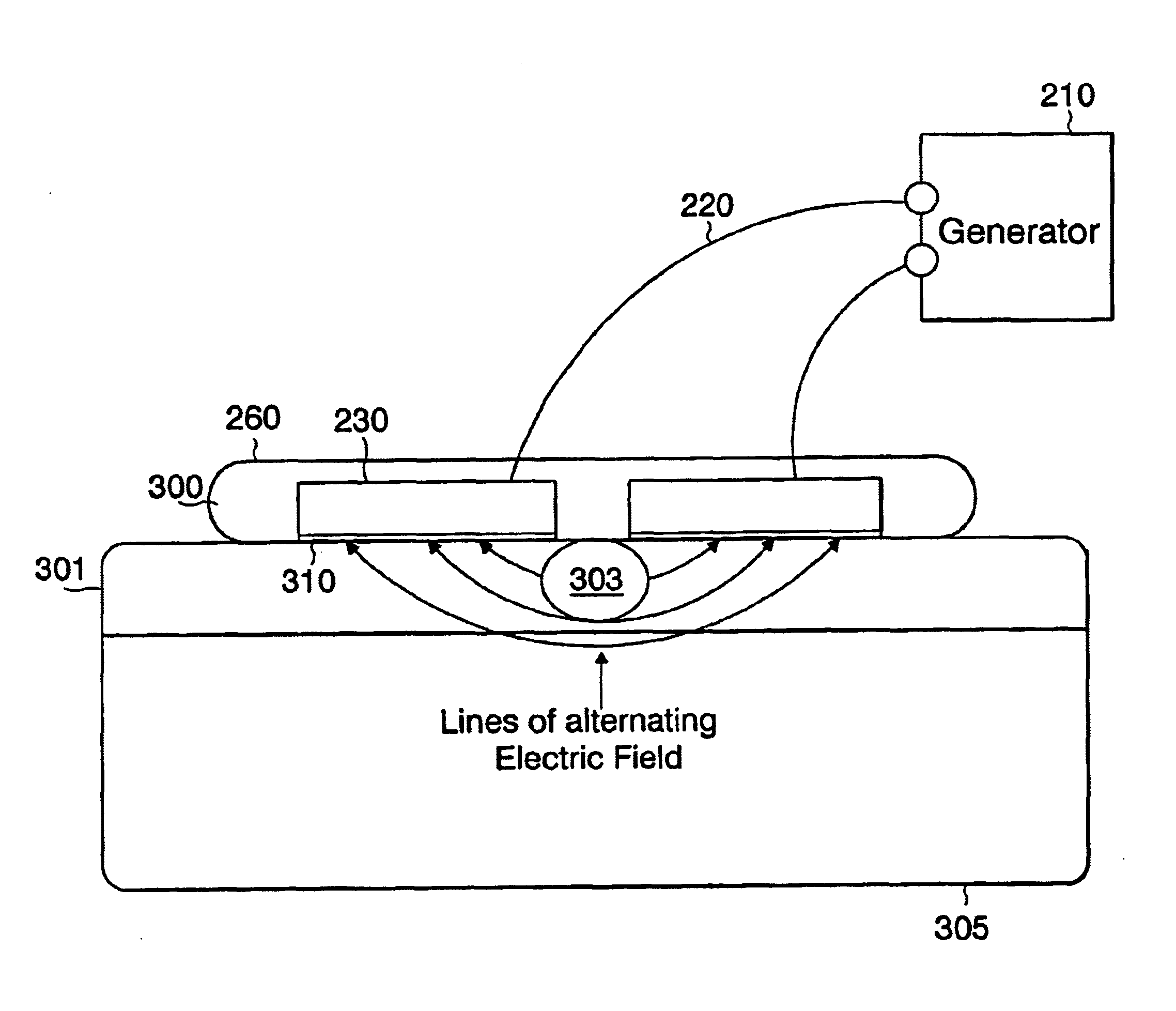
To demonstrate the effectiveness of electric fields having the above described properties (e.g., frequencies between 50 KHz and 500 KHz) in destroying tumor cells, the electric fields were applied to treat mice with malignant melanoma tumors. Two pairs of isolects 230 were positioned over a corresponding pair of malignant melanomas. Only one pair was connected to the generator 210 and 200 KHz alternating electric fields (TC fields) were applied to the tumor for a period of 6 days. One melanoma tumor was not treated so as to permit a comparison between the treated tumor and the non-treated tumor. After treatment for 6 days, the pigmented melanoma tumor remained clearly visible in the non-treated side of the mouse, while, in contrast, no tumor is seen on the treated side of the mouse. The only areas that were visible discernable on the skin were the marks that represented the points of insertion of the isolects 230. The fact that the tumor was eliminated at the treated side was furthe...


PUM


Abstract

An apparatus is provided for selectively destroying dividing cells in living tissue formed of dividing cells and non-dividing cells. The dividing cells contain polarizable intracellular members and during late anaphase or telophase, the dividing cells are connected to one another by a cleavage furrow. The apparatus includes insulated electrodes to be coupled to a generator for subjecting the living tissue to electric field conditions sufficient to cause movement of the polarizable intracellular members toward the cleavage furrow in response to a non-homogeneous electric field being induced in the dividing cells. The movement of the polarizable intracellular intracellular members towards the cleavage furrow causes the breakdown thereof which adversely impacts the multiplication of the dividing cells. Preferably, an intervening member is disposed between each insulated electrode and the skin surface and each intervening member includes a conductive “floating” plate that protects against the effects on the patient from a breakdown in the insulation of the electrode.

Description

TECHNICAL FIELDThis invention concerns selective destruction of rapidly dividing cells in a localized area, and more particularly, to an apparatus and method for selectively destroying dividing cells by applying an electric field having certain prescribed characteristics using an apparatus that is configured to be complimentary to a specific body part.BACKGROUNDAll living organisms proliferate by cell division, including cell cultures, microorganisms (such as bacteria, mycoplasma, yeast, protozoa, and other single-celled organisms), fungi, algae, plant cells, etc. Dividing cells of organisms can be destroyed, or their proliferation controlled, by methods that are based on the sensitivity of the dividing cells of these organisms to certain agents. For example, certain antibiotics stop the multiplication process of bacteria.The process of eukaryotic cell division is called “mitosis”, which involves nice distinct phases (see Darnell et al., Molecular Cell Biology, New York: Scientific ...

Claims


Application Information

Patent Timeline
Patent Type & Authority Patents(United States)
IPC IPC(8): A61N1/18A61N1/40A61B18/12A61B18/14A61B18/18
CPCA61B18/12A61N1/40A61N1/18A61B18/14A61B2017/00084
Inventor PALTI, YORAM
Owner NOVOCURE GMBH
Why Eureka
Try Eureka