
Method and apparatus for destroying dividing cells

Active Publication Date: 2006-03-21
NOVOCURE GMBH
View PDF49 Cites 148 Cited by
  • Summary
  • Abstract
  • Description
  • Claims
  • Application Information

AI Technical Summary

Benefits of technology

[0014]A major use of the method and apparatus of the present invention is in treatment of tumors by selective destruction of tumor cells with substantially no affect on normal tissue cells and, thus, the invention is described below in the context of selective destruction of tumor cells. It should be appreciated however that, for the purpose of the description that follows, the term “cell” may also refer to single-celled organisms (eubacteria, bacteria, yeast, protozoa), multi-celled organisms (fungi, algae, mold), and plants as or parts thereof that are not normally classified as “cells”. The method of the present invention enables selective destruction of tumor cells, or other organisms, by selective destruction of cells undergoing division in a way that is more effective and more accurate (e.g., more adaptable to be aimed at specific targets) than existing methods. Further, the method of the present invention causes minimal damage, if any, to normal tissue and, thus, reduces or eliminates many side-effects associated with existing selective destruction methods, such as radiation therapy and chemotherapy. The selective destruction of dividing cells in accordance with the method of the present invention does not depend on the sensitivity of the cells to chemical agents or radiation. Instead, the selective destruction of dividing cells is based on distinguishable geometrical characteristics of cells undergoing division, in comparison to non-dividing cells, regardless of the cell geometry of the type of cells being treated.
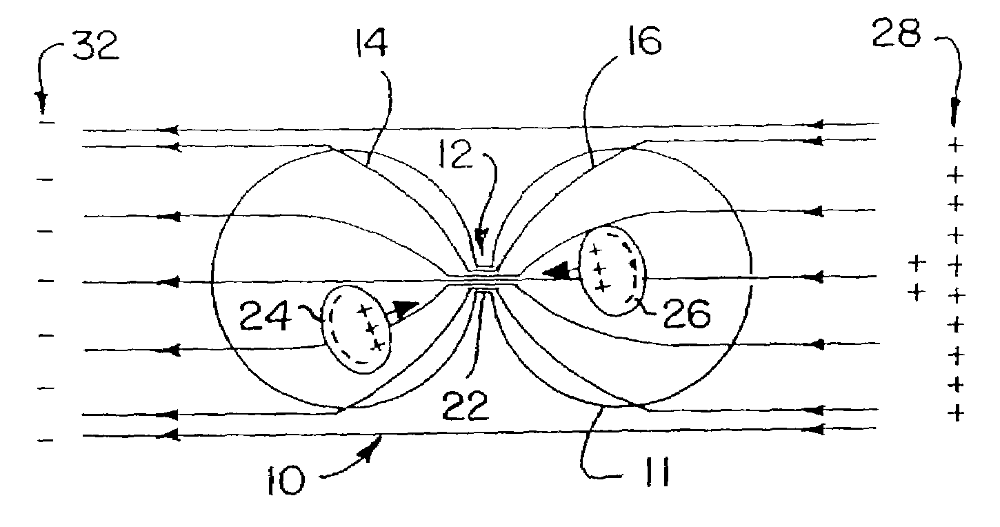
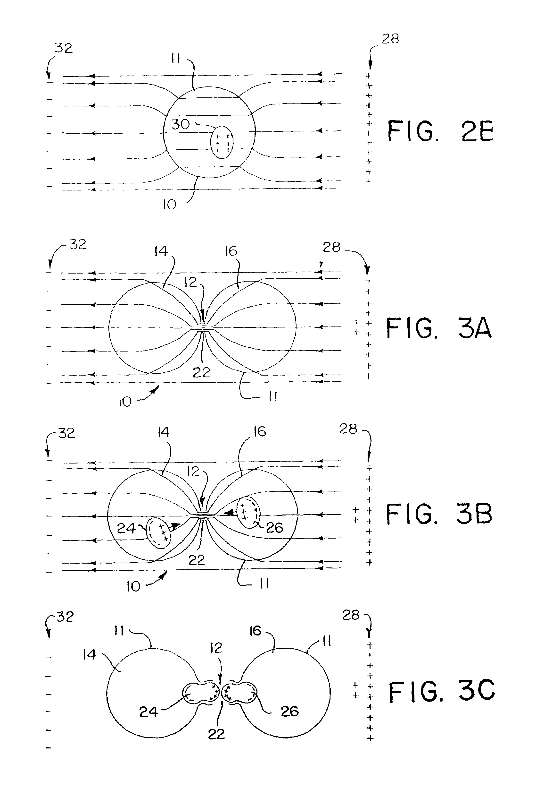
[0021]The movement of the cellular organelles towards the bridge disrupts the cell structure and results in increased pressure in the vicinity of the connecting bridge membrane. This pressure of the organelles on the bridge membrane is expected to break the bridge membrane and, thus, it is expected that the dividing cell will “explode” in response to this pressure. The ability to break the membrane and disrupt other cell structures can be enhanced by applying a pulsating alternating electric field, rather than a steady alternating electric field. When a pulsating electric field is applied to the tissue, the forces exerted on the intracellular organelles has a “hammering” effect, whereby force pulses (or beats) are applied to the organelles numerous times per second, enhancing the movement of organelles of different sizes and masses towards the bridge (or neck) portion from both of the sub-cells, thereby increasing the probability of breaking the cell membrane at the bridge portion. The forces exerted on the intracellular organelles also affect the organelles themselves and may collapse or break the organelles.

Problems solved by technology

Thus, unfortunately, the sensitivity of tumor cells is not sufficiently higher than that of many types of normal tissues.
This diminishes the ability to distinguish between tumor cells and normal cells and, therefore, existing cancer treatments typically cause significant damage to normal tissues, thus limiting the therapeutic effectiveness of such treatments.
Furthermore, the inevitable damage to other tissue renders treatments very traumatic to the patients and, often, patients are unable to recover from a seemingly successful treatment.
Also, certain types of tumors are not sensitive at all to existing methods of treatment.
The applied electrical current destroys substantially all cells in the vicinity of the target tissue.
Thus, this type of electrical method does not discriminate between different types of cells within the target tissue and results in the destruction of both tumor cells and normal cells.
One disadvantage of this method is that the ability to discriminate is highly dependent upon on cell type, for example, the size difference between normal cells and tumor cells is significant only in certain types of cells.
Another drawback of this method is that the voltages which are applied may damage some of the normal cells and may not damage all of the tumor cells because the differences in size and membrane dielectric properties are largely statistical and the actual cell geometries and dielectric properties may vary significantly.

Method used


Image

  • Method and apparatus for destroying dividing cells
  • Method and apparatus for destroying dividing cells
  • Method and apparatus for destroying dividing cells

Examples


Embodiment Construction

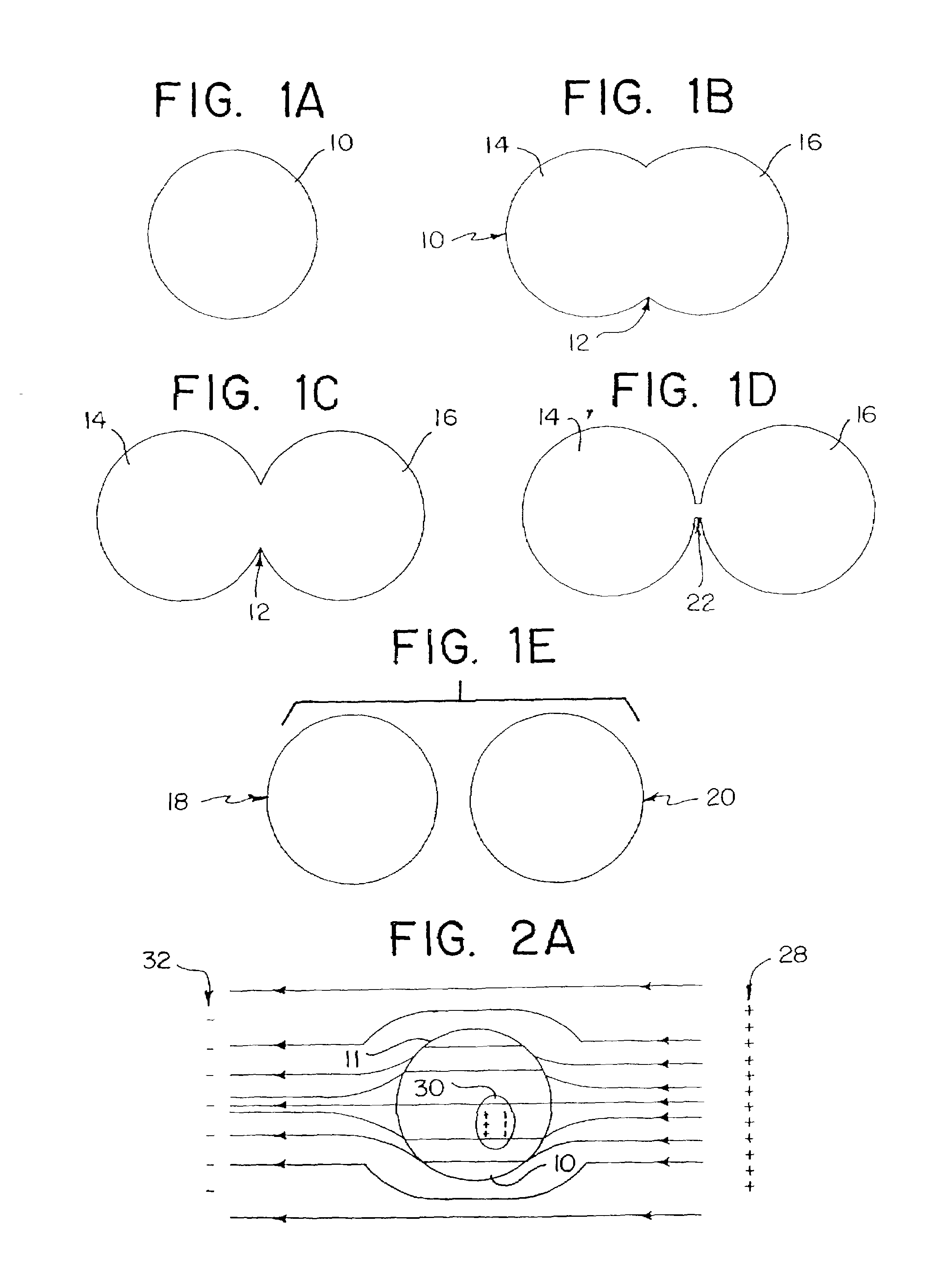
[0042]Reference is made to FIGS. 1A–1E which schematically illustrate various stages of a cell division process. FIG. 1A shows a cell 10 at its normal geometry, which may be generally spherical (as shown in the drawings), ellipsoidal, cylindrical, “pancake” like, or any other cell geometry, as is known in the art. FIGS. 1B–1D show cell 10 during different stages of its division process, which results in the formation of two new cells 18 and 20, shown in FIG. 1E.

[0043]As shown in FIGS. 1B–1D, the division process of cell 10 is characterized by a slowly growing cleft 12 which gradually separates cell 10 into two units, namely, sub-cells 14 and 16, which eventually evolve into new cells 18 and 20 (FIG. 1E). As shown specifically in FIG. 1D, the division process is characterized by a transient period during which the structure of cell 10 is basically that of the two sub-cells 14 and 16 interconnected by a narrow “bridge”22 containing cell material (cytoplasm surrounded by cell membrane)...


PUM


Abstract

The present invention provides a method and apparatus for selectively destroying dividing cells in living tissue formed of dividing cells and non-dividing cells. The dividing cells contain polarizable intracellular members and during late anaphase or telophase, the dividing cells are connected to one another by a cleavage furrow. According to the present method the living tissue is subjected to electric field conditions sufficient to cause movement of the polarizable intracellular members toward the cleavage furrow in response to a non-homogenous electric field being induced in the dividing cells. The non-homogenous electric field produces an increased density electric field in the region of the cleavage furrow. The movement of the polarizable intracellular members towards the cleavage furrow causes the break down thereof which results in destruction of the dividing cells, while the non-dividing cells of the living tissue remain intact.

Description

CROSS-REFERENCE TO RELATED APPLICATION[0001]This application claims the benefit of U.S. provisional patent application Ser. No.: 60 / 338,632, filed Nov. 6, 2001, which is hereby incorporated by reference in its entirety.FIELD OF THE INVENTION[0002]The present invention relates to selective destruction of rapidly dividing cells and, more particularly, to a method and device for selectively destroying dividing cells.BACKGROUND OF THE INVENTION[0003]It is known in the art that tumors, particularly malignant or cancerous tumors, grow very uncontrollably compared to normal tissue. Such expedited growth enables tumors to occupy an ever-increasing space and to damage or destroy tissue adjacent thereto. Furthermore, certain cancers are characterized by an ability to transmit cancerous “seeds”, including single cells or small cell clusters (metastasises), to new locations where the metastatic cancer cells grow into additional tumors.[0004]The rapid growth of tumors in general, and malignant t...

Claims


Application Information

Patent Timeline
IPC IPC(8): A61N1/32
CPCA61N1/326
Inventor PALTI, YORAM
Owner NOVOCURE GMBH
Learn More