Treatment of bioprosthetic tissues to mitigate post implantation calcification

a bioprosthetic tissue and post-implantation calcification technology, which is applied in the field of biomaterials, can solve the problems of undesirable stiffening or degradation of bioprosthetic materials, calcification of connective tissue proteins (i.e., collagen and elastin) within these materials, and achieve the effect of lessening the potential for untoward or undesirable reactions

Inactive Publication Date: 2005-04-12
EDWARDS LIFESCIENCES CORP
View PDF16 Cites 93 Cited by
  • Summary
  • Abstract
  • Description
  • Claims
  • Application Information

AI Technical Summary

Benefits of technology

Still further in accordance with this invention, the various embodiments of the method of mitigating post-implantation calcification of bioprosthetic tissues offer significant advantages over previous practices wherein glutaraldehyde was heated in the presence of the tissue, as the present invention allows the desirable features of the heat treatment to be achieved prior to any contact between the glutaraldehyde solution and the tissue, and also allows the temperature of the glutaraldehyde solution to be lowered to about 30 to 70° C., preferably about 40 to 60° C., or most preferably at about 50° C. prior to any contact with the tissue. This lessens the potential for untoward or undesirable reactions to the bioprosthetic tissue due to exposure to high free aldehyde concentrations and / or long term heat treatment at temperatures above 60° C. It also allows for treatment of the tissue within realistic manufacturing time frames.

Problems solved by technology

One problem associated with the implantation of many bioprosthetic materials is that the connective tissue proteins (i.e., collagen and elastin) within these materials can become calcified following implantation within the body.
Such calcification can result in undesirable stiffening or degradation of the bioprosthesis.
However, factors thought to influence the rate of calcification include the patient's age, the existence of metabolic disorders (i.e., hypercalcemia, diabetes, etc.), dietary factors, the presence of infection, parenteral calcium administration, dehydration, in situ distortion of the bioprosthesis (e.g., mechanical stress), inadequate anticoagulation therapy during the initial period following surgical implantation and immunologic host-tissue responses.

Method used


Image

  • Treatment of bioprosthetic tissues to mitigate post implantation calcification
  • Treatment of bioprosthetic tissues to mitigate post implantation calcification
  • Treatment of bioprosthetic tissues to mitigate post implantation calcification

Examples


Embodiment Construction

The following examples are provided for the purpose of describing and illustrating a few exemplary embodiments of the invention only. One skilled in the art will recognize that other embodiments of the invention are possible, but are not described in detail here. Thus, these examples are not intended to limit the scope of the invention in any way.

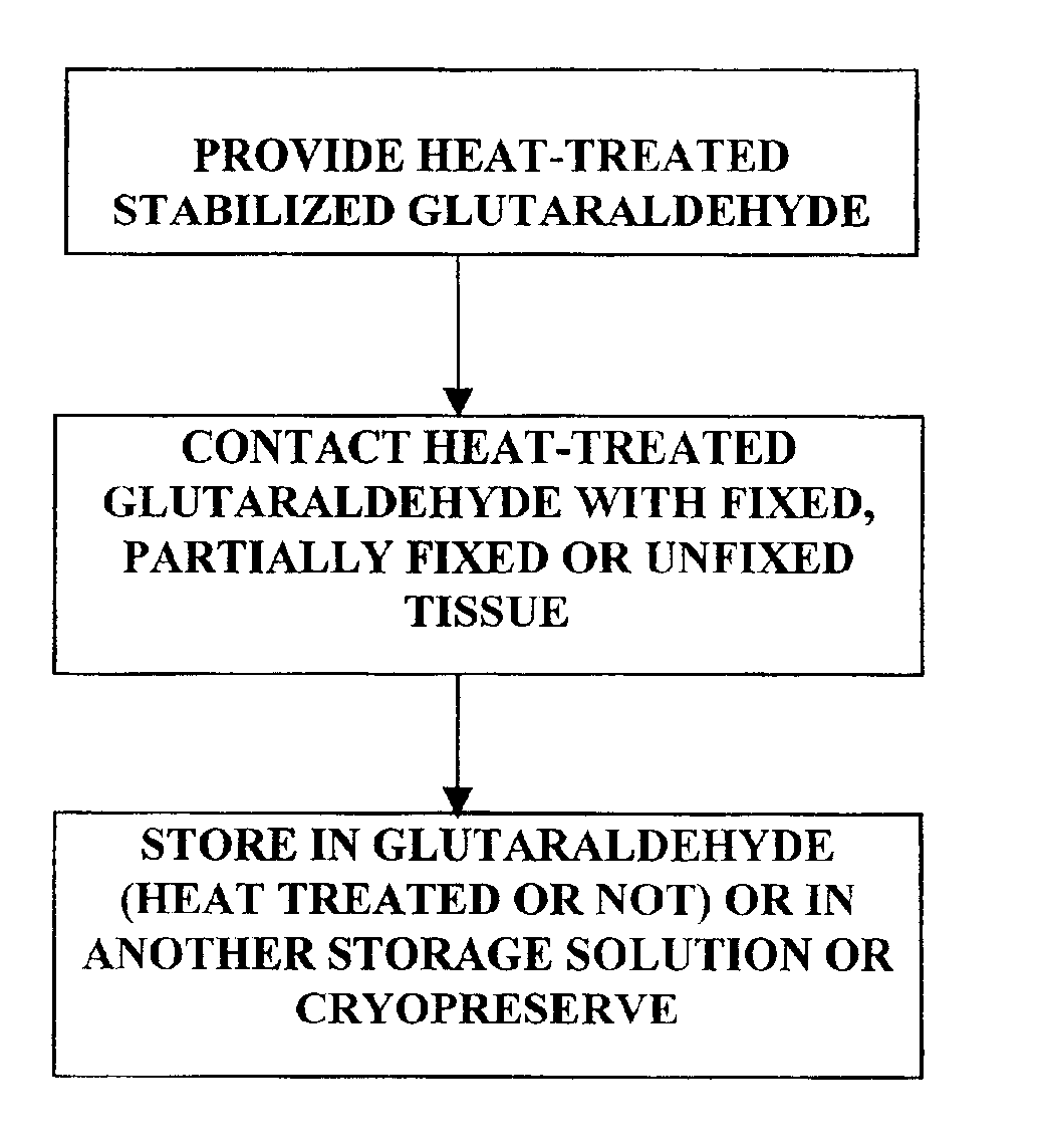
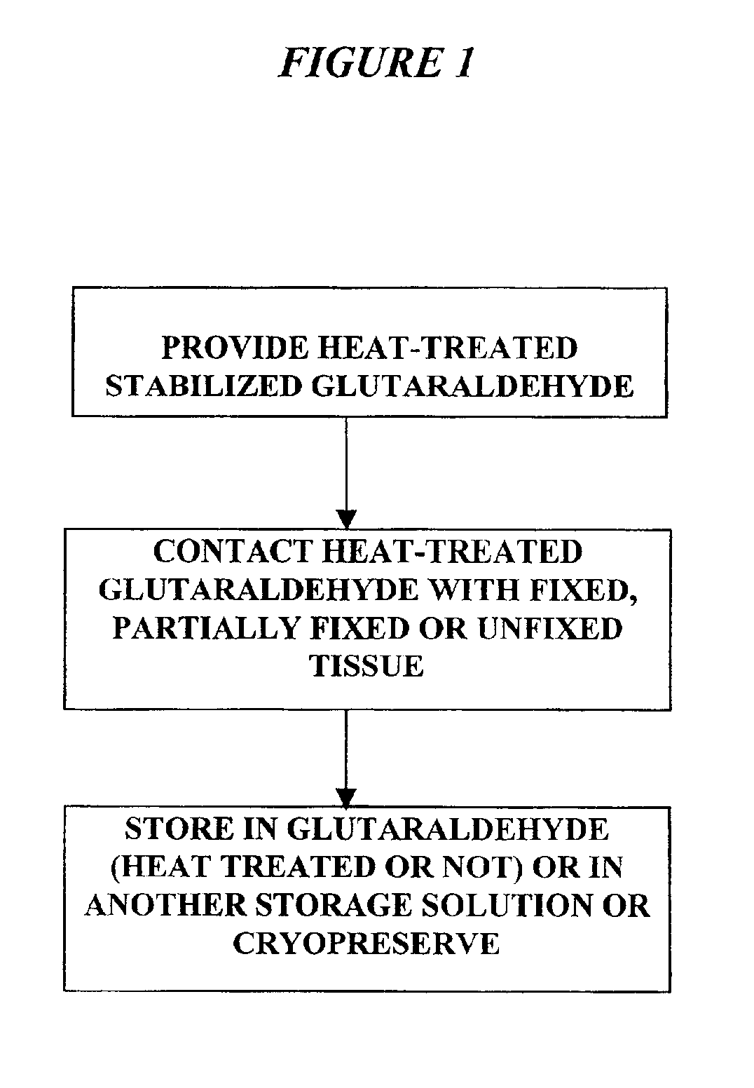
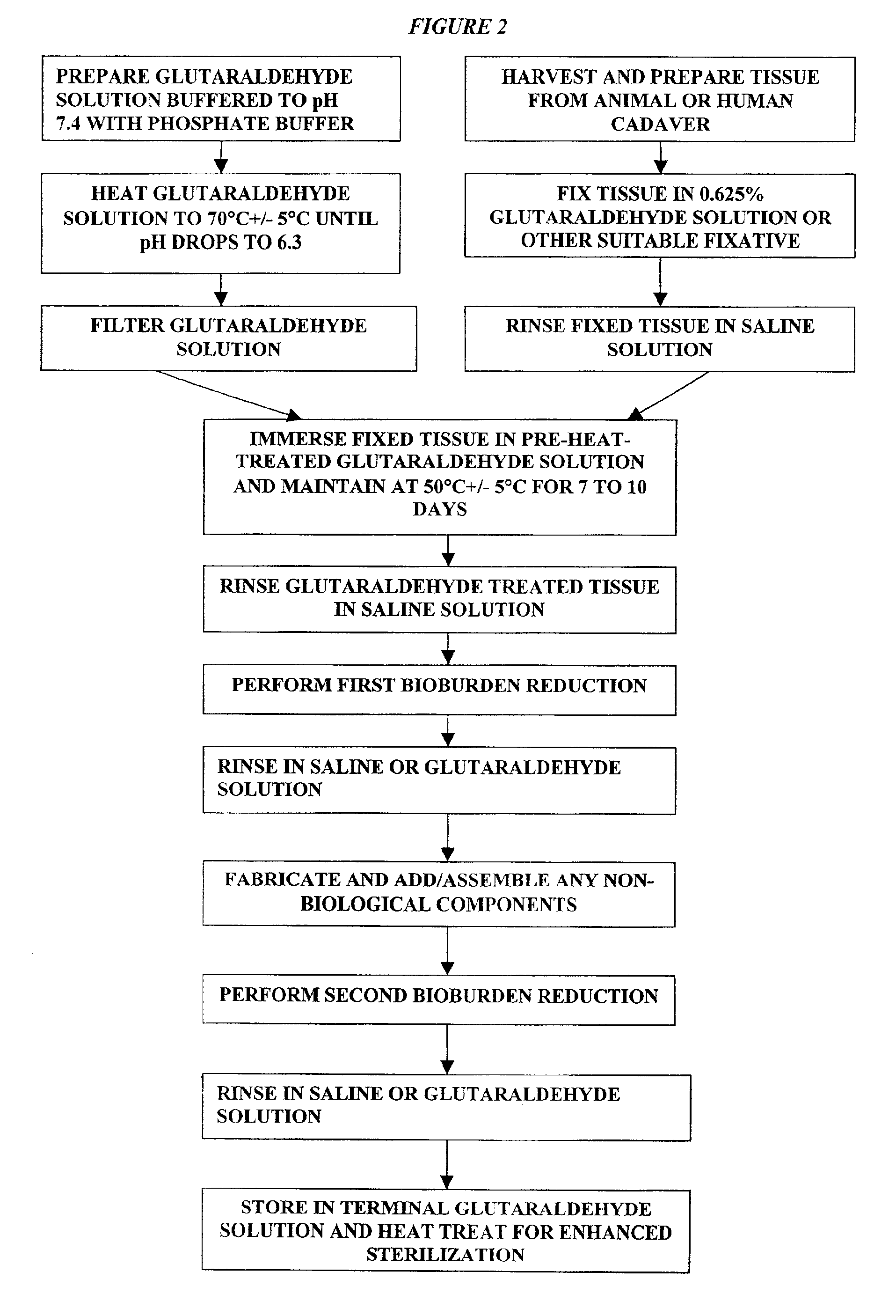
It has previously been reported that cross-linked bioprosthetic tissue post-treated in 0.625% glutaraldehyde phosphate solution for 2 months at 50° C., with fluid movement (e.g., shaking), exhibited less calcification in the rat subcutaneous and rabbit intramuscular implant models than control cross-linked bioprosthetic tissue fixed in 0.625% glutaraldehyde phosphate solution under typical conditions (i.e., room temperature for 1-14 days). See 66 Ann. Thoracic Surgery 264-6 (1998). Tissues treated under these conditions exhibited a characteristic tan to brown appearance. The heated 0.625% glutaraldehyde phosphate solution also darkened to a...


PUM

PropertyMeasurementUnit
temperatureaaaaaaaaaa
of timeaaaaaaaaaa
temperatureaaaaaaaaaa

Abstract

Bioprosthetic tissues are treated by immersing or otherwise contacting fixed, unfixed or partially fixed tissue with a glutaraldehyde solution that has previously been heat-treated or pH adjusted prior to its contact with the tissue. The prior heat treating or pH adjustment of the glutaraldehyde solution causes its free aldehyde concentration to decrease by about 25% or more, preferably by as much as 50%, and allows a “stabilized” glutaraldehyde solution to be obtained at the desired concentration and pH for an optimal fixation of the tissue at high or low temperature. This treatment results in a decrease in the tissue's propensity to calcify after being implanted within the body of a human or animal patient. The heat-treated or pH adjusted glutaraldehyde solution may, in some cases, also be used as a terminal sterilization solution such that the calcification-decreasing treatment with the previously treated glutaraldehyde and a terminal sterilization may be carried out simultaneously and / or in a single container.

Description

FIELD OF THE INVENTIONThis invention pertains generally to biomaterials and more particularly to methods for mitigating the post-implantation calcification of bioprosthetic materials and the bioprosthetic devices and articles produced by such methods.BACKGROUND OF THE INVENTIONImplantable biological tissues can be formed of human tissues preserved by freezing (i.e., cryopreserving) the so called homograft tissues, or of animal tissues preserved by chemically fixing (i.e., tanning) the so called bioprosthesis (Carpentier, Biological Tissues in Heart Valve Replacement, Butterworth (1972), Ionescu editor). The type of biological tissues used as bioprostheses include cardiac valves, blood vessels, skin, dura mater, pericardium, small intestinal submucosa (“SIS tissue”), ligaments and tendons. These biological tissues typically contain connective tissue proteins (i.e., collagen and elastin) that act as the supportive framework of the tissue. The pliability or rigidity of each biological ...

Claims


Application Information

Patent Timeline
Patent Type & Authority Patents(United States)
IPC IPC(8): A61L27/00A61L27/36
CPCA61L27/3604B65B63/08A61L27/3687A61L2400/02
Inventor CARPENTIER, SOPHIE M.CARPENTIER, ALAIN F.
Owner EDWARDS LIFESCIENCES CORP
Why Eureka
Try Eureka