Multiparameter FACS assays to detect alterations in exocytosis

a technology of exocytosis and facs assays, applied in the field of multiparameter facs assays to detect exocytosis alterations, can solve the problems of limited approach and limited allergy therapy

Inactive Publication Date: 2005-05-24
RIGEL
View PDF15 Cites 5 Cited by
  • Summary
  • Abstract
  • Description
  • Claims
  • Application Information

AI Technical Summary

Benefits of technology

[0017]In another preferred embodiment, a method for screening for a bioactive agent capable of modulating exocytosis in a cell is provided which comprises providing a population of cells wherein the cells have taken in a low pH concentration dye. The low pH concentration dye loaded cells are combined with at least one candidate bioactive agent and subjected to conditions that normally cause exocytosis. The release of the low pH concentration dye is detected. Alterations in the amount of released dye indicate that the agent modulates exocytosis.

Problems solved by technology

Therapy for allergy remains limited to blocking the mediators released by mast cells (anti-histamines), non-specific anti-inflammatory agents such as steroids and mast cell stabilizers which are only marginally effective at limiting the symtomatology of allergy.
Furthermore, it is widely believed that a wide array of psychiatric disorders are the result of an imbalance between neurotransmitter exocytosis and mediator reuptake.
However, this approach is limited by the fact that a single receptor blocker cannot overcome the effects of many diverse mediators.

Method used


Image

  • Multiparameter FACS assays to detect alterations in exocytosis
  • Multiparameter FACS assays to detect alterations in exocytosis
  • Multiparameter FACS assays to detect alterations in exocytosis

Examples


example 1

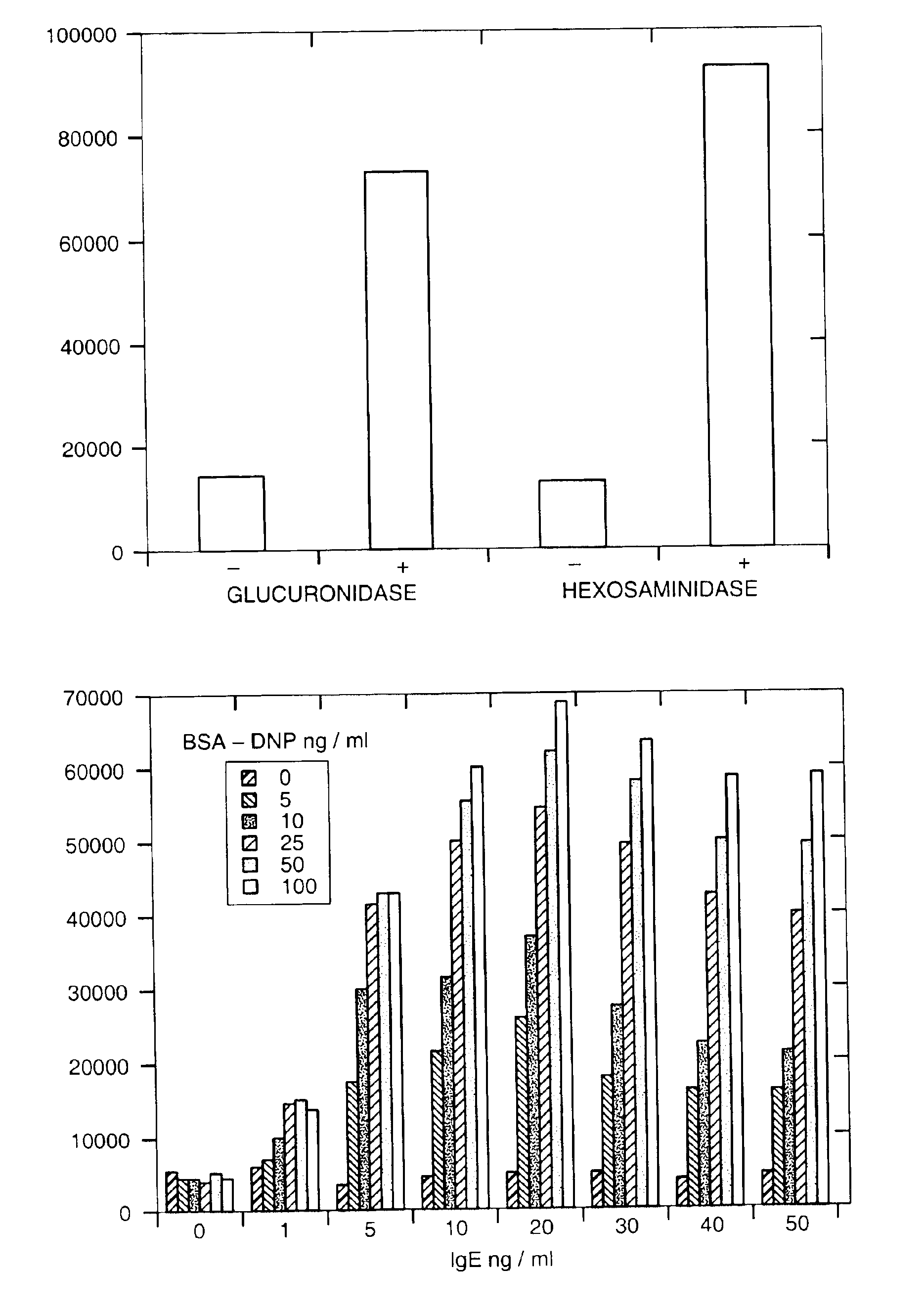
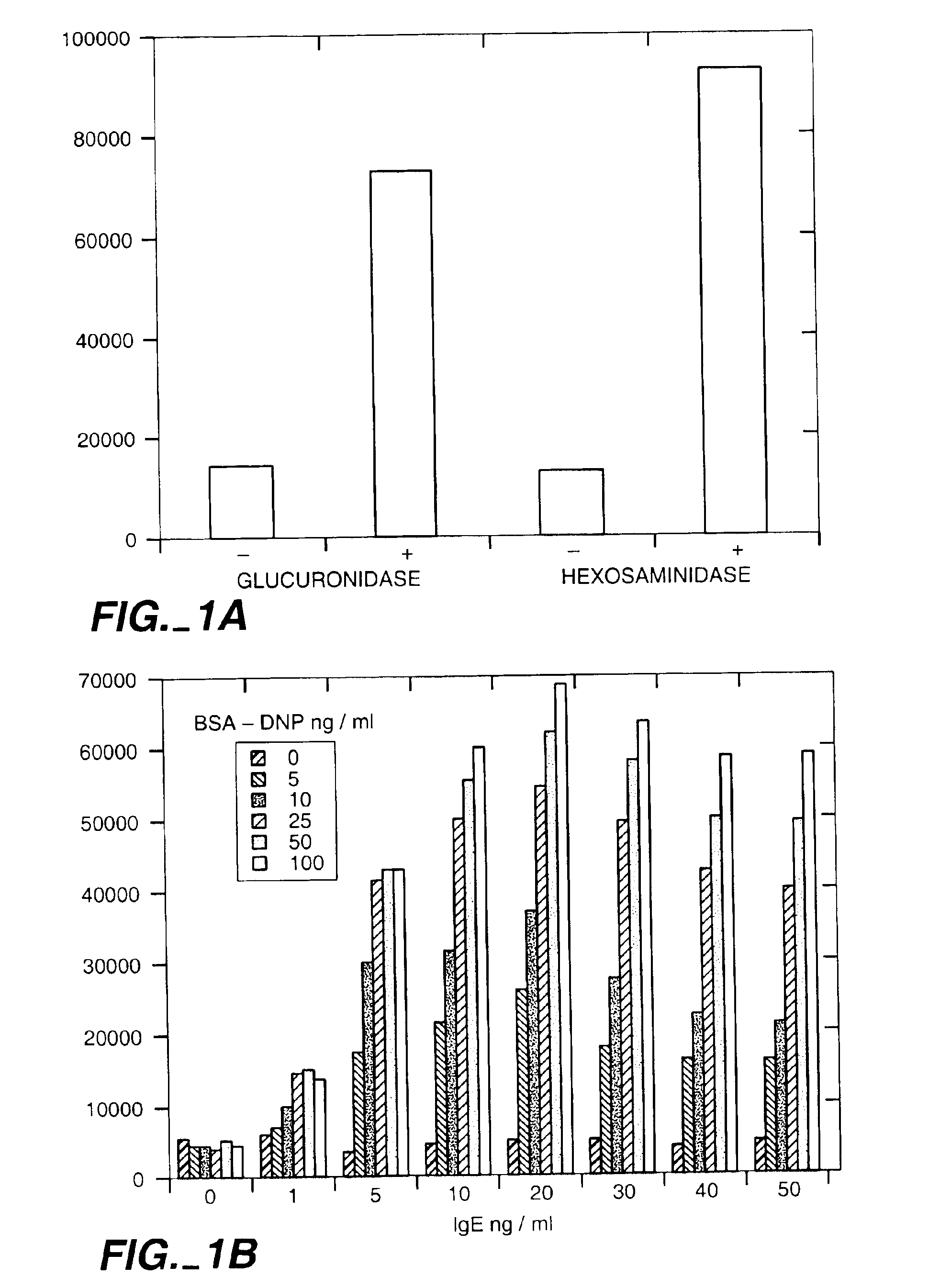
Population Based Exocytic Enzyme Activity Measurements

[0134]Materials: All chemicals were obtained from Sigma Chemical Co. Dyes and glucuronide were obtained from Molecular Probes, Inc. Cell lines MC-9 and RBL-2H3 were obtained from American Type Culture Collection (ATCC). Cell culture reagents were obtained from Fisher Scientific and molecular biology reagents from Clontech Inc.

[0135]Cell Culture: MC-9 cells were maintained as suspension cultures in flasks in media consisting of DMEM with L-arginine (116 mg / ml), L-asparagine (36 mg / ml), sodium pyruvate (1 mM), non-essential amino acids (0.1 mM), folic acid (6 mg / ml), 2-mercaptoethanol (0.05 mM), L-glutamine (2 mM), heat inactivated fetal bovine serum (10%), and 10% T-stim conditioned media (Collaborative Research, Inc.). The cells were kept at a density of between 0.25 and 2×10⁶ / ml. Experiments were only conducted on cells which were greater than 95% viable as determined by trypan blue exclusion. RBL-2H3 cells were maintained as ad...

example 2

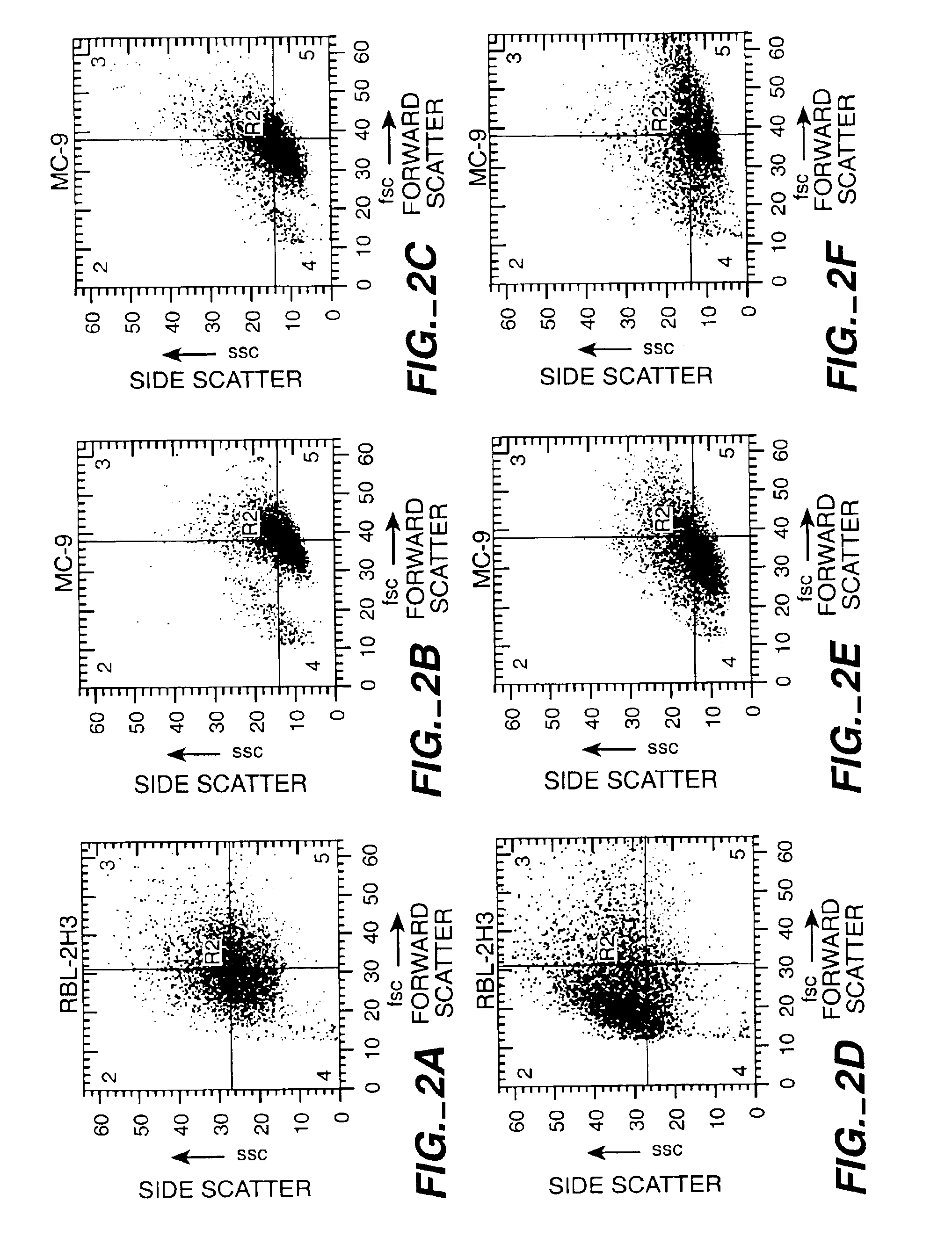
Mast Cell Exocytic Light Scatter Changes

[0140]The cells were prepared as described in Example 1, and light scatter properties were determined.

[0141]Results: The results are shown in FIG. 2. Light scatter changes observed on the flow cytometer (side scatter vs. forward scatter) are plotted as bivariate histograms for RBL-2H3 cells (A, D) and MC-9 cells (B, C, E, F). Cells were stimulated with the ionophore A23187 (0.5 ug / ml) and observed at various timepoints [minutes (A, C), 5 minutes (E), 10 minutes (D), and 30 minutes (B, F)]. Time dependent scatter changes are evident in both cell lines with significant changes occuring during the first 10 minutes which represents the major bolus of exocytosis in these cells.

example 3

Styryl Dyes Detect Mast Cell Exocytosis by FACS

[0142]Styrl Dye Straining: The cells were prepared as described above. Styryl dyes (FM1-43 or FM4-64; Molecular Probe s, Inc.) were diluted to a final concentration of 250 mM in MT and were incorporated into the stimulation buffer (see Example 1). After the stimulation protocol the cells were spun down, aspirated and resuspended in fresh ice cold MT. The cells were then ready for analysis in the flow cytometer (see Example 1).

[0143]Results: The results are shown in FIG. 3. MC-9 cells were stimulated (blue=DMSO, red=2 μM ionomycin) in the presence either FM 4-64 (A, B) or FM 1-43 (C,D,E). A) FM 4-64 labeled cells detected in the flow cytometer in fluorescence channel 1. B) FM 4-64 labeled cells detected in the flow cytometer in fluorescence channel 3. C) FM 1-43 labeled cells detected in the flow cytometer in fluorescence channel 1. D) FM 1-43 labeled cells detected in the flow cytometer in fluorescence channel 3. There is a clear stimul...


PUM

PropertyMeasurementUnit
concentrationaaaaaaaaaa

Abstract

Described is a method for screening for alterations in exocytosis of a population of cells. The cells are sorted by a FACS machine by assaying for alterations in at least three of the properties selected from the group consisting of light scattering, fluorescent dye uptake, fluorescent dye release, annexin granule binding, surface granule enzyme activity, and the quantity of granule specific proteins. Methods for screening for bioactive agents capable of modulating exocytosis in a cell are also described. The methods provide for reduced background and increased specificity without increasing the time or steps involved in assaying for exocytosis.

Description

FIELD OF THE INVENTION[0001]The invention relates to novel methods of detecting alterations in exocytosis in a cell or a cell population and screening for agents capable of modulating exocytosis through the use of multiparameter assays and a fluorescence-activated cell sorter (FACS) machine.BACKGROUND OF THE INVENTION[0002]Exocytosis is the fusion of secretory vesicles with the cellular plasma membrane, and has two main functions. One is the discharge of the vesicle contents into the extracellular space, and the second is the incorporation of new proteins and lipids into the plasma membrane itself.[0003]Exocytosis can be divided into two classes: constitutive and regulated. All eukaryotic cells exhibit constitutive exocytosis, which is marked by the continuous fusion of the secretory vesicles after formation. Regulated exocytosis is restricted to certain cells, including exocrine, endocrine and neuronal cells. Regulated exocytosis results in the accumulation of the secretory vesicle...

Claims


Application Information

Patent Timeline
Patent Type & Authority Patents(United States)
IPC IPC(8): G01N33/50G01N33/569
CPCG01N33/5008G01N33/5011G01N33/5014G01N33/502G01N33/5047G01N33/56966G01N2333/43595G01N2333/4718
Inventor FISHER, JOSEPHLORENS, JAMESPAYAN, DONALDROSSI, ALEXANDER
Owner RIGEL
Why Eureka
Try Eureka