Cell-based detection and differentiation of lung cancer

a cell-based, lung cancer technology, applied in the field of cell-based detection and differentiation of lung cancer, can solve the problems of poor sensitivity and specificity, many diagnostic tests have been criticized, and the test with poor sensitivity produces a high rate of false negatives

Inactive Publication Date: 2005-09-06
HOLOGIC INC
View PDF17 Cites 19 Cited by
  • Summary
  • Abstract
  • Description
  • Claims
  • Application Information

AI Technical Summary

Benefits of technology

[0041]FIG. 1. Molecular markers that are preferable markers to be included in a panel for identifying different histologic types of lung cancer. The column labeled “% ” indicates the percentage of tumor specimens that express a particular marker.

Problems solved by technology

Historically, many diagnostic tests have been criticized due to poor sensitivity and specificity.
A test having poor sensitivity produces a high rate of false negatives, i.e., individuals who have the disease but are falsely identified as being free of that particular disease.
The potential danger of a false negative is that the diseased individual will remain undiagnosed and untreated for some period of time, during which the disease may progress to a later stage wherein treatments, if any, may be less effective.
This type of test exhibits poor sensitivity because it fails to detect the presence of the virus until the disease is well established and the virus has invaded the bloodstream in substantial numbers.
A test having poor specificity produces a high rate
A test having poor specificity produces a high rate of false positives, i.e., individuals who are falsely identified as having the disease.
A drawback of false positives is that they force patients to undergo unnecessary medical procedures treatments with their attendant risks, emotional and financial stresses, and which could have adverse effects on the patient's health.
A feature of diseases which makes it difficult to develop diagnostic tests with high specificity is that disease mechanisms often involve a plurality of genes and proteins.
While it is the state-of-the-art in clinical diagnostic cytology, it is subjective and the diagnostic results are often not highly sensitive or reproducible, especially at early stages of cancer (e.g., ASCUS, LSIL).
A drawback to diagnostic tests that rely on morphological analyses is that cell morphology is a lagging indicator.
However, because the amount of genetic material in a sample of cells may be very low, only a very weak signal may be obtained.
Therefore, in situ hybridization tests that do not employ pre-amplification techniques will likely have a poor specificity, yielding many false negatives.
However, proteomics may have a poorer specificity than the cytogenetic marker detection technology since multiple pathologies may result in similar changes in protein over-expression or under-expression.
However, because other factors in addition to the disease state may cause the concentration of a disease marker to become raised or lowered, tests that rely on immunochemical analysis of a specific disease marker will likely have poor specificity, yielding a high rate of false positives.
As a result, different treatments are not equally effective in all cancers or even among the stages of a specific type of cancer.
However, in cases where the tumor has spread, surgery may provide, at best, only limited benefits.
While somewhat effective in prolonging life, treatment of patients with metastatic disease rarely produces a cure.
Even through there may be an initial response, with time the disease progresses and the patient ultimately dies from its effects and / or from the toxic effects of the treatments.
While there have been some successes such as mammography for breast cancer, PSA testing for prostate cancer, and the PAP smear for cervical cancer, in most cases cancer is detected at a relatively late stage where the patient is symptomatic and the disease is almost always fatal.
Some tests currently being used in the diagnosis and staging of cancer, however, either lack sufficient sensitivity or specificity, are too invasive, or are too costly to justify their use as a population-based screening test.
However, small lesions are often difficult to detect and although larger tumors are relatively easy to visualize on a chest film, at the time of detection most have already metastasized.
Thus, chest X-rays lack the necessary sensitivity for use as an early detection method.
However, the test has relatively low specificity with a 20% false positive rate [4].
Furthermore, while the cost of the initial test is relatively low ($300), the cost of follow-up can be high.
However, fluorescence bronchoscopy is unable to detect peripheral lesions, it takes a long time for bronchoscopists to examine a patient's airways, and it is an expensive procedure.
Additionally, the procedure is moderately invasive, creating an insurmountable barrier to its use as a population-based screening test.
The cost of establishing a testing facility is high and there is the need for a cyclotron on site or nearby.
This, coupled with the high cost of the test, has limited the use of PET scans to staging lung cancer patients rather than for early detection of the disease.
Although used for some time as a means of screening for lung cancer, sputum cytology has enjoyed only limited success due to its low sensitivity and its failure to reduce disease-specific mortality.
A number of factors including tumor size, location, degree of differentiation, cell clumping, inefficiency of clearing mechanisms to release cells and sputum to the external environment, and the poor stability of cells within the sputum contribute to the overall poor performance of the test.
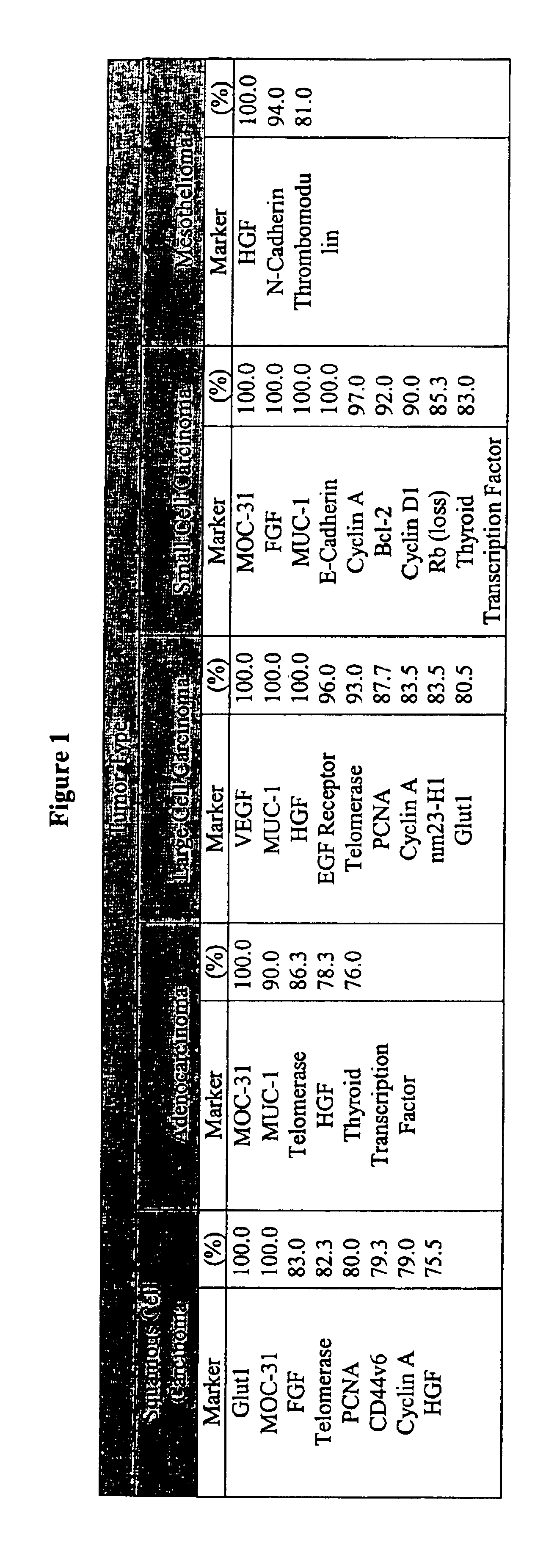
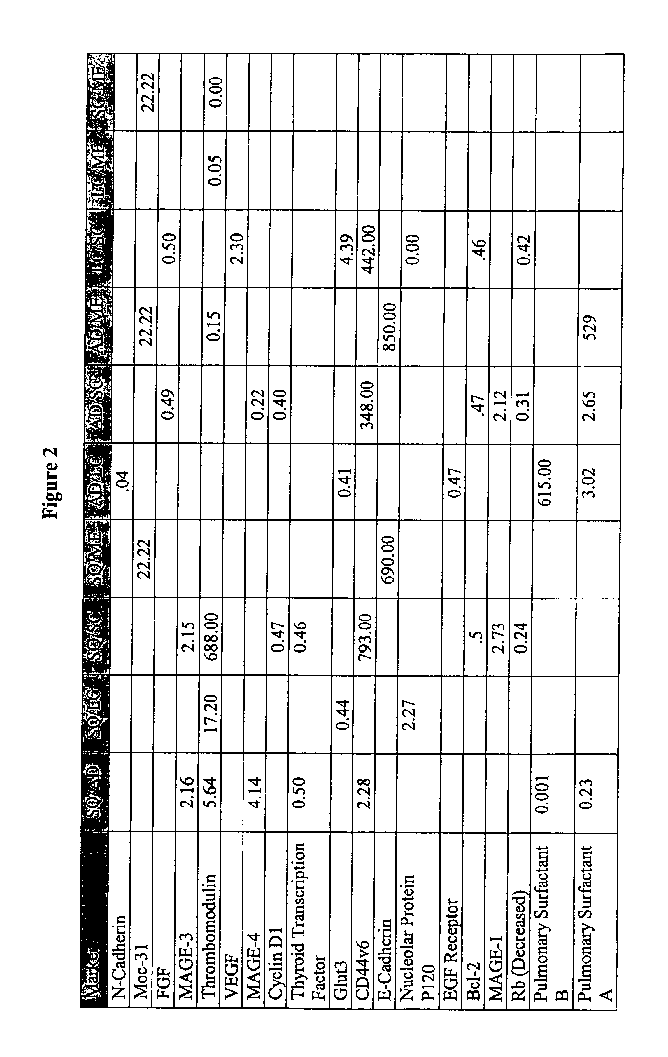
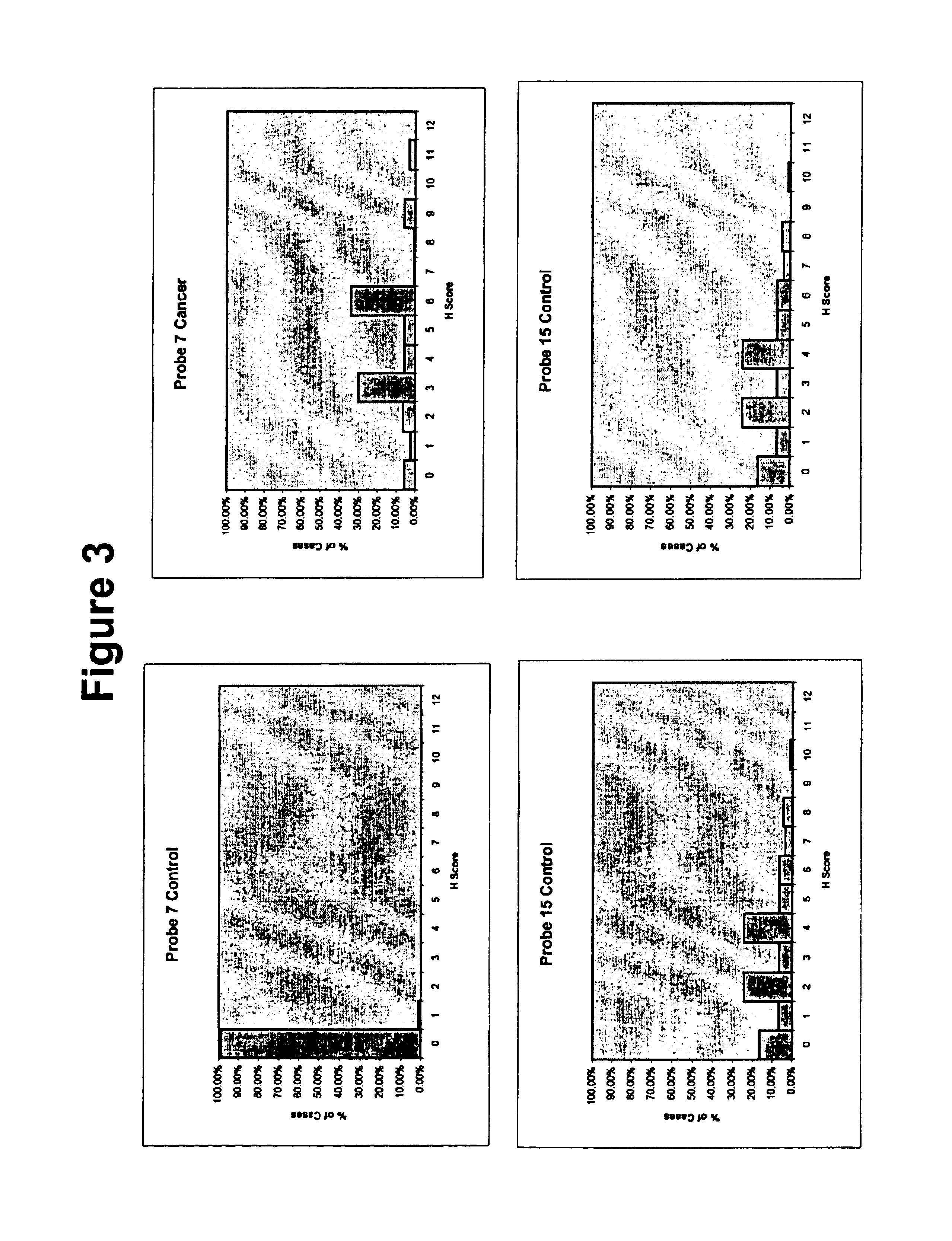
Unfortunately, cancer is a disease state in which single markers have typically failed to detect or differentiate many forms of the disease.
Thus, probes that recognize only a single marker have been shown to be largely ineffective.
Exhaustive searches for “magic bullet” diagnostic tests have been underway for many decades though no universal successful magic bullet probes have been found to date.

Method used


Image

  • Cell-based detection and differentiation of lung cancer
  • Cell-based detection and differentiation of lung cancer
  • Cell-based detection and differentiation of lung cancer

Examples


Embodiment Construction

1. Introduction

[0048]The present invention provides a noninvasive disease state detection and discrimination method with high sensitivity and specificity. The method involves contacting a cytological sample suspected of containing diseased cells with a panel comprising a plurality of agents, each of which quantitatively binds to a disease marker, and detecting a pattern of binding of the agents. This pattern includes the localization and density / concentration of binding of the component probes of the panel. The present invention also provides methods of making a panel for detecting a disease and also for discriminating between disease states as well as panels for detecting lung cancer in early stages and discriminating between different types of lung cancer. Panel tests have been used in medicine. For example, panels are used in blood serum analysis. However, because a cytology analysis involves imaging and localization of specific markers within individual cells and tissues, prior ...


PUM

PropertyMeasurementUnit
threshold correlation numberaaaaaaaaaa
thickaaaaaaaaaa
molecular structuresaaaaaaaaaa

Abstract

The present invention provides a method for detecting and differentiating disease states with high sensitivity and specificity. The method allows for a determination of whether a cell-based sample contains abnormal cells and, for certain diseases, is capable of determining the histologic type of disease present. The method detects changes in the level and pattern of expression of the molecular markers in the cell-based sample. Panel selection and validation procedures are also provided.

Description

[0001]This application is a Continuation-in-Part of provisional application No. 60 / 274,638, filed Mar. 12, 2001, the entire contents of which are incorporated by reference herein.BACKGROUND OF THE INVENTION[0002]The present invention relates to early detection of a general disease state in a patient. The present invention also relates to discrimination (differentiation) between specific disease states in their early stages.[0003]Early detection of a specific disease state can greatly improve a patient's chance for survival by permitting early diagnosis and early treatment while the disease is still localized and its pathologic effects limited anatomically and physiologically. Two key evaluative measures of any test or disease detection method are its sensitivity (Sensitivity=True Positives / (True Positives+False Negatives) and specificity (Specificity=True Negatives / (False Positives+True Negatives), which measure how well the test performs to accurately detect all affected individual...

Claims


Application Information

Patent Timeline
Patent Type & Authority Patents(United States)
IPC IPC(8): G01N33/574C12N5/08C12Q1/00C12Q1/02G01N33/50G01N33/53G01N33/566G01N33/569G01N33/571G01N33/58G01N33/60
CPCG01N33/56966G01N33/57423G01N33/57492G01N33/569
Inventor PRESSMAN, NORMAN J.HIRSCH, KENNETH S.HIRSCH, ADRIAN
Owner HOLOGIC INC
Why Eureka
Try Eureka