Radiation imaging apparatus, radiation imaging system, and radiation imaging method

a radiation imaging and radiation imaging technology, applied in the field of radiation imaging apparatuses, can solve the problems of reducing the efficiency of focus screening, affecting the detection of focus, and difficulty in detecting the shadow of focus overlapped in the transmitted image, so as to improve the detection ratio of abnormal regions, and improve the effect of abnormal region detection ratio

Inactive Publication Date: 2005-10-04
CANON KK
View PDF6 Cites 88 Cited by
  • Summary
  • Abstract
  • Description
  • Claims
  • Application Information

AI Technical Summary

Benefits of technology

[0018]According to the present invention, performing subtraction for two images sequentially radiographed can enhance parts that vary noticeably in black or white, compared with other parts. Furthermore, synchronizing the subtracted image with the original image that does not undergo subtraction to display them in the same screen in a display allows a doctor to recognize the parts that vary noticeably and to compare the subtracted image with the original image for reading them, thus improving the detection ratio of abnormal regions such as focus.
[0019]Synchronizing the energy-subtracted image with the original image that does not undergo the subtraction to display them in parallel in the display allows the doctor to compare and read the images, thus improving the detection ratio of abnormal regions such as focus, compared with a case where a single image is read.
[0020]Furthermore, displaying the motion of a patient (e.g., the motion of diaphragm or lung field due to breathing, the motion of heart, and the like) as moving pictures sometimes elicits lat

Problems solved by technology

However, because transmitted images are observed in the plain chest radiography, it can be difficult to detect the shadow of focus that is overlapped in the transmitted images when the focus to be observed exists, for example, behind a rib or diaphragm or in the shadow of a cardiovascular portion.
Accordingly, there is a problem that the efficiency of focus screening is decreased and the detection of focus can be delayed.
Although energy subtraction is useful for removing the shadows of bones, there is no guarantee that the shadows of the bones are entirely removed.
Particularly, a part of the shadows of bones is disadvantageously left depending on the body type or the physical constitution of a patient or on the kind of focus.
For example, focus does not always exist in the shadow of a

Method used


Image

  • Radiation imaging apparatus, radiation imaging system, and radiation imaging method
  • Radiation imaging apparatus, radiation imaging system, and radiation imaging method
  • Radiation imaging apparatus, radiation imaging system, and radiation imaging method

Examples


first embodiment

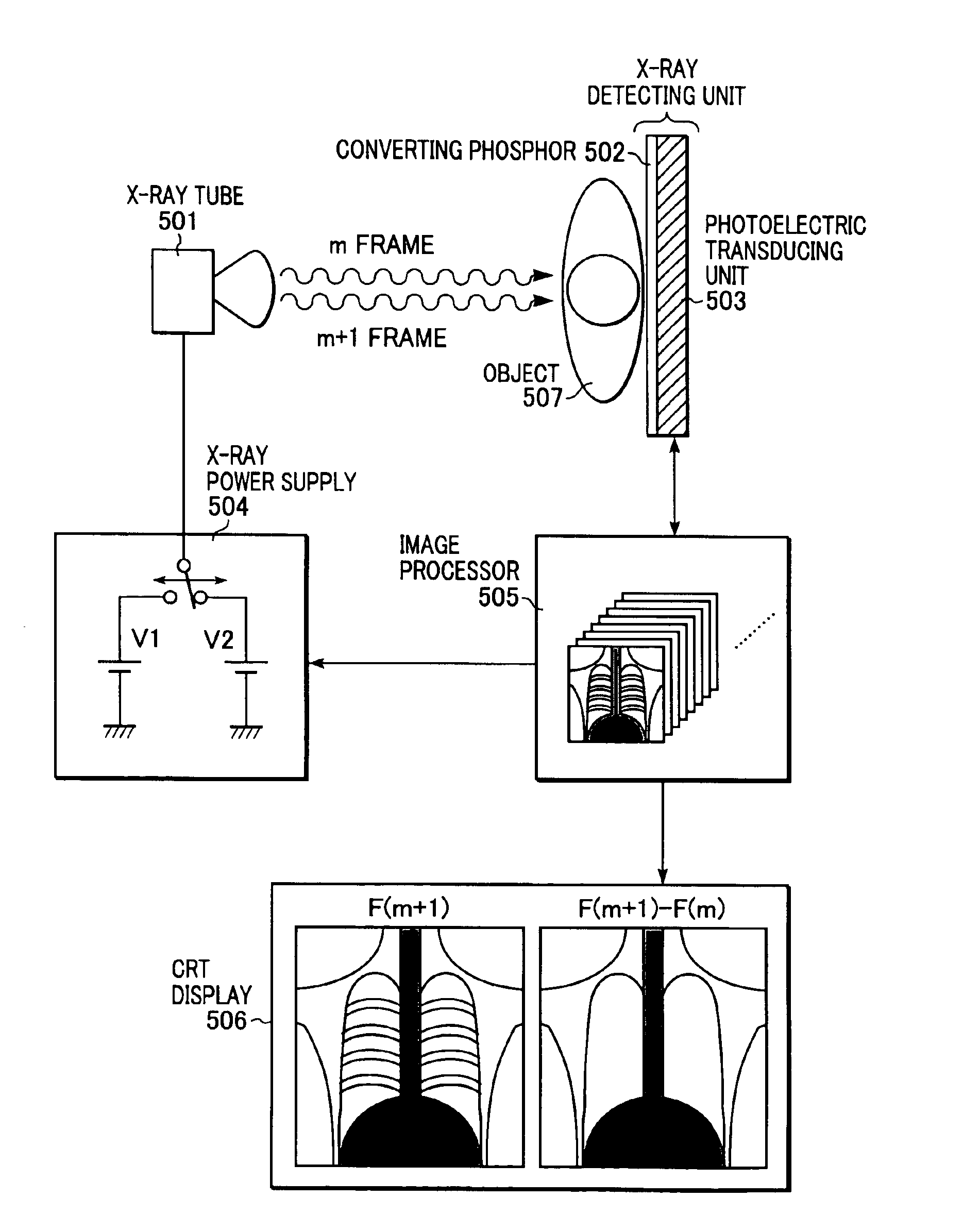
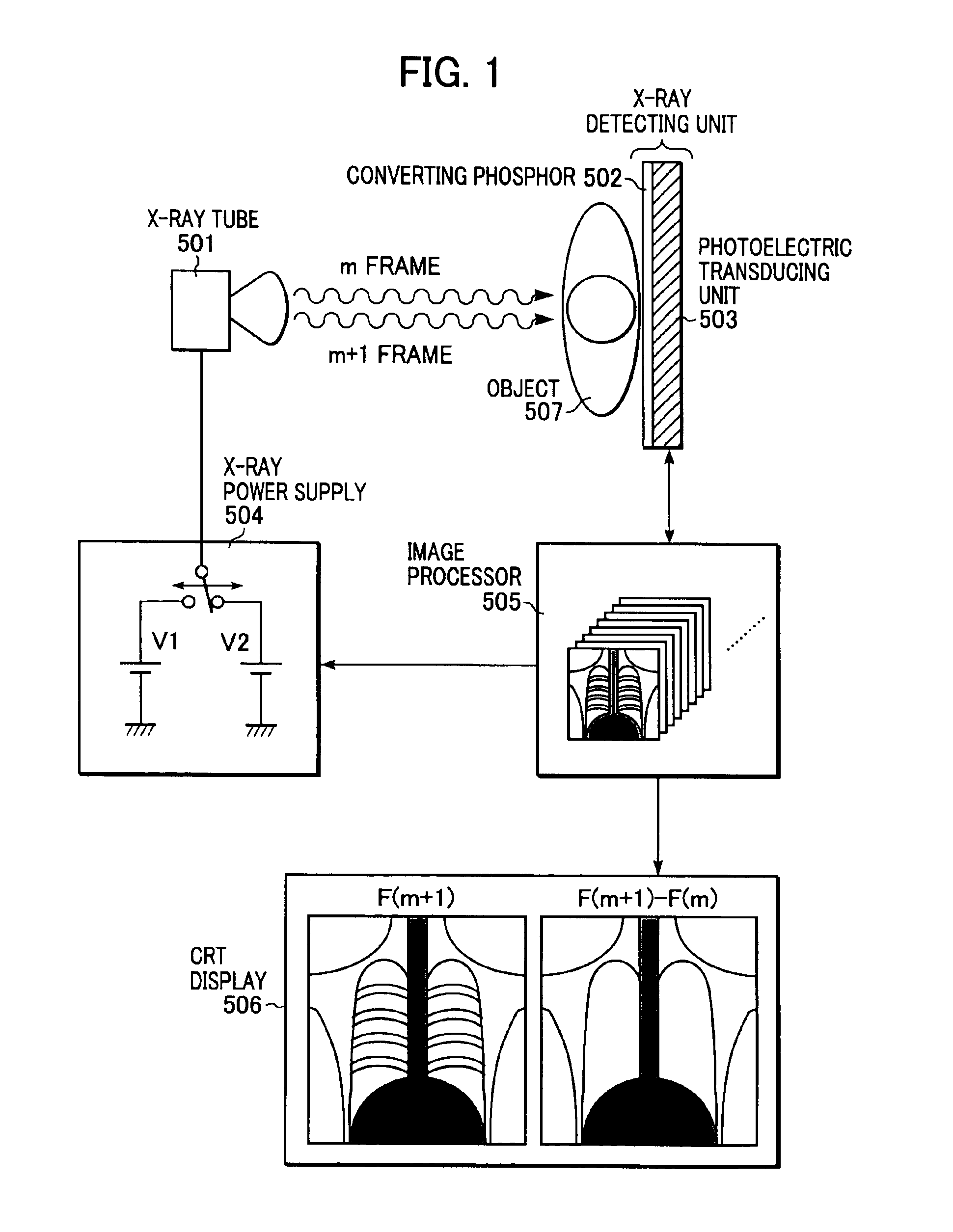
[0032]FIG. 1 is a diagram schematically showing an X-ray imaging system according to a first embodiment of the present invention.

[0033]An object 507 is irradiated with X-rays emitted from an X-ray tube 501. The object 507 is mainly a patient. The X-rays are transmitted through the patient and are converted into visible light by an X-ray to visible-light converting phosphor 502. The visible light supplied from the phosphor 502 is converted into an electrical signal by a photoelectric transducing unit 503. As a result, the radioscopic image of the object 507 (patient) is converted into the electrical signal. The X-ray to visible-light converting phosphor 502 is substantially adhered to the photoelectric transducing unit 503 by bonding or the like. The X-ray to visible-light converting phosphor 502 is combined with the photoelectric transducing unit 503 to form an X-ray detecting unit. An X-ray power supply 504 supplies a high voltage for accelerating electrons in the X-ray tube 501. T...

second embodiment

[0087]In an X-ray imaging system according to a second embodiment of the present invention, an image given by subtracting an image F(m) from an image F(m+1) is synchronized with an original image of the image F(m) (the original image of the image F(m+1) in the first embodiment) that does not undergo the subtraction to display the image F(m) and the image F(m+1) in parallel in the same screen in a display.

[0088]This subtraction provides difference images between frames. Images of parts that move noticeably or parts whose density significantly varies can be enhanced in black or white, compared with images of other parts. Synchronizing the subtracted image with the original image to display them allows a doctor to compare the subtracted image with the original image and to read them.

[0089]Table 2 shows the relationship between two kinds of frames to be displayed in the same screen in the display and their display, in the X-ray imaging system of the second embodiment.

[0090]

TABLE 2Number...


PUM


Abstract

A radiation imaging apparatus includes a radiation detecting unit and an image-display controlling unit. The radiation detecting unit has radiation detectors, arranged in a two-dimensional array, for detecting radiation transmitted through an object as electrical signals. The image-display controlling unit radiographs radiation images of the object, detected as the electrical signals by the radiation detecting unit, at a predetermined frame rate as continuous images in a plurality of frames and displays a processed image given by subtracting an m-th image from an (m+1)-th image in synchronous with either the m-th image or the (m+1)-th image that does not undergo the subtraction in a display, where m is a natural number.

Description

BACKGROUND OF THE INVENTION[0001]1. Field of the Invention[0002]The present invention relates to radiation imaging apparatuses for medical diagnoses or industrial nondestructive inspections and, more particularly, to a radiation imaging apparatus and a radiation imaging system suitable for taking moving pictures, where the radiation includes not only X-rays but also alpha-rays, beta-rays, and gamma-rays.[0003]2. Description of the Related Art[0004]Hitherto, X-ray imaging systems installed in hospitals or the like adopt two imaging technologies. A film imaging technology in which a patient is irradiated with X-rays and a film is exposed to the X-rays transmitted through the patient, and a digital imaging technology in which X-rays transmitted through a patient are converted into electrical signals, which are detected as digital values by an analog-to-digital converter to store the detected digital values in a memory. In the latter technology, a visible light emitted from a photostimu...

Claims


Application Information

Patent Timeline
IPC IPC(8): A61B6/00G01T1/164G01T1/161G01T1/24G01T1/00H01L27/14H05G1/64H05G1/00H04N5/32H04N5/321H04N5/325G01T1/29
CPCG01T1/24G01T1/2928
Inventor ENDO, TADAO
Owner CANON KK
Why Eureka
Try Eureka