Contrast enhancement agent for magnetic resonance imaging

a magnetic resonance imaging and contrast enhancement technology, applied in the field of contrast enhancement agents, can solve the problems of indistinguishable signal from background noise, increased life-threatening, and large linewidth

Inactive Publication Date: 2005-12-06
DUKE UNIV +1
View PDF1 Cites 19 Cited by
  • Summary
  • Abstract
  • Description
  • Claims
  • Application Information

AI Technical Summary

Benefits of technology

[0038]A contrast enhancement agent useful for providing an image of a biological sample is disclosed. The agent comprises at least one peptide comprising the amino acid sequence NXEQVSP (SEQ ID NO: 1), wherein X is any amino acid, at least one paramagnetic metal ion and at least one chelator. Preferably, the paramagnetic metal ion is selected from the group consisting of transition and inner transition elements. More preferably, the paramagnetic metal ion is selected from the group consisting of Gd(III), Mn(II), Cu(I

Problems solved by technology

In extreme situations, the linewidth can be so large that the signal is indistinguishable from background noise.
After a clot that has caused a myocardial infarction has been lysed, a second or third clot can reformat the same site, which can become more life-threatening than the formation of the first clot.
However, the patient still runs the risk of clot reformation and a subsequent occlusion of blood vessels.
The in vivo detection of blood clots, however, has been elusive.
The ability to monitor and image internal wound healing is vital, yet has been elusive.
If such a wound is not healing properly, a potentially life-threatening condition can develop in a subject.
When an internal operation is performed, however, it is much more difficult to assess wound healing.
Internal wounds, obviously, cannot be visually inspec

Method used


Image

  • Contrast enhancement agent for magnetic resonance imaging
  • Contrast enhancement agent for magnetic resonance imaging
  • Contrast enhancement agent for magnetic resonance imaging

Examples


Embodiment Construction

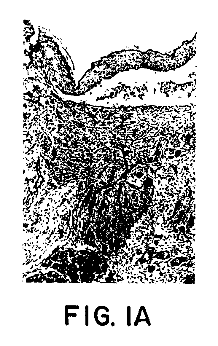
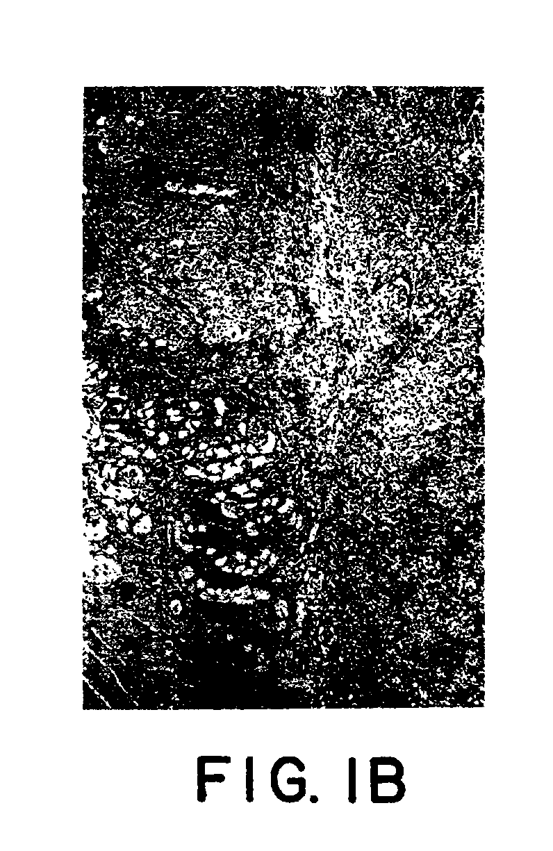
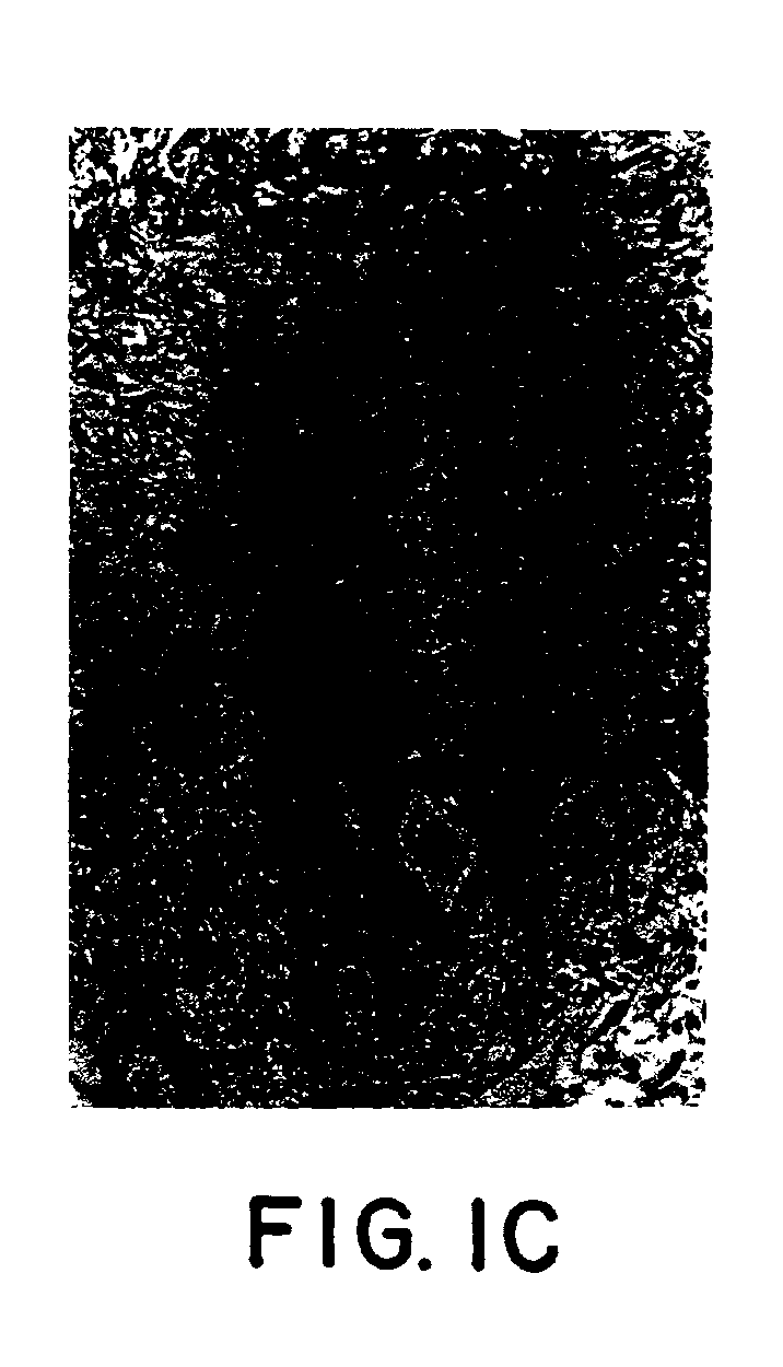
[0047]A contrast enhancement agent useful for providing an MR-based image of tumor boundary regions and blood clots is an aspect of the present invention. Notably, the formation (and the dissolution) of blood clots can be monitored in real time. The contrast enhancement agents of the present invention comprise at least one peptide comprising the amino acid sequence NXEQVSP (SEQ ID NO: 1), wherein X is any amino acid, at least one paramagnetic metal ion and at least one chelator. Most preferably, the peptide comprises the amino acid sequence NQEQVSP (SEQ ID NO: 2), the paramagnetic metal ion is gadolinium and the chelator is DTPA.

[0048]The present invention also discloses a method of non-invasively forming an image of a tumor boundary and allows real-time imaging of blood clot formation., These abilities are made possible by the contrast enhancement agents of the present invention. The method of generating an image comprises (a) providing a contrast enhancement agent comprising at le...


PUM

PropertyMeasurementUnit
Paramagnetismaaaaaaaaaa

Abstract

Imaging agents for magnetic resonance imaging are disclosed. Methods of monitoring angiogenesis and the growth and remission of tumor tissue are also disclosed. Methods of monitoring blood clot formation and dissolution are additionally disclosed. Methods of monitoring wound healing are further disclosed. A kit for obtaining an image of a biological structure is further disclosed.

Description

CROSS REFERENCE TO RELATED APPLICATIONS[0001]This application is based on and claims priority to U.S. Provisional Application Ser. No. 60 / 260,377, filed Jan. 5, 2001, herein incorporated by reference in its entirety.TECHNICAL FIELD[0002]The present invention relates to contrast enhancement agents for use in magnetic resonance (“MR”) imaging and methods of non-invasively detecting and monitoring tumor angiogenesis, tumor growth and regression, wound healing and blood clot formation and dissolution.[0003]Table of AbbreviationsAPalkaline phosphataseCTcomputerized tomographyDMSOdimethylsulfoxideDOTA1,4,7,10-tetraazacyclododecane-1,4,7,10-tetraacetic acidDTPAdiethylenetriamine-pentaacetic acidECMextracellular matrixEDTAethylenediaminetetraacetic acidELISAenzyme linked immunoadsorbent assayESRelectron spin resonancefXIIIfactor XIIIfXIIIafactor XIIIaHRPOhorseradish peroxidaseICPAECinductively coupled plasma atomic emission spectrometryMRmagnetic resonanceMRImagnetic resonance imagingNMRnuc...

Claims


Application Information

Patent Timeline
IPC IPC(8): A61K49/08A61K49/14G01N33/49
CPCA61K49/085A61K49/14G01N33/4905
Inventor HAROON, ZISHANDEWHIRST, MARK W.GREENBERG, CHARLES S.NEEMAN, MICHAL
Owner DUKE UNIV
Why Eureka
Try Eureka