Method of diagnosing and monitoring malignant breast carcinomas

a breast cancer and malignant technology, applied in the field of malignant breast cancer diagnosis and monitoring, can solve the problems of not being able to extend the scope of diagnosis innovations, and the diagnostic value of antigens is not high, so as to reduce morbidity and mortality and reduce overall national health care expenditures

Inactive Publication Date: 2005-12-06
MERIDIAN PROTEIN TECH
View PDF10 Cites 9 Cited by
  • Summary
  • Abstract
  • Description
  • Claims
  • Application Information

AI Technical Summary

Benefits of technology

[0033]The biomarkers and related inventive method can be used for detecting breast carcinoma and provide for an economical and logistical adjunct diagnostic test for mammography. Furthermore, these saliva

Problems solved by technology

However, such antigens are not diagnostic for breast cancer, and the aforementioned tumor biomarkers (e.g., CA 125, erb and CD) have not been tested for their

Method used


Image

  • Method of diagnosing and monitoring malignant breast carcinomas
  • Method of diagnosing and monitoring malignant breast carcinomas
  • Method of diagnosing and monitoring malignant breast carcinomas

Examples


example 1

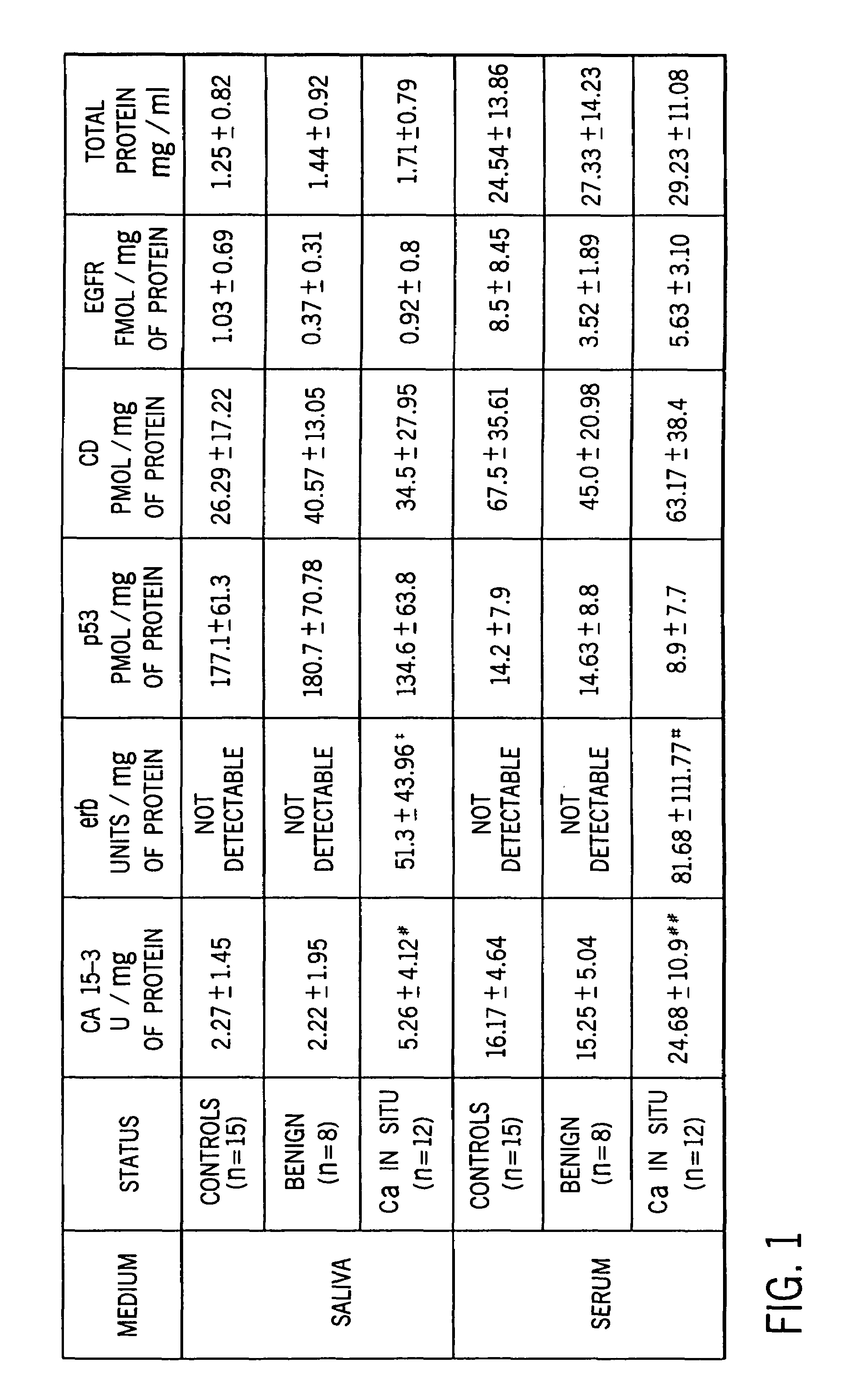
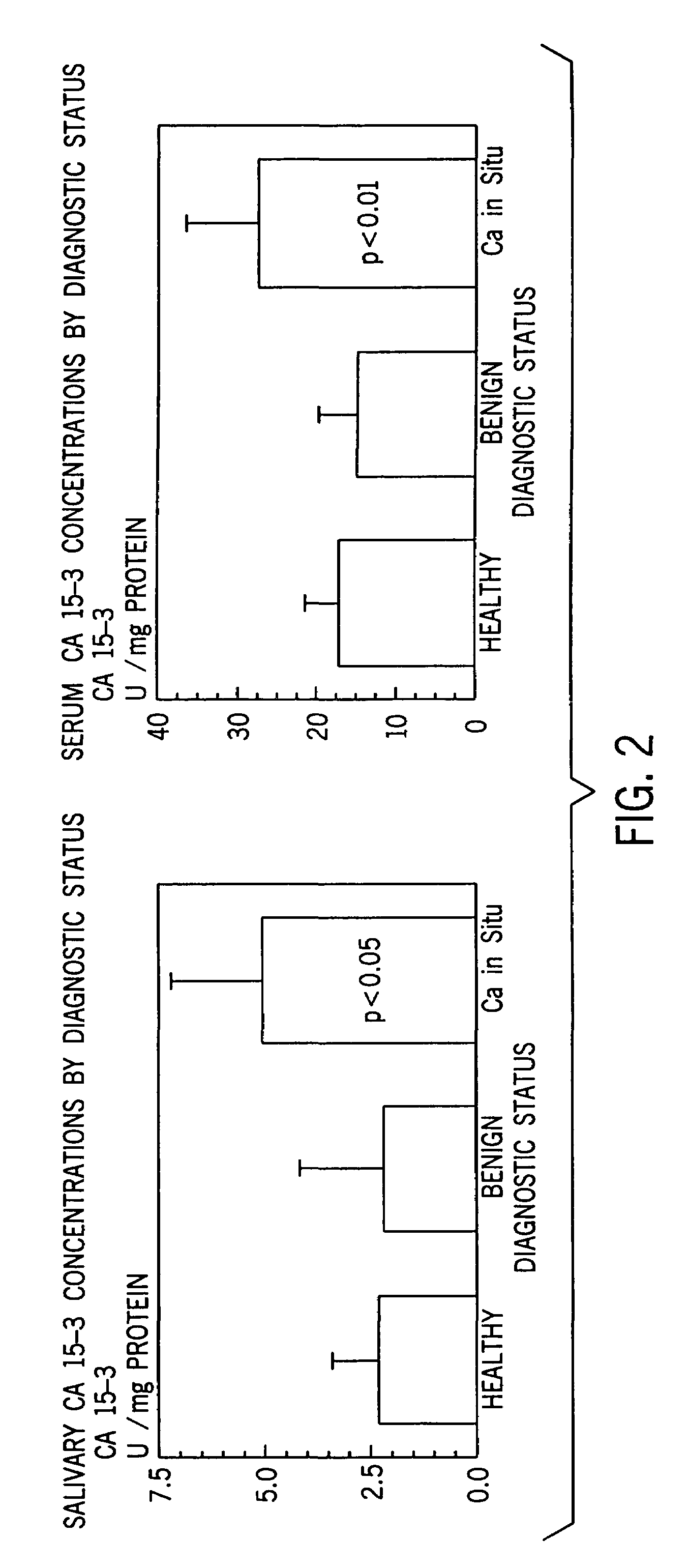
[0057]Statistical Analysis. Statistical analysis were performed using the SPSS statistical software package. A descriptive analysis was made comparing mean marker values for the controls, those with benign tumors, and carcinoma of the breast.

[0058]A one-way analysis of variance for unbalanced data, the general linear models procedure, was used to compare the mean values for the group with breast carcinoma with a non cancer groups. The polynomials formulated using the general linear models procedure are easy to interpret and are appropriate for all sample sizes including those too small to sustain an appropriate multivariate analysis. The Tukey post-hoc analysis was used for significant linear models.

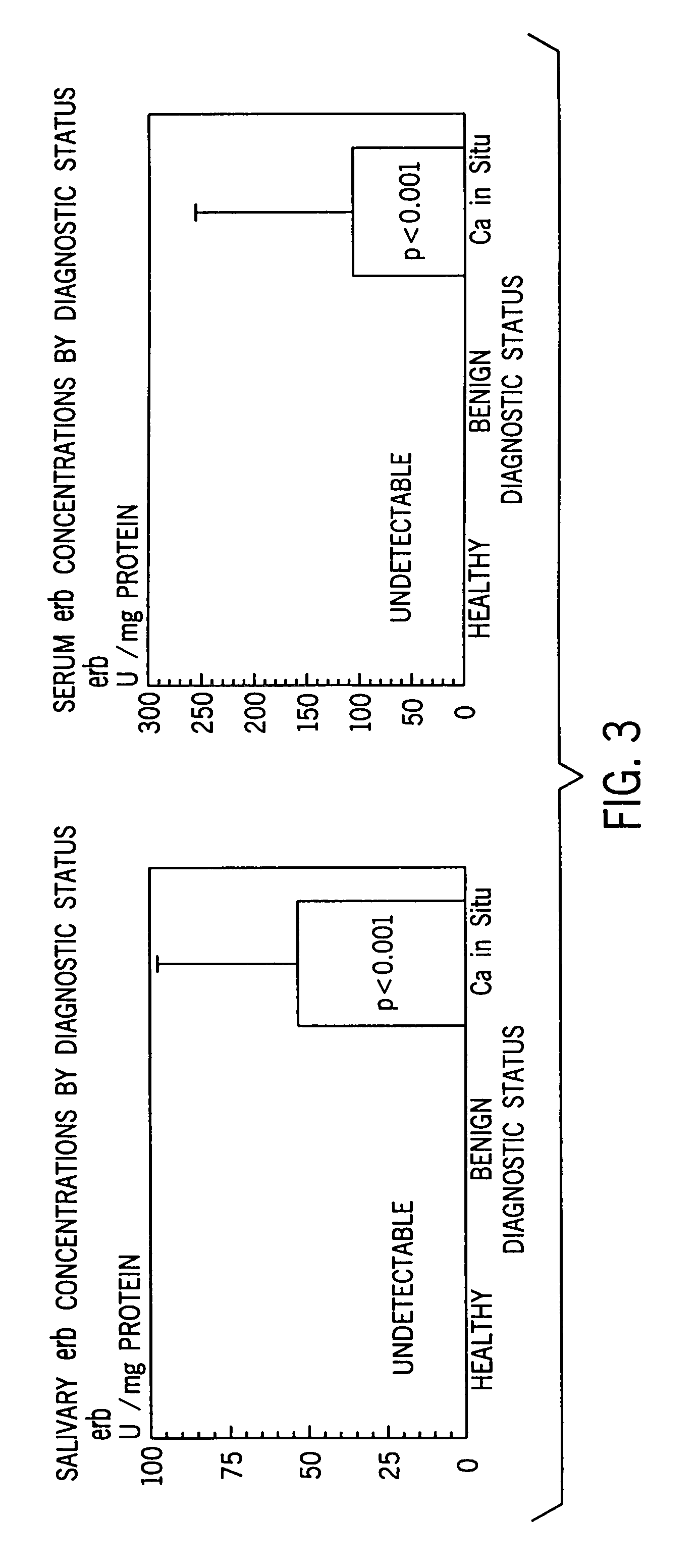
[0059]Considering that erb was undetected among controls and benign lesions for both saliva and serum, a one way sample t-test was performed. Due to the small sample size, issues concerning the specificity and sensitivity of the panel of markers were not addressed, but will be investigat...

example 2

[0060]Specimen Collection. Stimulated whole saliva specimens were collected for a 5 minute period using a cube of paraffin as a stimulant (Navasesh, 1982)17 Salivary flow rates were determined gravimetrically. All specimens were collected in the morning thereby controlling for any possible effects that circadian rhythm may produce in marker concentration. Samples can be frozen for future analysis. Blood was also drawn at the time of saliva collection by a phlebotormist. None of the participants exhibited cancerous or precancerous lesions in the oral cavity at the time the specimens were collected.

[0061]The frozen saliva samples were thawed and centrifuged at 500–1500 G for 20 min to precipitate cells and mucin in order to extract the bio-marker proteins. The clear saliva extract and the serum from the blood specimens were analyzed for total protein and the panel of biomarkers.

example 3

[0062]Total Protein. A colorimetric assay for measuring total protein concentration, based on the color change of Coomassie brilliant blue G-250 dye in response to various concentrations of proteins, was used (Bio-Rad Kit). Specimens were read on a spectrophotometer and absorbance measured at 595 nm. Total protein concentration of the samples was determined from a standard curve constructed with bovine gamma globulin standards.


PUM


Abstract

A panel of biomarkers for the diagnosis and treatment of breast cancer was examined in the saliva of a cohort of 1) healthy women, 2) women with benign lesions of the breast and 3) women with diagnosed breast cancer. Recognized tumor markers c-erbB-2 (erb), cancer antigen 15-3 (CA 15-3), and tumor suppresser oncogene protein 53 (p53) were found in the saliva of all three groups of women. The levels of erb and CA 15-3 in the cancer patients evaluated, however, were significantly higher than the salivary levels of healthy controls and benign tumor patients. Conversely, pantropic p53 levels were higher in controls as compared to those women with breast cancer and those with benign tumors.

Description

[0001]This application is a Continuation-in-Part of U.S. application Ser. No. 09 / 259,993 filed Mar. 1, 1999, now U.S. Pat. No. 6,294,349, and claims priority benefit therefrom.BACKGROUND OF THE INVENTION[0002]This invention relates generally to the use of salivary biomarkers to diagnose breast cancer and, more particularly, to diagnostically differentiate between women with carcinoma of the breast, women with benign tumors, and healthy controls.[0003]Breast cancer is the second leading cause of death among women in the United States. Approximately 1 woman in every 10 will develop breast cancer in her lifetime. Recent statistics estimate that 44,000 women will die of breast cancer, while 150,000 new female cases of breast cancer will be diagnosed in the next year.[0004]It has been shown that screening for breast cancer can reduce breast cancer mortality. Among women aged 50 and older, studies have demonstrated a 20% to 40% reduction in breast cancer mortality for women screened by ma...

Claims


Application Information

Patent Timeline
IPC IPC(8): G01N33/574
CPCG01N33/57415
Inventor STRECKFUS, CHARLES F.BIGLER, LENORA G.THIGPEN, JAMES TATE
Owner MERIDIAN PROTEIN TECH
Why Eureka
Try Eureka