Device and method for the examination of samples in a non-vacuum environment using a scanning electron microscope

a scanning electron microscope and environment technology, applied in the direction of instruments, heat measurement, machines/engines, etc., can solve the problems of vacuum preservation of samples, failure to successfully view wet objects, and failure to achieve the effect of high resolution

Inactive Publication Date: 2006-01-31
EL MUL TECH +1
View PDF47 Cites 32 Cited by
  • Summary
  • Abstract
  • Description
  • Claims
  • Application Information

AI Technical Summary

Benefits of technology

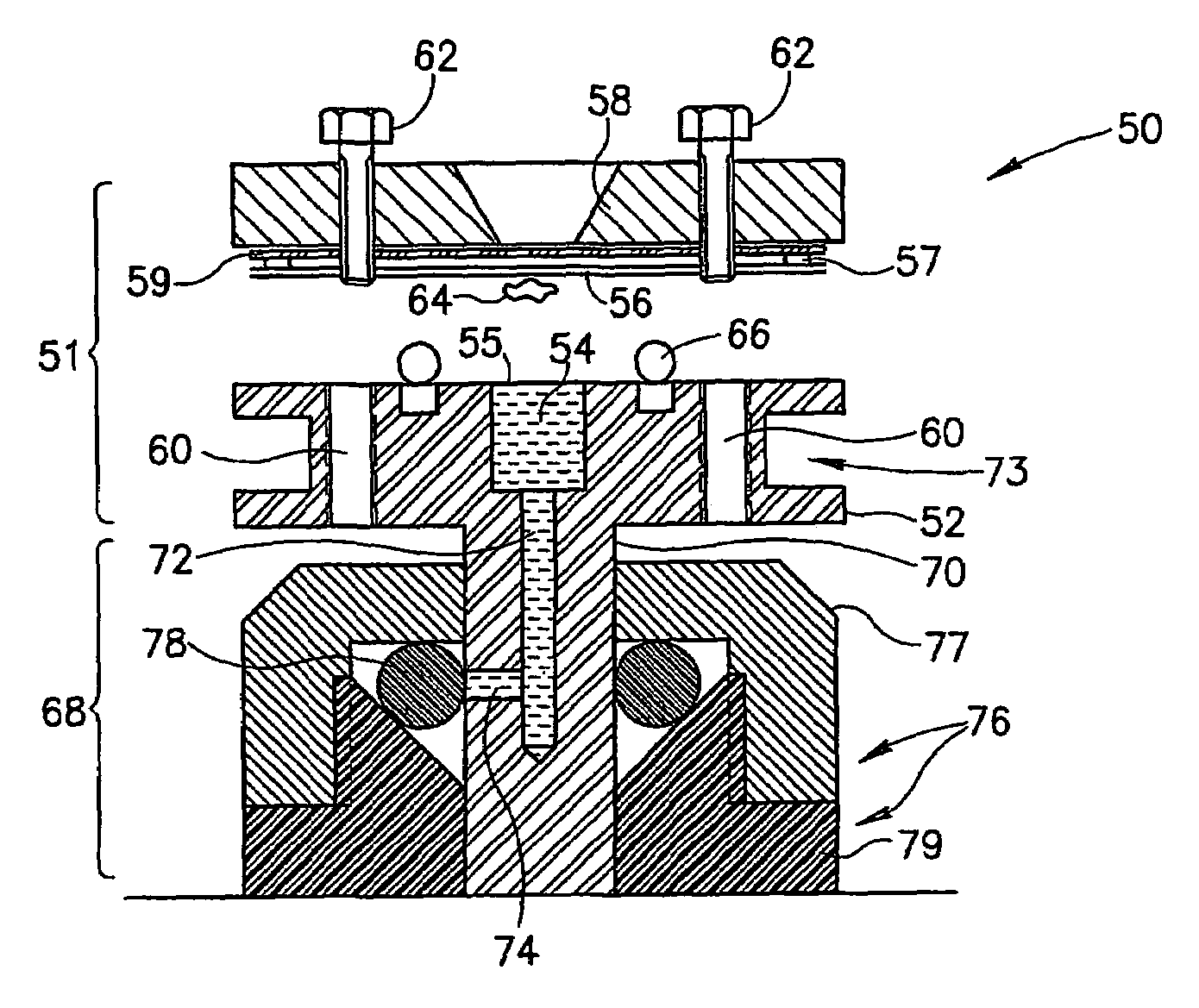
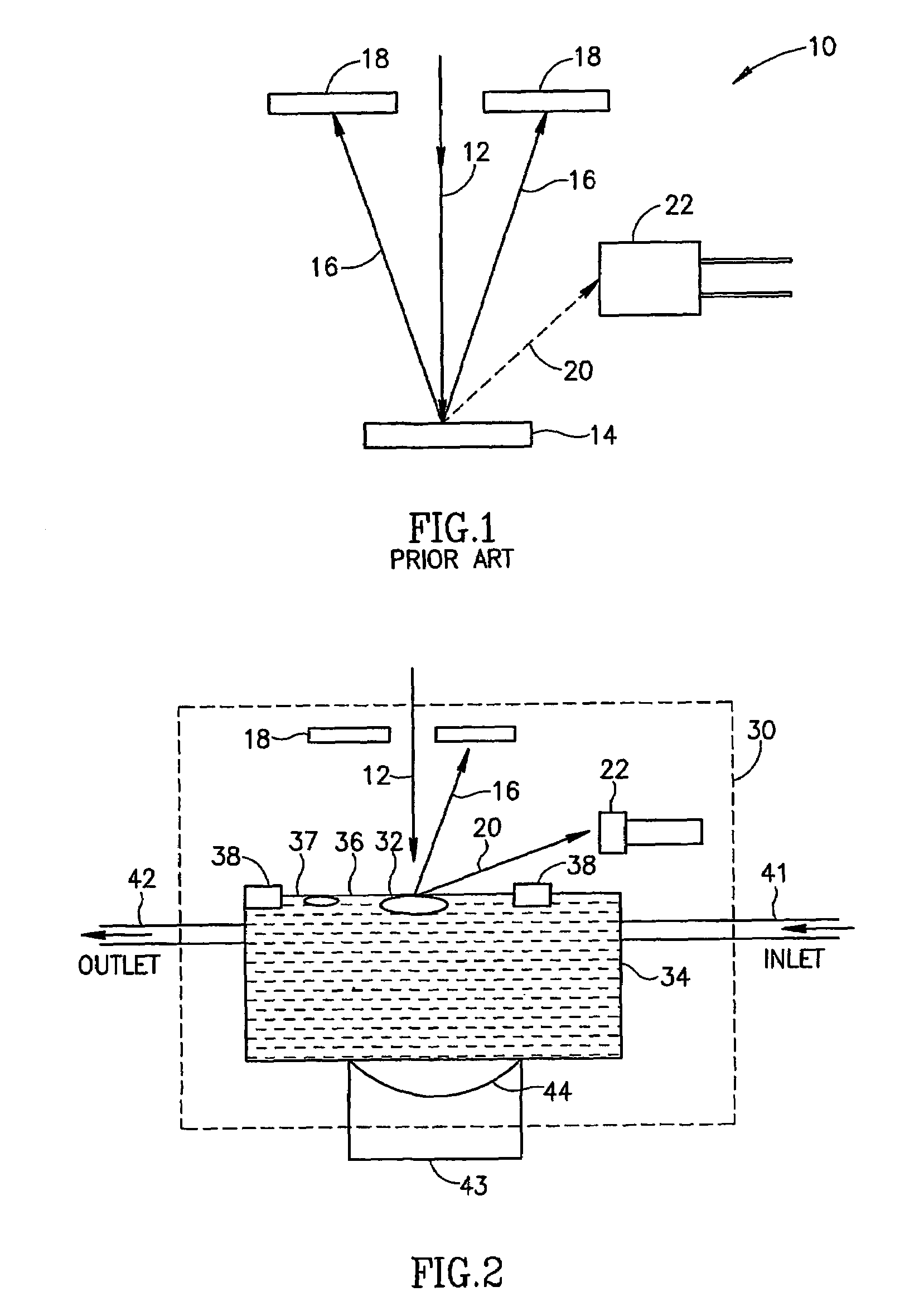
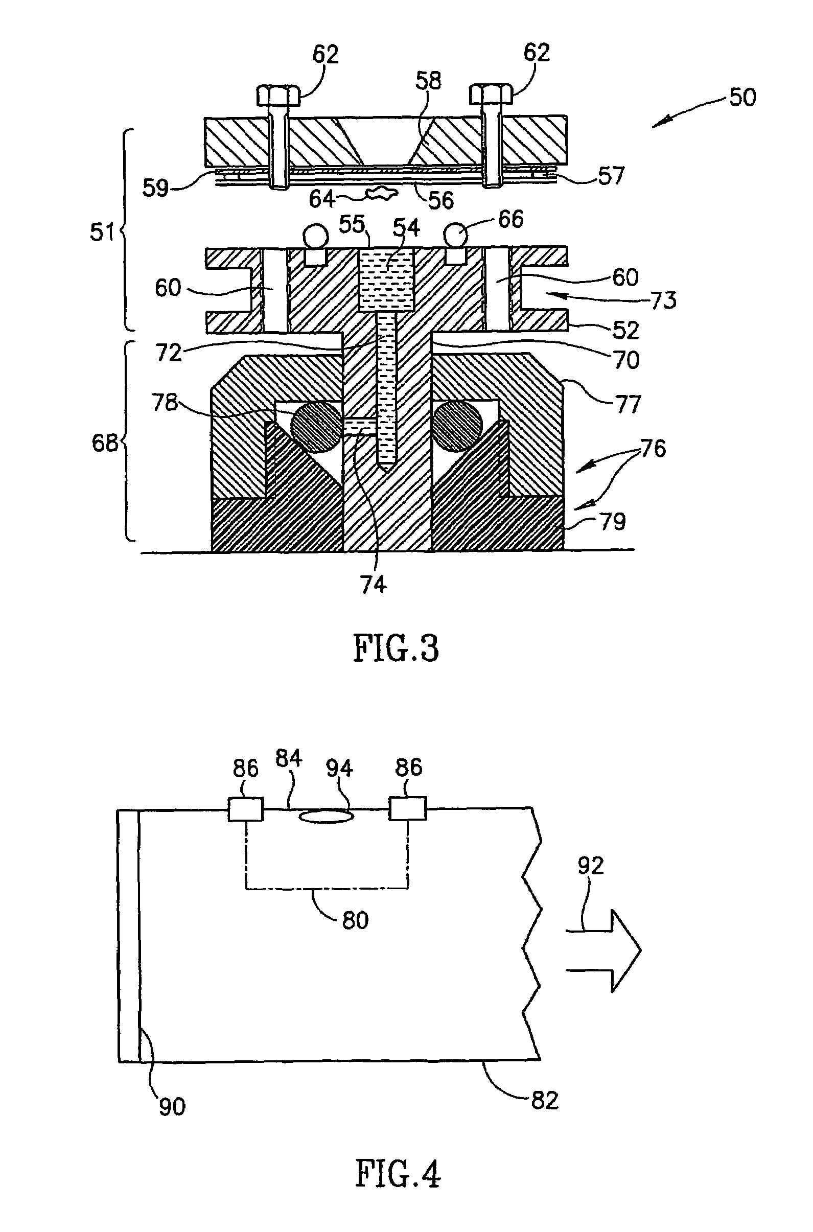
[0018]Preferably, a sample is placed in proximity to the membranes. Alternatively a sample is mounted in contact with said membrane and this gives the advantage that the electron beam has to traverse a minimal distance through matter (as opposed to vacuum) to reach the sample. The membrane is preferably supported by a supporting grid placed between the membrane and a vacuum.
[0020]According to a second aspect of the present invention there is provided apparatus for high resolution inspection of an object in a chamber using a scanning electron beam, comprising a vacuum path for the electron beam and a vacuum-resistant barrier placed in association with the object, said vacuum resistant barrier being placed such as to isolate the object from the vacuum. The object may have thereon a fluorescent and an electro-luminescent marker, whereby a beam of electrons are able to excite photo-emission. The marker may be associated with a particular molecule of interest or the like and this method allows far light based images containing information about individual molecules. Such an embodiment preferably comprises an optical sensing unit and the chamber may be built as part of a light guide. The optical sensing unit may also comprise a photo-multiplier tube.

Problems solved by technology

One of the disadvantages of SEMs is that samples have to be maintained in vacuum, precluding the study of in-vivo processes or the study of wet materials.
Neither of these attempts succeeds, however, in successfully viewing wet objects.
However, so far there is no evidence that wet and / or living objects can be viewed at resolutions of 10 nm and below.
A very high energy beam travels through the specimen and surrounding fluid leading to a poor signal to noise ratio, as well as considerable damage to the sample.
The main reason for the prevalent underutilization of EM is the complexity of sample preparation, that is not only labor intensive and time consuming, but also raises concerns regarding the biological relevance of the results.
At present, therefore, despite a long-felt need, there is no microscope that permits the study of wet samples at resolutions showing macro-molecular and molecular levels of detail.

Method used


Image

  • Device and method for the examination of samples in a non-vacuum environment using a scanning electron microscope
  • Device and method for the examination of samples in a non-vacuum environment using a scanning electron microscope
  • Device and method for the examination of samples in a non-vacuum environment using a scanning electron microscope

Examples


examples

Growing Cells on the Partition Membrane

[0222]The following are exemplary procedures for growing cells on the membrane given as nonlimiting examples. The polyimide partition membranes are first coated with fibronectin (0.1 mg / ml) for 15 minutes. After washing with PBS and culture medium cells are plated in the usual ways at concentrations of 800–1500 cells in 12–15 μl per chamber. After 24 hours cells are ready for manipulation / labeling. Most cell types can grow on the partition membrane itself without the support of an additional matrix like fibronectin, but under such conditions they would be more likely to be washed off during the extensive washing procedures. The viability of the cells is not affected by any of these procedures.

[0223]The current invention is compatible with both adherent and non-adherent cells (as exemplified hereinbelow). For the purpose of imaging non-adherent cells (lymphocytes for example), cells are first labeled in suspension, and then allowed to adhere for...


PUM

PropertyMeasurementUnit
pressureaaaaaaaaaa
pressuresaaaaaaaaaa
thicknessesaaaaaaaaaa

Abstract

A chamber suitable for use with a scanning electron microscope. The chamber comprises at least one aperture sealed with a membrane. The membrane is adapted to withstand a vacuum, and is transparent to electrons and the interior of the chamber is isolated from said vacuum. The chamber is useful for allowing wet samples including living cells to be viewed under an electron microscope.

Description

[0001]This is a continuation of international application Ser. No. PCT / IL01 / 01108, filed Nov. 30, 2001, which claims priority of U.S. Provisional Application No. 60 / 250,879, filed Dec. 1, 2000 and U.S. Provisional Application No. 60 / 306,458, filed Jul. 20, 2001, the entire disclosure of which is hereby incorporated by reference.FIELD OF THE INVENTION[0002]The present invention related to a device and method for the examination of samples in a non-vacuum environment using a scanning electron microscope, and more particularly but not exclusively to the use of such an apparatus and method for inspection of objects in a wet environment.BACKGROUND OF THE INVENTION[0003]Optical microscopy is limited, by the wavelength of light, to resolutions in the range of a hundred and usually hundreds of nanometer. Scanning electron microscopes (SEMs) do not have this limitation and are able to attain a considerably higher resolution in the range of a few nanometers.[0004]One of the disadvantages of S...

Claims


Application Information

Patent Timeline
Patent Type & Authority Patents(United States)
IPC IPC(8): G21K5/08G01N23/00G01N23/225G01N1/28G01N23/227G21K7/00H01J37/16H01J37/20H01J37/28
CPCH01J37/20H01J37/28H01J2237/2807H01J2237/2003H01J2237/006
Inventor MOSES, ELISHAZIK, ORYTHIBERGE, STEPHAN
Owner EL MUL TECH
Why Eureka
Try Eureka