Semi-automatic segmentation algorithm for pet oncology images

a segmentation algorithm and pet oncology technology, applied in the field of medical images, can solve the problems of insufficient accuracy of supervised segmentation methods, inability to accurately segment images, and inability to accurately measure 3d images, etc., and achieve the effect of eliminating noise voxels

Inactive Publication Date: 2006-02-28
GENERAL ELECTRIC CO
View PDF7 Cites 51 Cited by
  • Summary
  • Abstract
  • Description
  • Claims
  • Application Information

AI Technical Summary

Benefits of technology

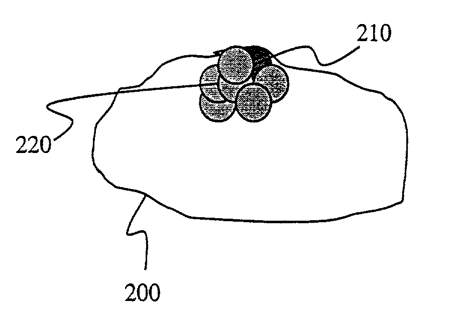
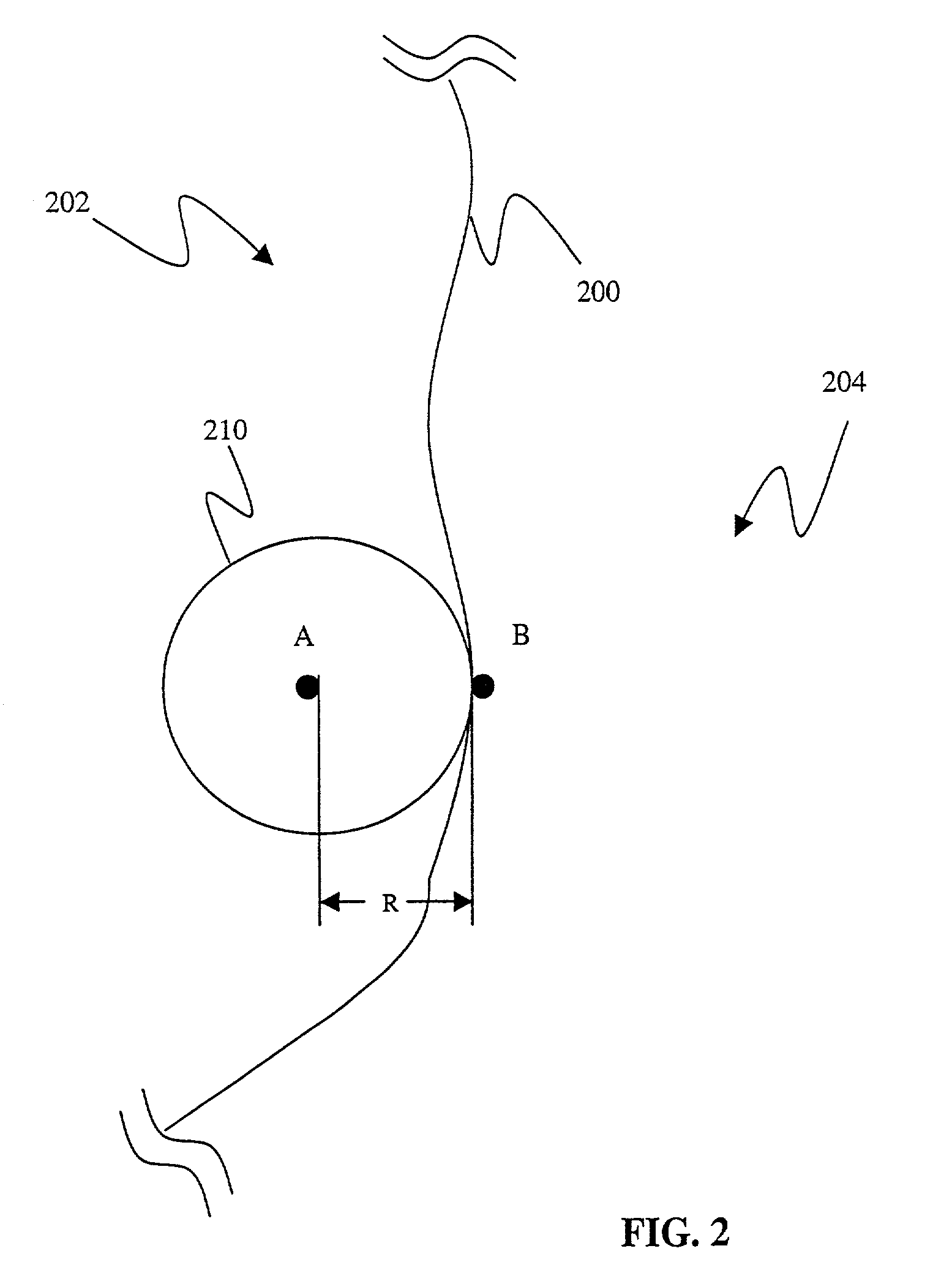
[0006]A method and apparatus for segmenting three-dimensional (3D) medical images containing a region of interest is provided that includes the identification of a first set of seed points within the region of interest and a second set of seed points outside the region of interest. A processor uses the identified seed points to classify all the voxels in the data set using a spatially constrained fuzzy clustering algorithm. Herein, the 3D image set is transformed into a fuzzy partition domain where each voxel is assigned a probability of belonging to the desired region of interest based on a homogeneity function. A processor constructs a set of first spheres within the region of interest centered on the first set of seed points. A plurality of second spheres is generated by the processor. Ones of the plurality of second spheres are accepted that satisfy the homogeneity function threshold as defined by the spatial constrained fuzzy clustering algorithm. A three-dimensional area defining the region of interest is adaptively grown based on the accepting of the ones of the plurality of second spheres. A radius of curvature of the spheres is determined to eliminate noise voxels on the interface between an inside of the region of interest and outside the region of interest. The region of interest defined by the adaptively growing of the three-dimensional area and selecting the radius of curvature is displayed to a human user.

Problems solved by technology

Measurements of 3D images either by registration methods or by segmentation methods are typically tedious because manual editing is required in a 3D representation.
Supervised segmentation methods are not sufficiently accurate because of inter-observer error.
However, most of the available techniques encounter some degree of error.

Method used


Image

  • Semi-automatic segmentation algorithm for pet oncology images
  • Semi-automatic segmentation algorithm for pet oncology images
  • Semi-automatic segmentation algorithm for pet oncology images

Examples


Embodiment Construction

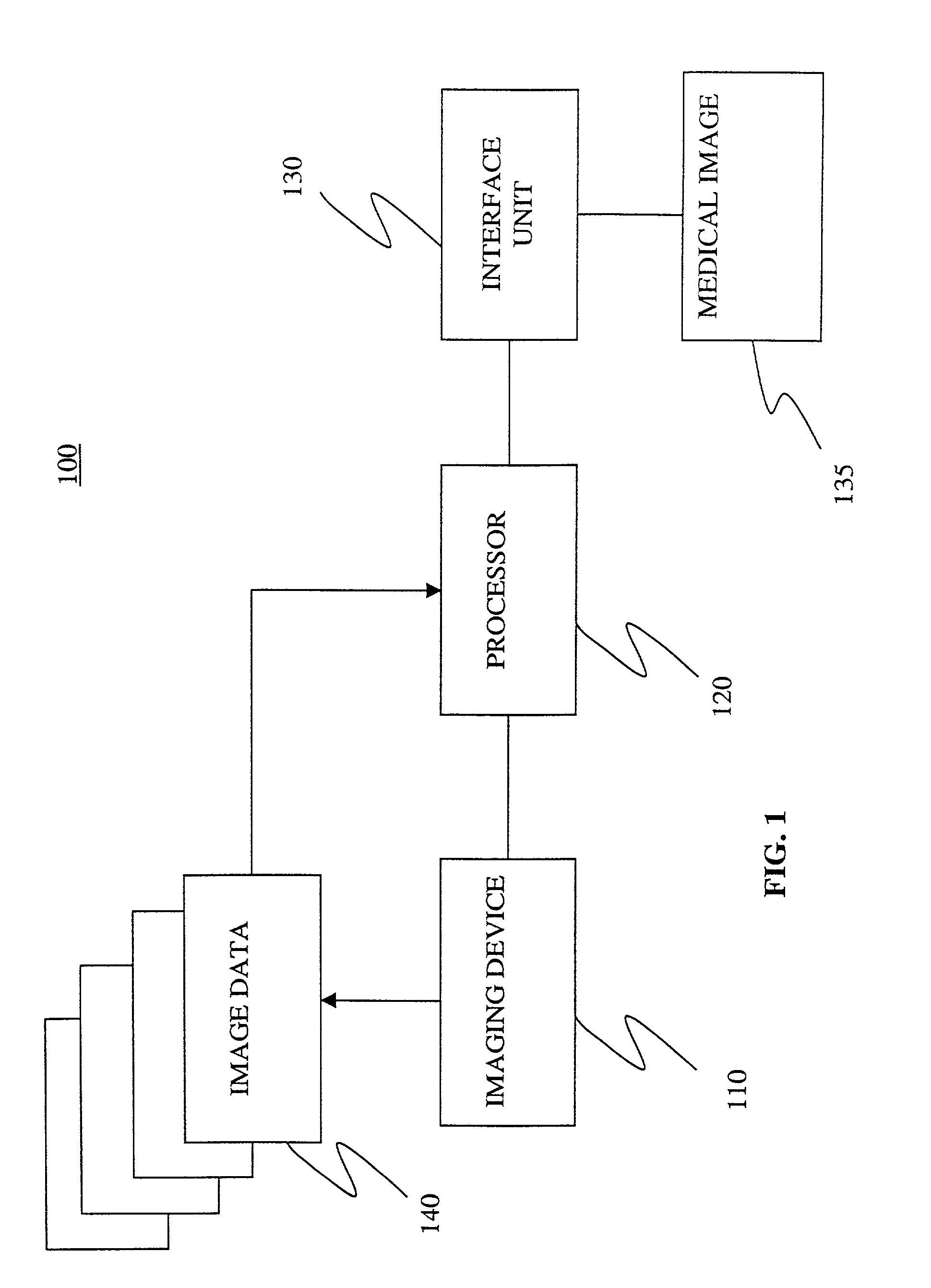
[0011]Referring to FIG. 1, a block diagram of a system 100 for semi-automatic segmentation of a medical image 135. The system 100 includes an imaging device 110 that can be selected from various medical imaging devices for generating a plurality of medical images 135. In one embodiment, medical imaging techniques, such as, for example, positron emission tomography (PET) and magnetic resonance imaging (MRI) systems, are used to generate the medical images 135.

[0012]In one embodiment, during a PET imaging session, a patient is injected with a radio-labeled ligand that specifically targets metabolically hyperactive regions like tumors. The patient horizontally lies inside the scanner. Photon emissions from the decay of the radio-active ligand are detected by a series of photon detectors. The detectors measure the amount radio-active emissions from inside the patient's body. This information is used to compute the concentration of the radio-labeled ligand for sample points in the body. ...


PUM


Abstract

An apparatus and method for segmenting three-dimensional (3D) medical images containing a region of interest is provided that identifies a first set of seed points within the region of interest and a second set of seed points outside the region of interest. A first sphere is constructed within the region of interest. Voxels contained within the medical image are classified using a spatial constrained fuzzy clustering algorithm. A plurality of second spheres is generated. Ones of the plurality of second spheres are accepted that satisfy the homogeneity function threshold as defined by the spatial constricted fuzzy clustering algorithm. A three-dimensional area is grown that defines the region of interest. The region of interest defined by the three-dimensional area is displayed.

Description

BACKGROUND OF THE INVENTION[0001]This invention relates to segmentation of medical images. More particularly, the invention relates to a method and system for segmenting an region of interest in three-dimensional medical images for use in volumetric measurement.[0002]It is well-known to obtain three-dimensional (3D) arrays of data representing one or more physical properties within an interior of a solid body, for example, anatomical structures. In medical imaging, such data is obtained by a variety of non-invasive methods such as positron emission tomography (PET), computed tomography (CT), magnetic resonance imaging (MRI), ultrasound, x-ray or a combination thereof. Regardless of the image data acquisition method, the 3D array of data typically consists of a plurality of sets of three-dimensional coordinates distributed at regular positions about the body of interest. There are a variety of techniques available to generate a three-dimensional model or structure. Typically, a seed ...

Claims


Application Information

Patent Timeline
Patent Type & Authority Patents(United States)
IPC IPC(8): G06K9/00G01R33/32A61B5/00A61B5/055A61B6/03G01T1/161G06T1/00G06T5/00G06T7/00G06T7/40
CPCG06K9/342G06T7/0081G06K2009/366G06T2207/10104Y10S128/922G06T2207/20141G06T2207/20156G06T2207/30016G06T2207/20104G06T7/11G06T7/187G06V10/248G06V10/267
Inventor MANJESHWAR, RAVINDRA MOHANCLINE, HARVEY ELLIS
Owner GENERAL ELECTRIC CO
Why Eureka
Try Eureka