
Method for determining specific nucleotide variations

a specific nucleotide and variation technology, applied in the field of specific nucleotide variations, to achieve the effect of easy automation and small and easy-to-perform procedures

Inactive Publication Date: 2006-04-11
LAB OF AMERICA HLDG
View PDF58 Cites 3 Cited by
  • Summary
  • Abstract
  • Description
  • Claims
  • Application Information

AI Technical Summary

Benefits of technology

The present invention is a method for detecting nucleotide variations, which has several advantages over prior art methods. The method is easy to perform and is especially suitable for routine determinations of point mutations and nucleotide variations. It allows quantification of the proportion of mutated cells in a sample and identifies mutations present in as little as about 0.5% of the analyzed cell population. The method is also easily automated, which is becoming increasingly important in routine diagnostics. The detection step primers are selected from the region immediately adjacent to the variable nucleotide and are dependent on the nucleotide sequence of interest. The method also includes a method for amplification and affinity modification of the target nucleic acid to be detected. The invention also includes individual primer reagents and reagent combinations or kits for practicing the method.

Problems solved by technology

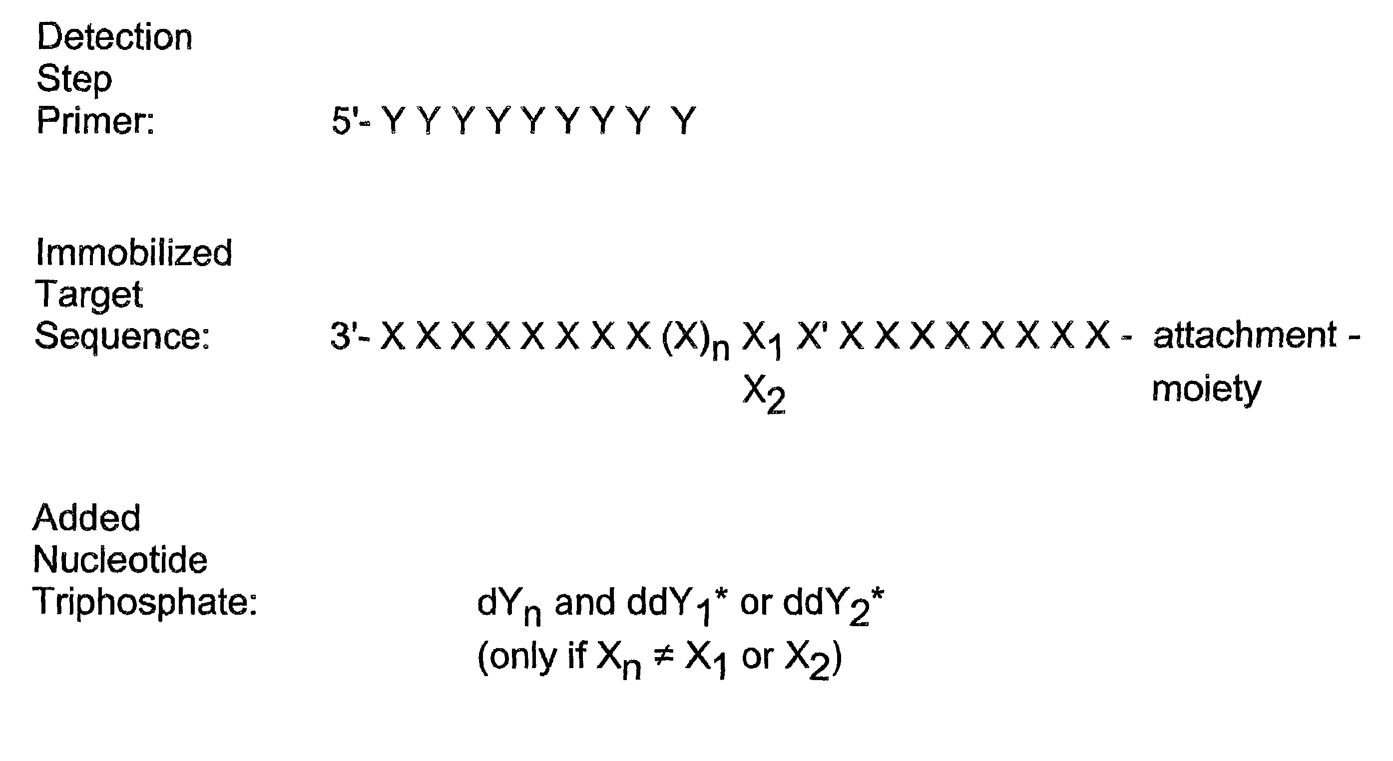
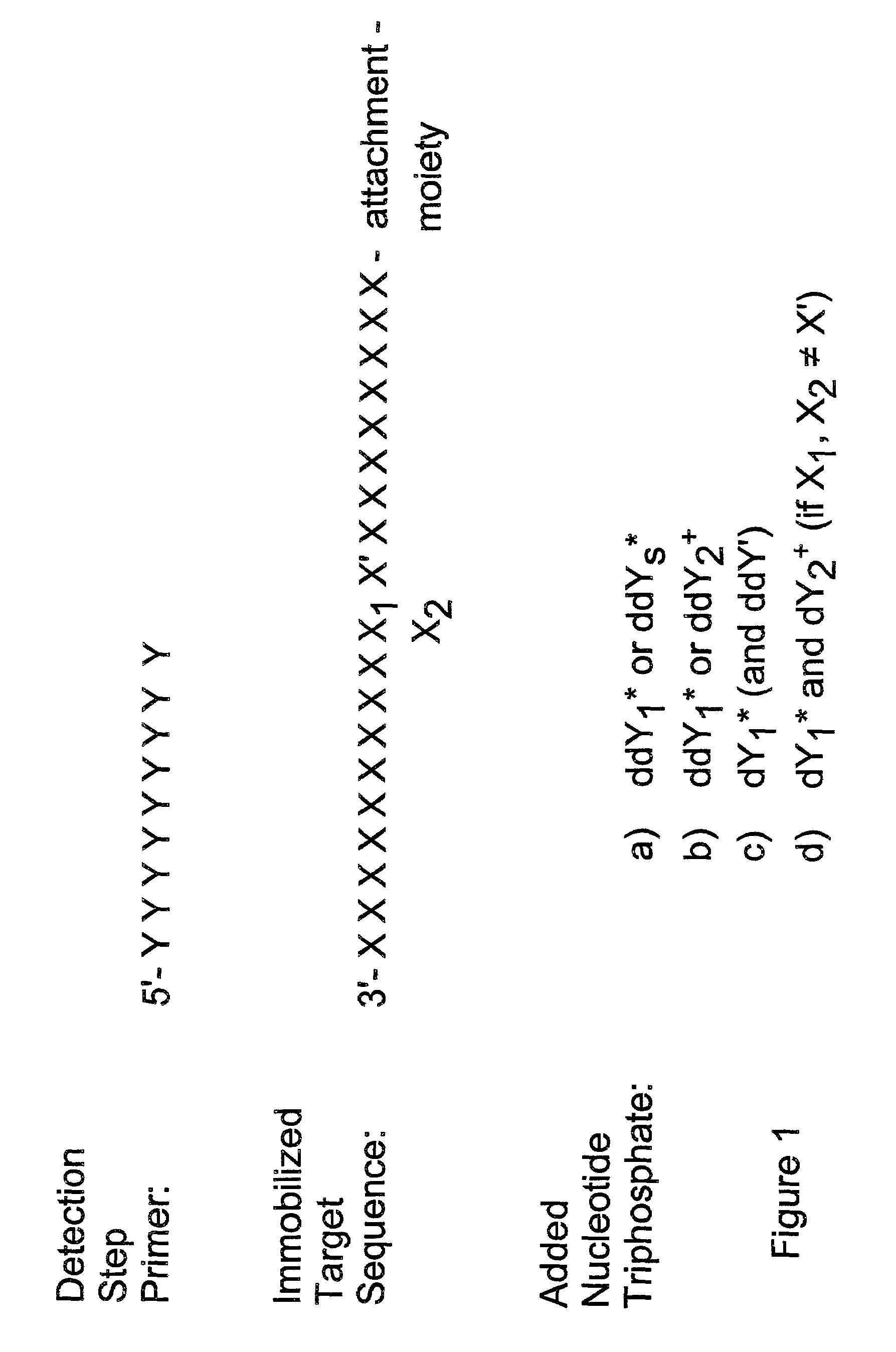
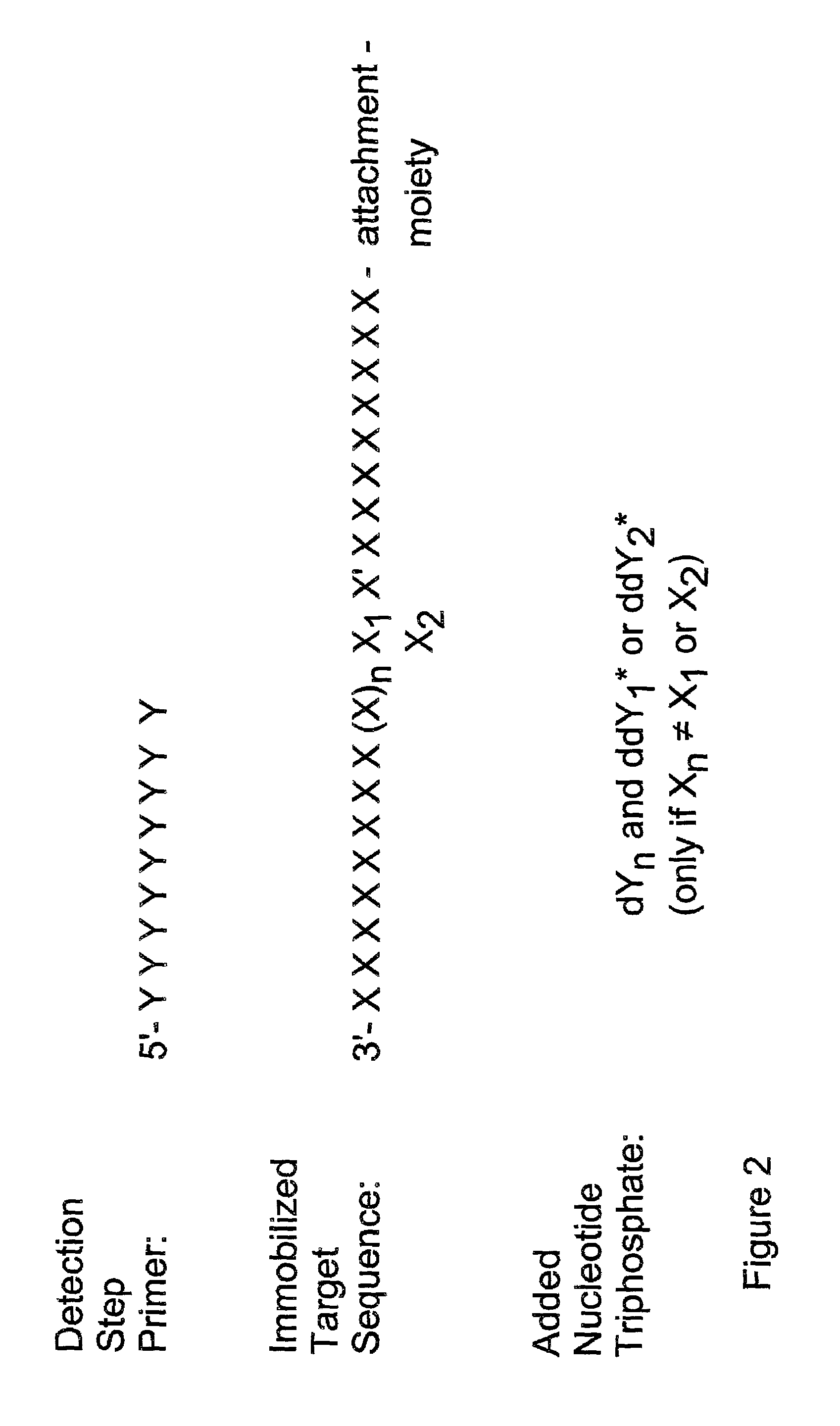
The only limitation concerning the position of the detection step primers is that the sequence between the 3′ end of the detection step primer and the variable nucleotide to be detected must not contain a nucleotide residue of the same type as the one to be detected.

Method used


Image

  • Method for determining specific nucleotide variations
  • Method for determining specific nucleotide variations
  • Method for determining specific nucleotide variations

Examples


example 1

[0075]Identification of the apolipoprotein E polymorphism in the codons for amino acid residues 112 and 158 using [35S] as label

Synthesis of Oligonucleotides

[0076]Four PCR primers (P1–P4) and two detection step oligonucleotide primers (D1 and D2) were synthesized on an Applied Biosystems 381A DNA synthesizer. The nucleotide sequence of the oligonucleotides, and their location (given as nucleotide numbers) on the apolipoprotein E gene (Apo E) were:

[0077]

P1: 5′-AAG GAG TTG AAG GCC TAC AAA T(3616–3637)P3: 5′-GAA CAA CTG AGC CCG GTG GCG G(3649–3670)D1: 5′-GCG CGG ACA TGG AGG ACG TG (3725–3744)D2: 5′-ATG CCG ATG ACC TGC AGA AG (3863–3882)P2: 5′-TCG CGG GCC CCG GCC TGG TAC A(3914–3893)P4: 5′-GGA TGG CGC TGA GGC CGC GCT C(3943–3922)

[0078]A 5′-aminogroup was added to the primer P2 with the aminolink II reagent (Applied Biosystems). The amino group was biotinylated using sulfo-NHS-biotin (Pierce Chemical Co.) and purified by reverse phase HPLC.

The DNA Samples

[0079]Venous blood samples were o...

example 2

[0090]Identification of the variable nucleotide in codon 112 of the apolipoprotein E gene using double labelling ([3H] and [32P]) in one reaction.

Oligonucleotides and DNA Samples

[0091]The PCR primers P1, biotinylated P2, P3 and P4, and the detection step primer D1 described in Example 1 were used in this example. The DNA was extracted from blood samples as described in Example 1.

Polymerase Chain Reaction-Amplification and Affinity-Capture

[0092]The PCR amplification and the affinity-capture of the amplified fragments were carried out as described in Example 1. In the last washing step each sample was divided into two aliquots.

Identification of the Variable Nucleotide at Codon 112

[0093]The particles were suspended in 10 μl of buffer (see Example 1) containing 2 pmol of the detection step primer D1, which hybridizes immediately 3′ to the variable nucleotide in codon 112. The annealing reaction was carried out as in Example 1. One μl of 0.1 M DTT was added.

[0094]The identification of th...

example 3

[0098]Identification of the variable nucleotide in codon 158 of the apolipoprotein E gene after a single polymerase chain reaction amplification.

Oligonucleotides and DNA Samples

[0099]In this example the biotinylated primer P2 and the primer P3 were used in the PCR amplification. The detection step primer was D2 (see Example 1). The DNA was extracted from venous blood as described in Example 1.

Polymerase Chain Reaction-Amplification and Affinity-Capture

[0100]The DNA (100 ng per sample) was amplified with the biotinylated P2 and the P3 primer (final concentration 1 μM) at the conditions described in Example 1 with the following exception: Only one amplification process consisting of 30 cycles of 1 min. at 96° C., 1 min. at 55° C. and 1 min. 72° C. was carried out.

[0101]The affinity-capture on avidin-coated polystyrene particles was done as in Example 1. Each sample was divided into two parts in the last washing step.

Identification of the Variable Nucleotide in Codon 158

[0102]The parti...


PUM

PropertyMeasurementUnit
concentrationaaaaaaaaaa
pHaaaaaaaaaa

Abstract

Detection of variable nucleotide(s) is based on primer extension and incorporation of detectable nucleoside triphosphates. By selecting the detection step primers from the region immediately adjacent to the variable nucleotide, this variation can be detected after incorporation of as few as one nucleoside triphosphate. Labelled nucleoside triphosphates matching the variable nucleotide are added and the incorporation of a label into the detection step primer is measured. The selection of the detection step primer is important to the method according to this invention and is dependent on the nucleotide sequence of interest. The detection step primers are preferably selected so as to span the region immediately toward the 3′ end from the variable nucleotide to be detected. The detection step primers can also be complementary to a sequence beginning several nucleotides removed from the variable nucleotide. The only limitation concerning the position of the detection step primers is that the sequence between the 3′ end of the detection step primer and the variable nucleotide to be detected must not contain a nucleotide residue of the same type as the one to be detected. The detection step primers can equally well be chosen to be complementary to either the coding or non-coding strands of the nucleotide sequence of interest.

Description

[0001]This application is a continuation of application Ser. No. 08 / 162,376 filed Dec. 2, 1993, now U.S. Pat. No. 6,013,431, which is a continuation of application Ser. No. 07 / 656,575 filed Feb. 15, 1991, abandoned, which is a continuation in part of application Ser. No. 07 / 482,005 filed Feb. 16, 1990, abandoned.TECHNICAL FIELD[0002]The present invention relates to a method and reagents for determining specific nucleotide variations in a defined polynucleotide region, and to the use of this method in identifying specific point mutations and genetic variations.BACKGROUND OF THE INVENTION[0003]The genetic information of living organisms is carried in the nucleotide sequence of their genome. In the process of gene expression the nucleotide sequence is translated to amino acid sequences, i.e. proteins. Minor changes in the nucleotide sequence, even a single base substitution, may result in an altered protein product. The altered quality or quantity of given proteins changes the phenotyp...

Claims


Application Information

Patent Timeline
Patent Type & Authority Patents(United States)
IPC IPC(8): C12Q1/68C07H21/04C12P19/34
CPCC12Q1/6869C12Q1/6834C12Q1/6886C12Q2563/101C12Q2535/125C12Q2535/101
Inventor SODERLUND, HANS E.SYVANEN, ANNE-CHRISTINE
Owner LAB OF AMERICA HLDG
Learn More