Elimination of endogenous porcine retrovirus

a technology of endogenous retrovirus and elimination, which is applied in the field of compositions, can solve the problems of insufficient human organs and cells for use as donor tissues, inability to prevent infection by endogenous retroviruses, and insufficient perv sequences to indicate infectious potential, so as to reduce the risk of perv transmission

Inactive Publication Date: 2006-05-02
MAYO FOUND FOR MEDICAL EDUCATION & RES
View PDF5 Cites 6 Cited by
  • Summary
  • Abstract
  • Description
  • Claims
  • Application Information

AI Technical Summary

Benefits of technology

[0017]Therefore, the compositions of the present invention also provide methods for reducing the risk of transmission of PERV from a xenotransplant tissue to a host or recipient.

Problems solved by technology

However, there are insufficient resources of human organs and cells for use as donor tissue.
Although risks associated with some pathogens can be reduced by breeding for and using specific-pathogen-free (SPF) animal colonies, this approach is not feasible for preventing infection from endogenous retroviruses, because these pathogens exist in the germine of all pigs.
However, other studies of cells cultured directly from pig tissue (primary culture) showed no evidence of infectious potential.
Keeping in mind that all pig genomes have PERV sequences, but that not all PERV are infectious, prescreening of a transplant tissue merely for presence of a PERV sequence is not a sufficient indicator of its infectious potential.
Therefore, current methods for detecting the presence of PERV are limited by their inability to determine which PERV loci are infectious.

Method used


Image

  • Elimination of endogenous porcine retrovirus
  • Elimination of endogenous porcine retrovirus
  • Elimination of endogenous porcine retrovirus

Examples


example 1

[0062]The following is provided for exemplary purposes only and to further aid in the understanding of the invention. As is apparent from the disclosure provided herein, one skilled in the art can make, use or obtain any clone or clones to unique flanking sequences of PERV loci using the methods as exemplified below and described herein.

Cloning of Unique Flanking Sequences

Construction of Pig Genomic Library

[0063]All unique 3′ flanking sequence and unique 5′ flanking sequence clones were isolated from a genomic pig library constructed as follows:

[0064]High molecular weight (>100 kb) genomic liver DNA from a transgenic (CD59 / DAF) male pig (for example, an F2 outcross consisting of Large White, Landrace, and Duroc) was partially digested with Sau3Al, and 9–24 KB fragments were isolated by sucrose gradient ultracentrifugation. The fragments were cloned into a Lambda FIX II vector (Stratagene, Calif.) to construct a pig genomic library.

[0065]The amplified library titer was 5.2×10⁹ pfu / ml...

example 2

Cloning of a Unique 3′ Flanking Sequence, G3–25 (SEQ ID NO:8)

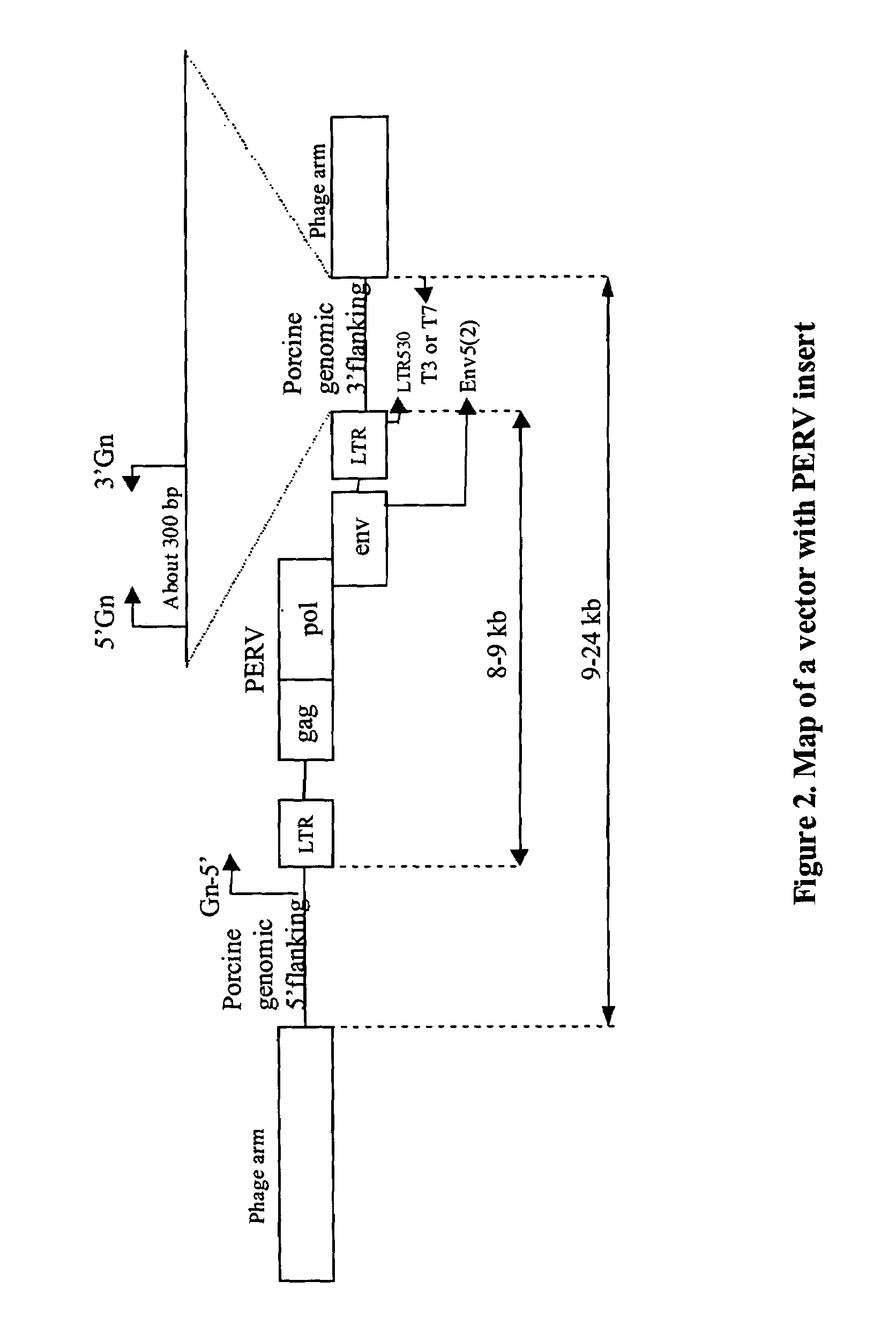
[0087]The following illustrates how one skilled in the art would make or obtain a PERV clone having a unique 3′ flanking sequence. Given the following description and teachings, one skilled in the art can apply the following methods to obtain any PERV clone described herein and is not intended to be limited to isolation of a specific PERV clone. A lambda library was constructed as described in Example 1. A PERV clone was isolated from a lambda FIX II vector using the env-cons probe (SEQ ID NO:1). In order to distinguish a 3′ flanking sequence as distinct and unique (i.e. as a PERV locus), the PERV lambda clone was amplified using the primer pairs env5(2) and T3 or T7 (see FIG. 8) in order to generate a product corresponding from the end of the env region of PERV to porcine genomic sequence flanking the 3′ region of PERV LTR. The purified PCR product was then sequenced with the LTR530 (located 100 bp upstream from the end o...

example 3

Cloning of a Unique 3′ Flanking Sequence, G19-A45 (SEQ ID NO:24)

[0090]The following illustrates how one skilled in the art would make or obtain a PERV clone having a unique 3′ flanking sequence. Given the following description and teachings, one skilled in the art can apply the following methods to obtain any PERV clone described herein and is not intended to be limited to isolation of a specific PERV clone. A lambda library was constructed as described in Example 1. A PERV clone was isolated from a lambda FIX II vector using the PERV-A probe (SEQ ID NO:4). In order to distinguish a 3′ flanking sequence as distinct and unique (i.e. as a PERV locus), the PERV lambda clone was amplified using the primer pairs env5(2) and T3 or T7 (see FIG. 8) in order to generate a product corresponding from the end of the env region of PERV to porcine genomic sequence flanking the 3′ region of PERV LTR. The purified PCR product was then sequenced with the LTR530 (located 100 bp upstream from the end ...


PUM

PropertyMeasurementUnit
temperatureaaaaaaaaaa

Abstract

Porcine nucleic acid sequences flanking potentially infectious porcine endogenous retroviral (PERV) insertion sites have been identified and isolated. The unique flanking sequences include porcine nucleic acid sequences that flank the 3′ end and porcine nucleic acid sequences that flank the 5′ end of PERV insertion sites. The present invention provides compositions and methods for detecting presence of PERV in a sample, particularly those with infectious potential. In addition, the invention relates to breeding of pigs or selection of porcine tissue that is free of infectious PERV for use as a xenotransplant tissue.

Description

CROSS-REFERENCE TO RELATED APPLICATIONS[0001]The present application claims priority to U.S. Ser. No. 60 / 279,337, filed Mar. 28, 2001, herein incorporated by reference in its entirety.STATEMENT AS TO RIGHTS IN INVENTIONS MADE UNDER FEDERALLY SPONSORED RESEARCH AND DEVELOPMENT[0002]Not applicable.BACKGROUND OF THE INVENTION[0003]1. Field of the Invention[0004]This invention relates to compositions and methods for detecting the presence of endogenous retroviruses, in particular, the detection of porcine endogenous retroviruses (PERV) in tissues useful as a xenograft.[0005]2. General Background and State of the Art[0006]The most common source of tissue used today as donor tissue for transplantation is the allograft (same species, different person). However, there are insufficient resources of human organs and cells for use as donor tissue. The shortage of human donor material has resulted in alternative solutions for transplantation. Xenotranplantation, which is the use of living tissu...

Claims


Application Information

Patent Timeline
Patent Type & Authority Patents(United States)
IPC IPC(8): A01K67/033A01K67/027C12N15/00A01K67/02C07K14/15C12N1/15C12N1/19C12N1/21C12N5/10C12N15/09C12Q1/68C12Q1/70
CPCA01K67/027C12Q1/702C12Q1/701C07K14/005A01K2227/108A01K2267/02C12N2740/10022
Inventor CUI, CUNQIDIAMOND, LISA E.LOGAN, JOHN S.
Owner MAYO FOUND FOR MEDICAL EDUCATION & RES
Why Eureka
Try Eureka