Peptidoglycan recognition proteins

a technology of peptidoglycans and recognition proteins, applied in the direction of depsipeptides, peptide/protein ingredients, fungi, etc., can solve the problems of weight loss in cancer patients, and achieve the effect of regulating the growth activity of keratinocytes and preventing septic shock

Inactive Publication Date: 2006-05-09
HUMAN GENOME SCI INC
View PDF6 Cites 131 Cited by
  • Summary
  • Abstract
  • Description
  • Claims
  • Application Information

AI Technical Summary

Benefits of technology

[0025]In another aspect, a method for identifying PGRP-K, PGRP-W, or PGRP-C receptors is provided, as well as a screening assay for agonists and antagonists using such receptors. This assay involves determining the effect a candidate compound has on PGRP-K, PGRP-W, or PGRP-C binding to the PGRP-K, PGRP-W, or PGRP-C receptor. In particular, the method involves contacting a PGRP-K, PGRP-W, or PGRP-C receptor with an PGRP-K, PGRP-W, or PGRP-C polypeptide and a candidate compound and determining whether PGRP-K, PGRP-W, or PGRP-C polypeptide binding to the PGRP-K, PGRP-W, or PGRP-C receptor is increased or decreased due to the presence of the candidate compound. The antagonists may be employed to prevent septic shock, inflammation, and to regulate the growth activity of keratinocytes.
[0026]The present inventors have discovered that PGRP-K, PGRP-W, and PGRP-C is expressed in keratinocytes, wound healing tissues, and chondrosarcomas, respectively. For a number of disorders of these tissues and cells, such as tumor and tumor metastasis, infection of bacteria, viruses and other parasites, immunodeficiencies, septic shock, apoptosis or proliferation of these tissues, and proper antigen processing and presentation, it is believed that significantly higher or lower levels of the PGRP-K, PGRP-W, or PGRP-C gene expression can be detected in certain tissues (e.g., keratinocytes, wound-healing tissues, and chondrosarcoma) or bodily fluids (e.g., serum, plasma, urine, synovial fluid or spinal fluid) taken from an individual having such a disorder, relative to a “standard” PGRP-K, PGRP-W, or PGRP-C gene expression level, i.e., the PGRP-K, PGRP-W, or PGRP-C expression level in tissue or bodily fluids from an individual not having the disorder. Thus, the invention provides a diagnostic method useful during diagnosis of a disorder, which involves: (a) assaying PGRP-K, PGRP-W, or PGRP-C gene expression levels in cells or body fluid of an individual; (b) comparing the PGRP-K, PGRP-W, or PGRP-C gene expression level with a standard PGRP-K, PGRP-W, or PGRP-C gene expression level, whereby an increase or decrease in the assayed PGRP-K, PGRP-W, or PGRP-C gene expression level compared to the standard expression level is indicative of a disorder.

Problems solved by technology

A major problem in cancer patients is weight loss, usually associated with anorexia.

Method used


Image

  • Peptidoglycan recognition proteins
  • Peptidoglycan recognition proteins
  • Peptidoglycan recognition proteins

Examples


example 1

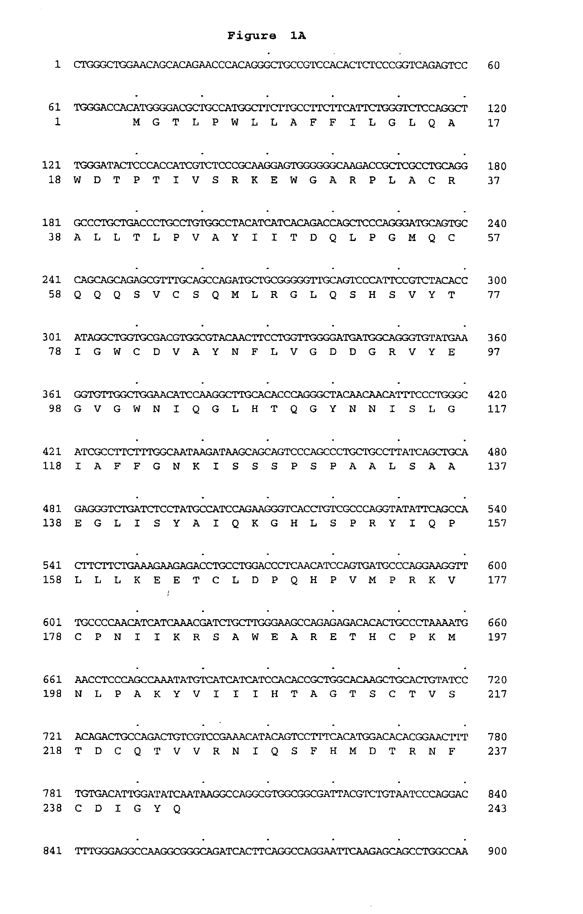
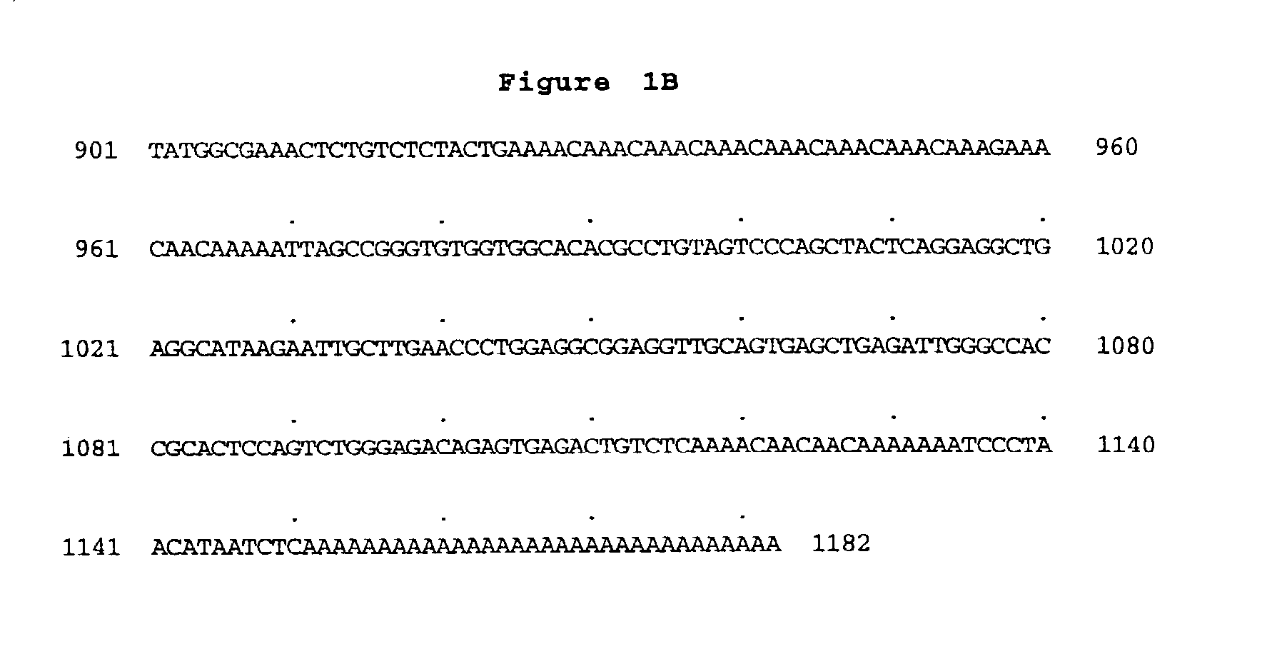
Isolation of PGRP-K, PGRP-W, and / or PGRP-C cDNA Clone(s) from the Deposited Sample(s)

[0305]The cDNA for PGRP-K (ATCC Accession No: 203564) is inserted into the Sal I and Not I multiple cloning site of pCMVSport 2.0 (Life Technologies). pCMVSport 2.0 contains an ampicillin resistance gene and may be transformed into E. coli strain DH10B, available from Life Technologies. (See, for instance, Gruber, C. E., et al., Focus 15:59-(1993).)

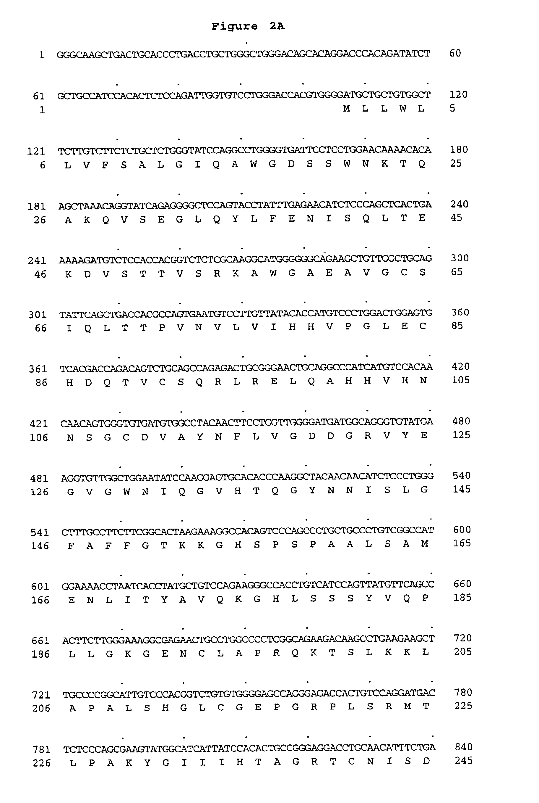
[0306]The cDNA for PGRP-W (ATCC Accession No: 203563) is inserted into the Sal I and Not I multiple cloning site of pCMVSport 3.0 (Life Technologies). pCMVSport 3.0 contains an ampicillin resistance gene and may be transformed into E. coli strain DH10B, available from Life Technologies. (See, for instance, Gruber, C. E., et al., Focus 15:59-(1993).)

[0307]The cDNA for PGRP-C (ATCC Accession No: 209683) is inserted into the EcoRI and Xho I multiple cloning site of Uni-Zap XR (Stratagene). Uni-Zap XR contains an ampicillin resistance gene and may be transfor...

example 2

Isolation of PGRP-K, PGRP-W, or PGRP-C Genomic Clones

[0315]A human genomic P1 library (Genomic Systems, Inc.) is screened by PCR using primers selected for the cDNA sequences corresponding to SEQ ID NO:1, SEQ ID NO:3, or SEQ ID NO:5, according to the method described in Example 1. (See also, Sambrook.)

example 3

Chromosomal Mapping of PGRP-K, PGRP-W, or PGRP-C

[0316]An oligonucleotide primer set is designed according to the sequence at the 5′ end of SEQ ID NO:1, SEQ ID NO:3, or SEQ ID NO:5. This primer preferably spans about 100 nucleotides. This primer set is then used in a polymerase chain reaction under the following set of conditions: 30 seconds, 95 degree C.; 1 minute, 56 degree C.; 1 minute, 70 degree C. This cycle is repeated 32 times followed by one 5 minute cycle at 70 degree C. Human, mouse, and hamster DNA is used as template in addition to a somatic cell hybrid panel containing individual chromosomes or chromosome fragments (Bios, Inc). The reactions is analyzed on either 8% polyacrylamide gels or 3.5% agarose gels. Chromosome mapping is determined by the presence of an approximately 100 bp PCR fragment in the particular somatic cell hybrid.


PUM

PropertyMeasurementUnit
lengthaaaaaaaaaa

Abstract

The present invention relates to three novel peptidoglycan recognition binding proteins expressed by keratinocytes, wound-healing tissues and chondrosarcoma tissue. More specifically, isolated nucleic acid molecules are provided encoding human peptidoglycan recognition protein-related proteins, referred to herein as PGRP-K (Keratinocytes), PGRP-W (Wound-healing), and PGRP-C (Chondrosarcoma) of FIGS. 1A–B, FIGS. 2A–C, and FIG. 3, respectively, each having homology to both human peptidoglycan recognition protein (PGRP) as well as murine Tag-7. PGRP-K, PGRP-W, and PGRP-C polypeptides are also provided. Further provided are vectors, host cells and recombinant methods for producing the same. The invention also relates to both the inhibition and enhancement of activities of PGRP-K, PGRP-W, and PGRP-c polypeptides and diagnostic methods for detecting PGRP-K, PGRP-W, and PGRP-C gene expression.

Description

[0001]This application is a Divisional of U.S. patent application Ser. No. 09 / 469,242, filed Dec. 22, 1999, now U.S. Pat. No. 6,444,790, which claims benefit under 35 U.S.C. § 119(e) of U.S. application Ser. No. 60 / 113,809, filed Dec. 23, 1998, all of which are incorporated herein by reference in their entirety.FIELD OF THE INVENTION[0002]The present invention relates to three novel peptidoglycan recognition binding proteins expressed by keratinocytes, wound-healing tissues and chondrosarcoma tissue. More specifically, isolated nucleic acid molecules are provided encoding human peptidoglycan recognition protein-related proteins, referred to herein as PGRP-K (Keratinocytes), PGRP-W (Wound-healing), and PGRP-C (Chondrosarcoma) of FIGS. 1A–B, FIGS. 2A–C, and FIG. 3, respectively, each having homology to both human peptidoglycan recognition protein (PGRP) as well as murine Tag-7. PGRP-K, PGRP-W, and PGRP-C polypeptides are also provided. Further provided are vectors, host cells and reco...

Claims


Application Information

Patent Timeline
Patent Type & Authority Patents(United States)
IPC IPC(8): C07K16/00C12N5/06C12P21/08G01N33/53G01N33/50A61K31/7088A61K38/00A61K48/00A61P31/04A61P31/12A61P35/00A61P35/04A61P37/02A61P43/00C07K14/47C07K16/18C12N1/15C12N1/19C12N1/21C12N5/10C12N15/09C12P21/02C12Q1/02C12Q1/68G01N33/15G01N33/566
CPCC07K14/47A61K38/00A61P31/04A61P31/12A61P35/00A61P35/04A61P37/02A61P43/00
Inventor YOUNG, PAUL E.RUBEN, STEVEN M.ROSEN, CRAIG A.OLSEN, HENRIK S.
Owner HUMAN GENOME SCI INC
Why Eureka
Try Eureka