Wide field method for detecting pathogenic microorganisms

a microorganism and wide field technology, applied in the field of chemical and biological analysis, can solve the problems of ineffective microorganisms, biovariability which complicates optical analysis, and organic biological molecules are far more complicated materials, so as to reduce the effect of biovariability, reduce the intensity of irradiation, and reduce optical radiation

Inactive Publication Date: 2006-06-06
CHEMIMAGE CORP
View PDF27 Cites 64 Cited by
  • Summary
  • Abstract
  • Description
  • Claims
  • Application Information

AI Technical Summary

Benefits of technology

[0008]Current analytical optical measurements of materials typically fall into either of two categories. One in which a pure batch of the material is available to be measured as a bulk macroscopic sample, i.e. in a large sampling volume, and a second which utilizes micro-analysis techniques to analyze a single small object. In practical situations, sampling of a macroscopic volume does not have the sensitivity to see small concentrations of pathogens, a particularly important need for evaluating BWAs and CWAs. On the other hand, micro-analysis methods allow one to focus on the analysis of an individual microscopic object by using a highly focused excitation beam. Such focused beam optical methods are effective for inorganic and organic particulates but can be ineffective for microorganisms due to their bio-variability and sensitivity to irradiation. Bio-variability comes in two forms: one from variations in the signal from different locations in the microorganism, i.e. the sampling probe is smaller than the organism, and the other from small variations between the same type of organism. In addition, biological microorganisms can decompose or ‘burn’ under the high power density of the highly focused incident beam. This requires reducing the optical radiation on the microorganism so as to not destroy any one organism and thereby collecting optical emission for longer times to compensate for the reduced irradiation intensity. To reduce the effects of bio-variability, sampling many small objects is also needed. Thus use of optical microanalysis methods to sequentially sample many organisms thereby becomes very time consuming. Our approach removes these limitations by using a wide field of view and a wide field of optical excitation to average the optical emission from a small plurality of pathogenic microorganisms. The resulting sensitivity allows us to detect and reliably identify a few microorganisms even in the presence of a matrix of foreign material, i.e. so called ‘masking agents’.
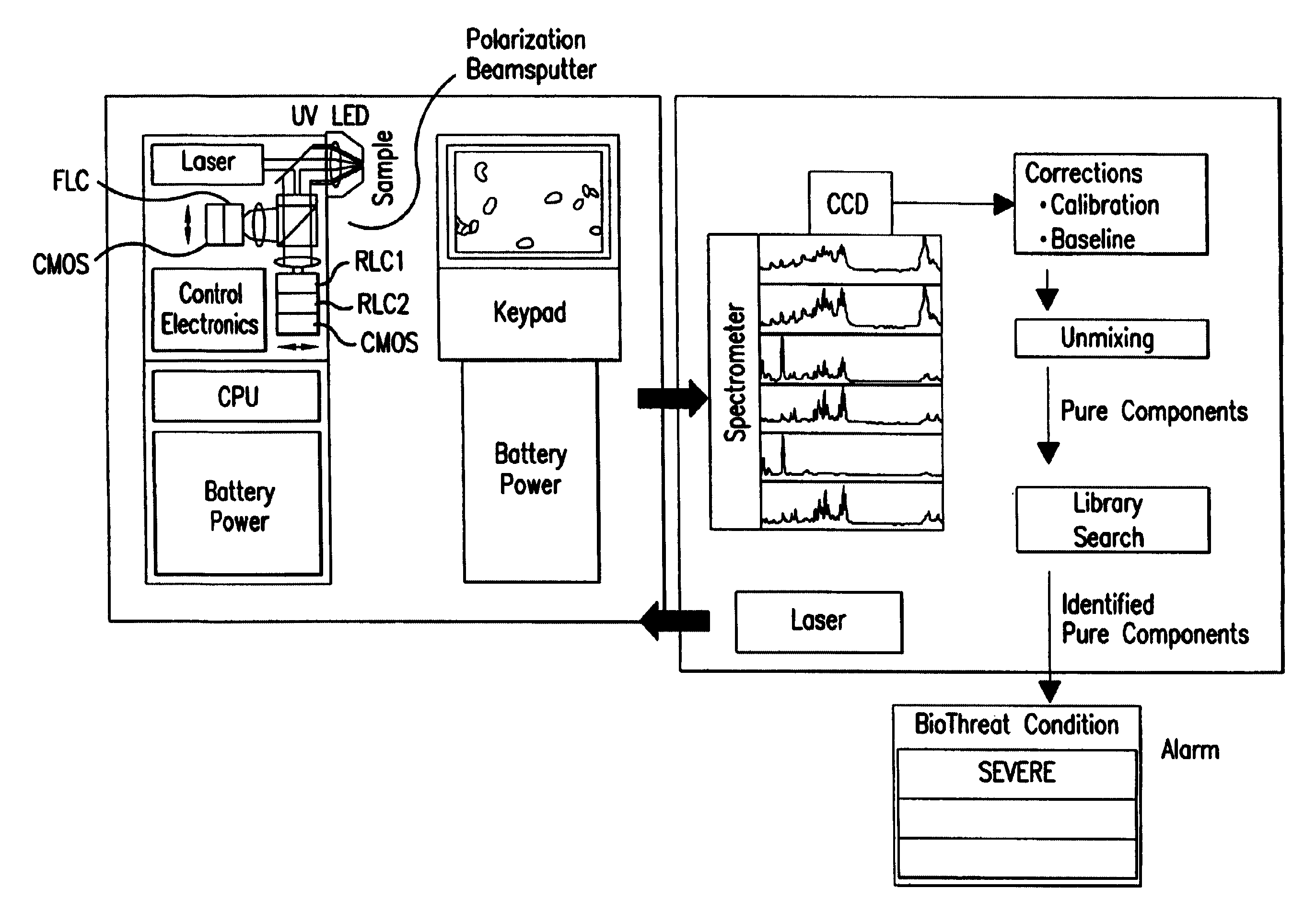
[0029]The system and methodology described above are applied in a variety of modes. The system is applied as a laboratory or transportable field Raman microscope which integrates a dispersive Raman spectrometer and digital data analysis module. The system is also applied as a UV / Vis / NIR fluorescence, Raman, or UV / Vis / NIR / Mid-IR absorption / reflectance macroscope system such as ChemImage's CONDOR Macroscope. Alternatively, the system is applied as a laboratory or field fiberscope such as ChemImage's RAVEN endoscope. Each of the modes of application are used separately or in combination with one another to achieve the desired speed and results. In addition, the properties of the wide area detection method are scalable down to the size of the wide area being investigated thereby allowing the miniaturization of the required hardware to enable a hand held device with local sampling and / or remote analysis capabilities.

Problems solved by technology

Organic biological molecules tend to be far more complicated materials.
These complex structures and structural / chemical variations gives rise to bio-variability which complicates optical analysis.
Such focused beam optical methods are effective for inorganic and organic particulates but can be ineffective for microorganisms due to their bio-variability and sensitivity to irradiation.
In addition, biological microorganisms can decompose or ‘burn’ under the high power density of the highly focused incident beam.
Thus use of optical microanalysis methods to sequentially sample many organisms thereby becomes very time consuming.
Infrared, Raman, visible reflectance, fluorescence and near Infrared optical spectroscopies has been applied to detect microorganisms with limited success.

Method used


Image

  • Wide field method for detecting pathogenic microorganisms
  • Wide field method for detecting pathogenic microorganisms
  • Wide field method for detecting pathogenic microorganisms

Examples


Embodiment Construction

[0060]Methods of Raman chemical imaging and spectroscopy are extensively covered in the following United States patents and patent applications assigned to the assignee of the present invention: U.S. Pat. No. 6,002,476; U.S. Non-Provisional Application Ser. No. 09 / 619,371 filed Jul. 19, 2000; U.S. Non-Provisional Application Ser. No. 09 / 800,953 filed Mar. 7, 2001; U.S. Non-Provisional Application Ser. No. 09 / 976,391 filed Oct. 12, 2001; U.S. Non-Provisional Application Ser. No. 10 / 185,090 filed Jun. 28, 2002; U.S. Non-Provisional Application Ser. No. 10 / 184,580 filed Jun. 28, 2002; U.S. Provisional Application 60 / 144,518 filed Jul. 19, 1999; U.S. Provisional Application No. 60 / 347,806 filed Jan. 10, 2002; U.S. Provisional Application No. 60 / 187,560 filed Mar. 7, 2000; U.S. Provisional Application No. 60 / 239,969 filed Oct. 13, 2000; U.S. Provisional Application No. 60 / 301,708 filed Jun. 28, 2001; U.S. Provisional Application No. 60 / 422,604 filed Oct. 31, 2002.

[0061]The above identifi...


PUM

PropertyMeasurementUnit
wavelengthaaaaaaaaaa
with wavelengthaaaaaaaaaa

Abstract

Pathogenic microorganisms are detected in a wide field of view and classified by Raman light scattered light from these organisms together with digital pattern recognition of their spectral patterns.

Description

CROSS REFERENCE TO RELATED APPLICATIONS[0001]This application is a continuation-in-part application of U.S. application Ser. No. 10 / 339,807, filed on Jan. 10, 2003, now issued as U.S. Pat. No. 6,765,668, which itself claims priority from and is related to the now abandoned U.S. Provisional Patent application 60 / 347,806, filed Jan. 10, 2002.FIELD OF THE INVENTION[0002]The present invention relates to the field of chemical and biological analysis and more specifically to the use of wide field Raman and fluorescence spectroscopy to quickly identify biological agents and pathogens.BACKGROUND OF THE INVENTION[0003]Terrorist deployment of chemical and / or infectious biological agents as weapons of mass destruction threatens the welfare of the human populace. Public concern has grown, especially in our nation, as terrorist uses of biothreat agents, such as Anthrax, become reality. Nightmare images of tens of thousands of infected and dying innocent victims strike fear in the hearts of nearl...

Claims


Application Information

Patent Timeline
Patent Type & Authority Patents(United States)
IPC IPC(8): G01J3/44G01N21/65G01J3/02G01J3/10G01N21/35G01N21/64
CPCG01J3/02G01J3/0272G01J3/10G01J3/44G01N21/35G01N21/6458G01N21/65G01N21/6486G01N21/3577G01N2021/6417G01N2021/6423G01N2021/656G01N2201/0221
Inventor GARDNER, JR., CHARLES W.MAIER, JOHN S.NELSON, MATTHEW P.SCHWEITZER, ROBERT C.TREADO, PATRICK J.VANNI, G. STEVENWOLFE, JULIANNE
Owner CHEMIMAGE CORP
Why Eureka
Try Eureka