Methods for introducing genes into mammalian subjects

Inactive Publication Date: 2006-06-27
ANTICANCER
View PDF10 Cites 30 Cited by
  • Summary
  • Abstract
  • Description
  • Claims
  • Application Information

AI Technical Summary

Benefits of technology

[0007]The invention resides in the discovery that histocultured tissues, including tissues containing hair follicles, can be successfully modified genetically ex vivo and then transp

Problems solved by technology

However, skin lacking hair follicles wa

Method used


Image

  • Methods for introducing genes into mammalian subjects
  • Methods for introducing genes into mammalian subjects
  • Methods for introducing genes into mammalian subjects

Examples


example 1

Genetic Modification of Hair Follicles of Histocultured Skin

[0039]C57BL / 10 and albino mice were used in this study. The hair follicles when modified were in anagen phase. The hair follicles of six-day-old mice used in the study were naturally in anagen phase. Eight-week-old mice used in the study, judged to be in telogen by their pink color, were anesthetized and treated to place the hair follicles in synchronized anagen by depilation with wax in a 3×5 cm dorsal area; i.e., the hair follicles in eight-week-old mice were natively in telogen phase, but converted to anagen phase six days after depilation from the dorsal area.

[0040]In each case, subcutaneous tissue was removed and cut into pieces of 1 mm×2 mm. The skin pieces were histocultured. Some of the histocultured specimens were treated with collagenase by incubating in 2 mg / ml collagenase solution in culture medium (RPMI 1640, 10% FBS) for 1–2 hours at 37° C. The histocultures treated with collagenase were then rinsed in PBS.

[00...

example 2

Transfer of Modified Histoculture to a Recipient

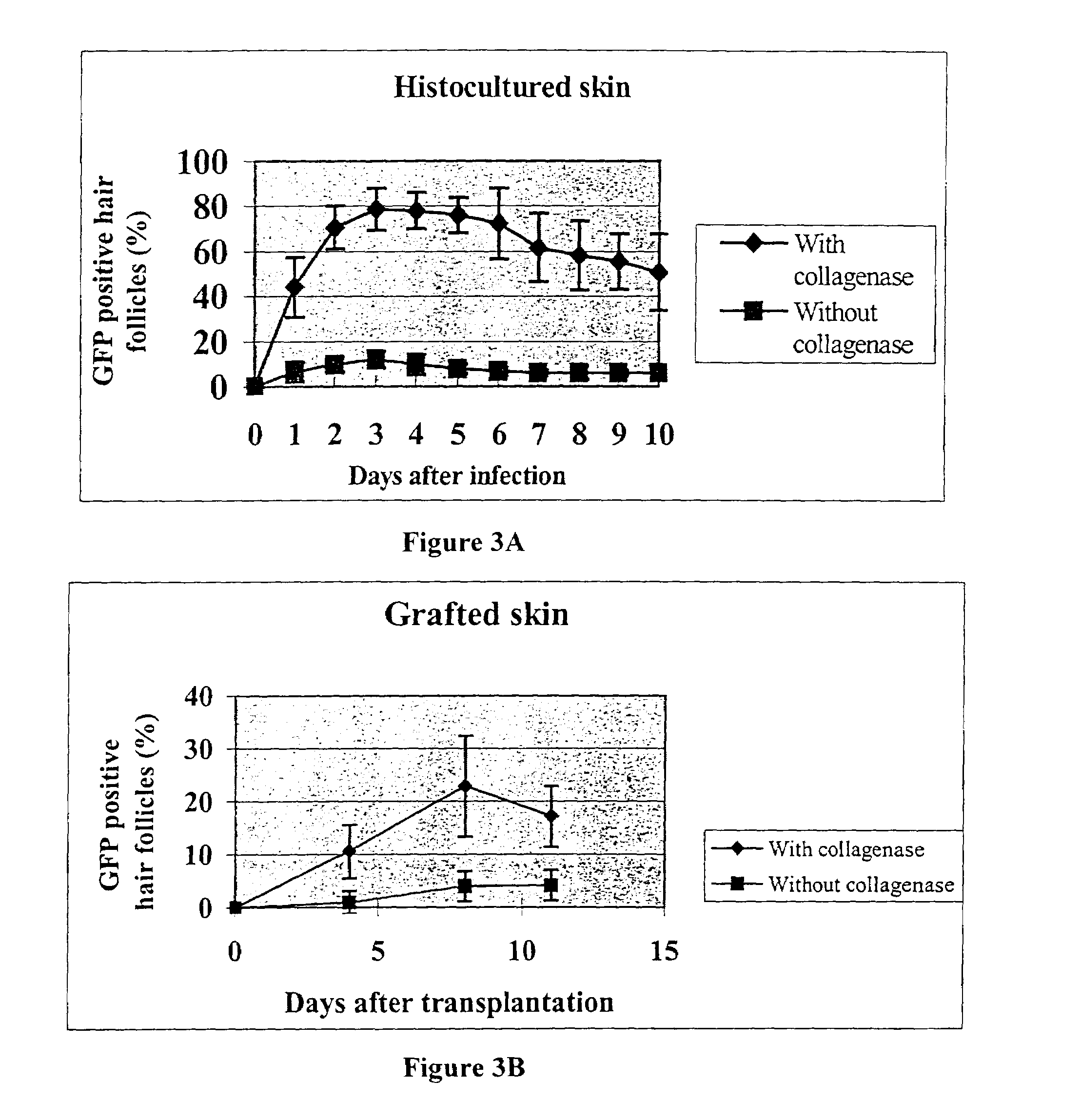
[0050]Eight-week-old female C57BL / 10 mice whose hair follicles were in telogen phase were used as donors. The dorsal area was depilated with wax to induce anagen which occurred six days after depilation. Tissue was removed from the dorsal area and cut into small pieces, cultured, treated with collagenase, and infected with adenovirus containing GFP as described in Example 1. The skin pieces were then transplanted to the dorsal area of seven-week / three-day-old female nude mice. Observation of the grafted skin after transplant showed extensive expression of GFP in the hair follicles which expression was enhanced by the pretreatment with collagenase.

[0051]In more detail, to visualize the expression of the transgene in vivo, histocultured skin was grafted to nude mice or C57BL / 10 mice after viral GFP transduction. Histocultured specimens were grafted within 24 hrs of harvest. Grafting surgery was performed in a laminar-flow hood using stei...

example 3

Cloning of the Streptomyces Tyrosinase Gene, the Upstream ORF-438 and the Internal Ribosome Entry Site (IRES)

[0054]The sequences encoding the Streptomyces antibioticus tyrosinase gene and ORF-438 were amplified by PCR from plasmid pIJ702 obtained from the American Type Culture Collection (ATCC #35287). Katz, E., et al,. J. Gen. Microbiol. (1983) 129:2703–2714.

[0055]Oligomers for PCR amplification were designed according to the sequence of S. antibioticus tyrosinase gene and ORF-438 cDNA. Bernan, V., et al., Gene, (1985) 37:101–110. In order to enhance expression of the bacterial gene in mammalian cells, the ORF-438 and tyrosinase-gene TGA termination codons were altered to TAA. The Kozak consensus sequence, GCCGCCACC, was added upstream, immediately preceding the ATG initiator codon in each case to facilitate translation efficiency.

[0056]The sequence of the ORF-438 upstream primer, which included the Kozak consensus sequence was

[0057]5′-CGGAATTCGCCGCCACCATGCCGGAACTCACCCGTC-3′ (SEQ I...


PUM


Abstract

Methods to obtain genetic modifications of cells in histoculture are described. Modification is assisted by treating the histoculture with collagenase prior to contacting the histoculture with the delivery vehicle for the desired gene. Hair follicles and other organized tissues can be modified in this was and then transplanted into intact recipients.

Description

CROSS-REFERENCE TO RELATED APPLICATION[0001]This application claims priority under 35 U.S.C. § 119(e) from provisional application Ser. No. 60 / 170,166, filed Dec. 10, 1999, the contents of which are incorporated herein by reference.TECHNICAL FIELD[0002]The invention relates to modifying mammalian subjects to contain heterologous genes. More particularly, the invention concerns treating histocultured tissue, including hair follicles, ex vivo and re-implanting tissue or hair follicles into a recipient.BACKGROUND ART[0003]Somatic modification of the genetic complement of mammalian subjects, including humans, has been attempted using a variety of techniques. For example, adenoviral vectors containing a desired gene can be used directly to infect tissues and organs in situ. More typically, perhaps, cell cultures or suspensions of cells are modified ex vivo and then returned to the intact subject via the bloodstream. For example, an RNA-DNA oligonucleotide (RDO) designed to correct the al...

Claims


Application Information

Patent Timeline
IPC IPC(8): A61K31/7088C12N15/63C12N5/06A61K35/12C12N15/09A61K35/26A61K35/36A61K38/00A61K38/22A61K39/00A61K48/00A61P1/16A61P5/00A61P5/06A61P5/10A61P5/22A61P7/02A61P7/06A61P17/14A61P31/04A61P31/12A61P31/20A61P33/02A61P37/04A61P43/00C07K14/36C12N5/071C12N5/10C12N9/02C12Q1/37
CPCC12N5/0627C12N9/0059C07K14/36A61K35/12A61K48/00C12N2509/00A61P1/16A61P17/14A61P31/04A61P31/12A61P31/20A61P33/02A61P37/04A61P43/00A61P5/00A61P5/06A61P5/10A61P5/22A61P7/02A61P7/06C12N15/00
Inventor SAITO, NORIMITSUZHAO, MING
Owner ANTICANCER
Why Eureka
Try Eureka