
Monoclonal antibody and method and kit for immunoassay of soluble human ST2

a human st2 and monoclonal antibody technology, applied in the field of monoclonal antibody and method and kit for immunoassay of soluble human st2, can solve the problems of not being able to recognize a specific non-denatured human st2, no satisfactory biological data have been obtained, and the physiological function of this antibody has not been clarified. , to achieve the effect of rapid and convenien

Inactive Publication Date: 2006-08-08
MEDICAL & BIOLOGICAL LAB CO LTD
View PDF3 Cites 81 Cited by
  • Summary
  • Abstract
  • Description
  • Claims
  • Application Information

AI Technical Summary

Benefits of technology

[0041]An objective of the present invention is to provide a monoclonal antibody which binds specifically to a non-denatured human ST2. Another objective of the present invention is to provide a rapid and convenient method for determining a non-denatured soluble (secretion type) human ST2 in a biological sample employing the monoclonal antibody described above. Still another objective of the invention is to provide an effective means for the detection, diagnosis, research, follow-up and prognosis of an abnormality in an immune system such as an allergic disease by the method for determining a soluble human ST2 described above.

Problems solved by technology

This theory was considered to be applicable only to the mouse long term subcultured cell line, and to be difficult to be applied to an in vivo cell.
Nevertheless, the physiological function of this antibody has not been clarified.
Nevertheless, no satisfactory biological data have been obtained currently with regard to the secretion type ST2.
However, such antibodies were obtained using as an immunogen a human ST2 protein obtained by expressing a human ST2 gene in E.coli, and bind specifically to a denatured human ST2 but could not recognize a non-denatured human ST2 specifically.
On the other hand, Werenskiold et al. disclosed a method for measuring a human ST2 by means of a PCR method for detecting an mRNA or by means of an immunohistological staining (WO98 / 430990), but such a method was only qualitative or semi-quantitative, and was not intended to measure the human ST2 quantitatively.

Method used


Image

  • Monoclonal antibody and method and kit for immunoassay of soluble human ST2
  • Monoclonal antibody and method and kit for immunoassay of soluble human ST2
  • Monoclonal antibody and method and kit for immunoassay of soluble human ST2

Examples


example 1

Preparation of Monoclonal Antibody Binding Specifically to Non-denatured Human ST2

(1-1) Preparation of Antigen

[0092]Two different antibodies were employed to obtain a monoclonal antibody binding specifically to a non-denatured human ST2 (hereinafter referred to as hST2). The first antigen employed was a soluble recombinant hST2 which was produced and secreted in a COS7 cell into which an hST2 gene had been transduced. The second antigen employed was a COS7 cell expressing a chimera molecule HMS whose extracellular region corresponded to a human ST2L (hereinafter referred to as hST2L) and whose transmembrane region and intracellular region corresponded to a mouse ST2L. The method for preparing each antigen is described below.

a. Preparation of Soluble Recombinant hST2 (rhST2)

[0093]An expression vector containing all coding region of a human whole length ST2 cDNA (Sequence ID No.1) (pEF-BOS-ST2H) was constructed and transfected into a COS7 cell (see Tominaga S., et al.,: Biochim. Bioph...

example 2

Examination of Monoclonal Antibody Activity

a. Immunoblotting Method

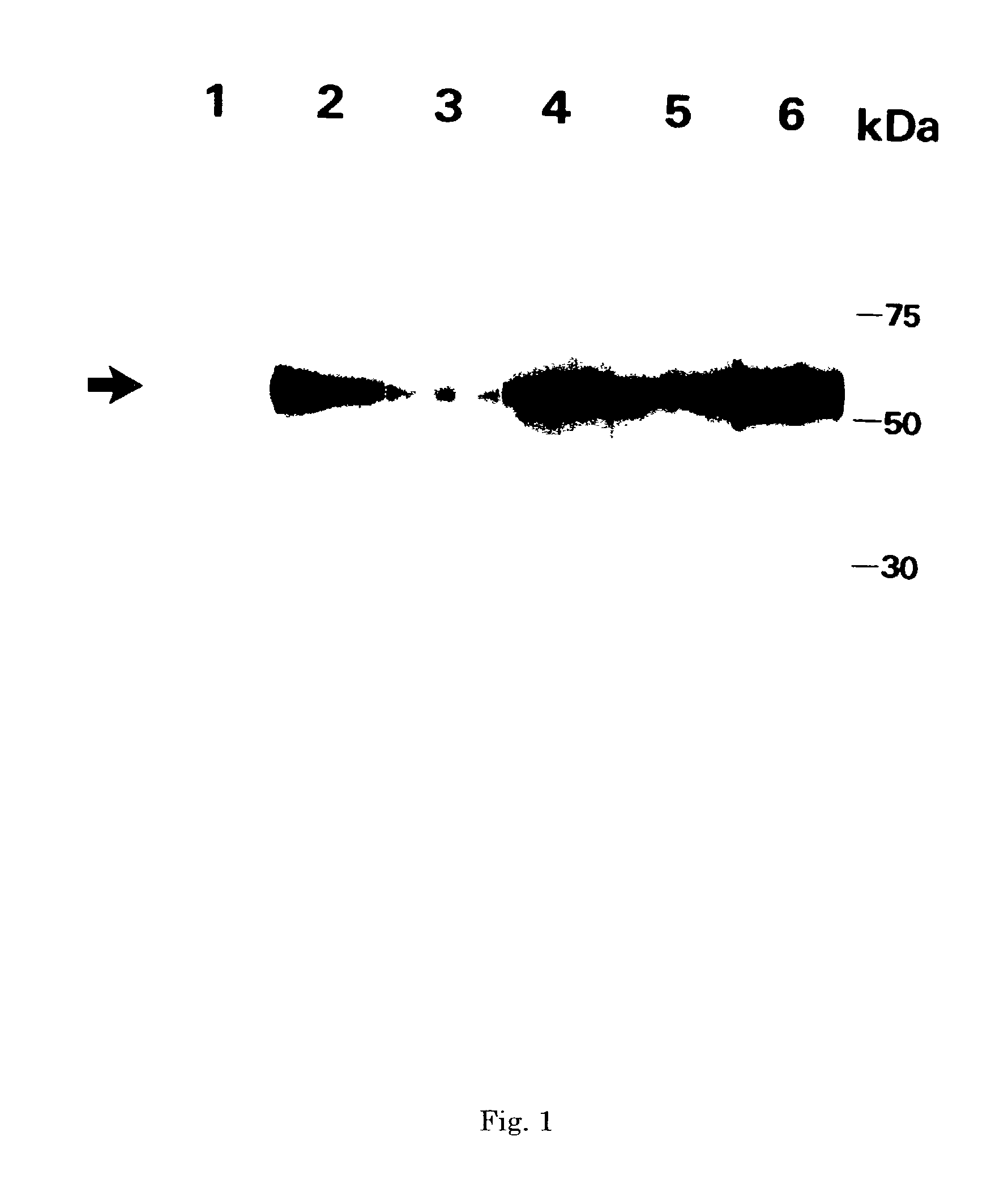
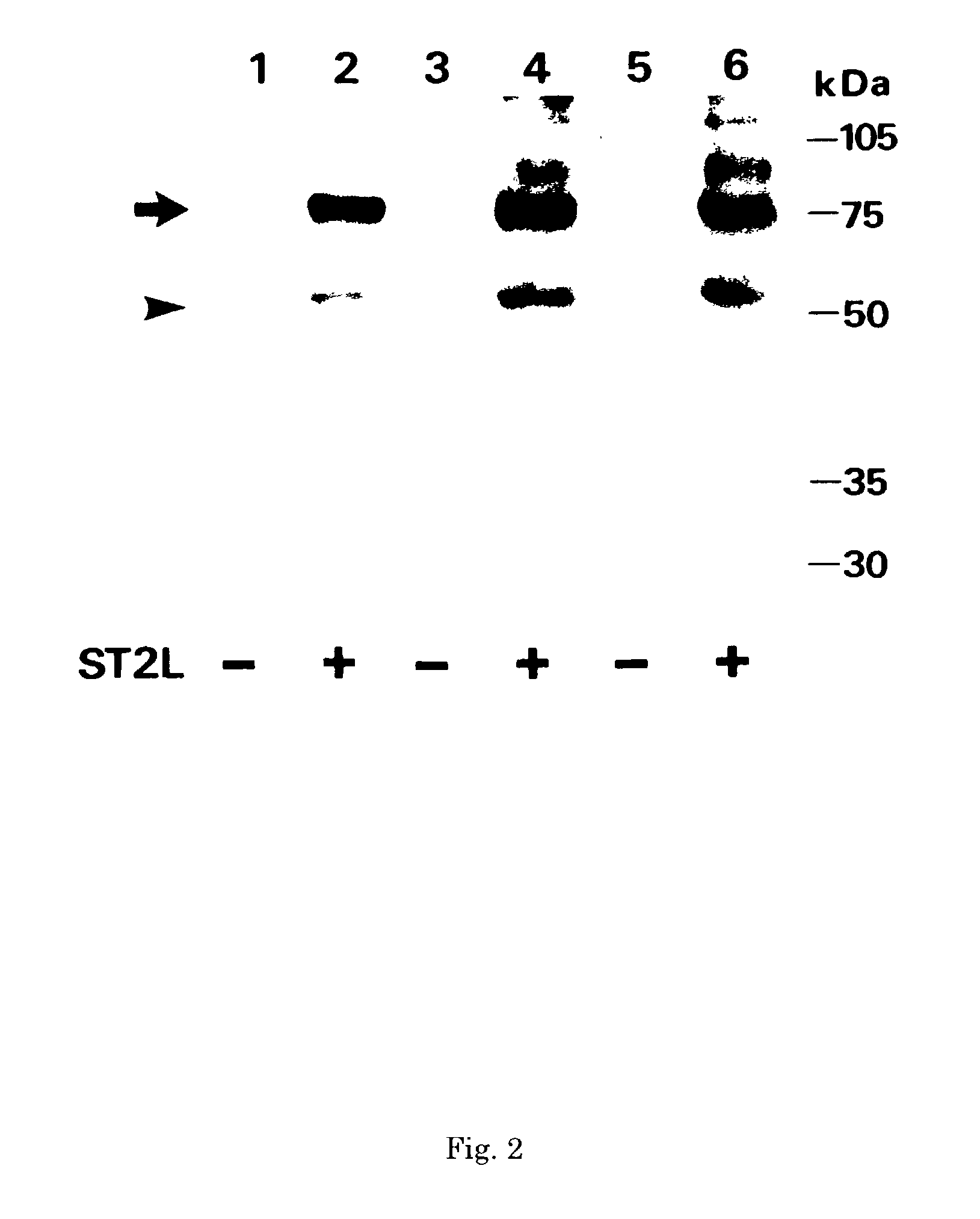
[0112]In order to examine the monoclonal antibodies 2A5, FB9 and HB12 obtained as described above for their activities on the hST2, an immunoblotting method was employed.

[0113]First, the rhST2 purified partially on a heparin agarose column in Section a in (1-1) described above was developed by a 10% SDS-PAGE and then transferred onto a PVDF membrane filter. Subsequently, the filter was cut into strips, and each filter was blocked with 10 mg / ml skim milk dissolved in a 0.05% (v / v) solution of Tween 20 in a tris buffered physiological saline (T-TBS; pH 7.5). Then each filter was reacted with the purified monoclonal antibody obtained in (1-5) described above dissolved in a skin milk / T-TBS. After reacting for 1 hour, the filter was washed three times with the skim milk / T-TBS, and reacted with an HRP-binding goat anti-mouse IgG dissolved in the skim milk / T-TBS for 30 minutes. After washing three times, the bands of the pr...

example 3

Reactivity of Monoclonal Antibody with Non-denatured hST2 and Epitope

(3-1) Cell ELISA Method

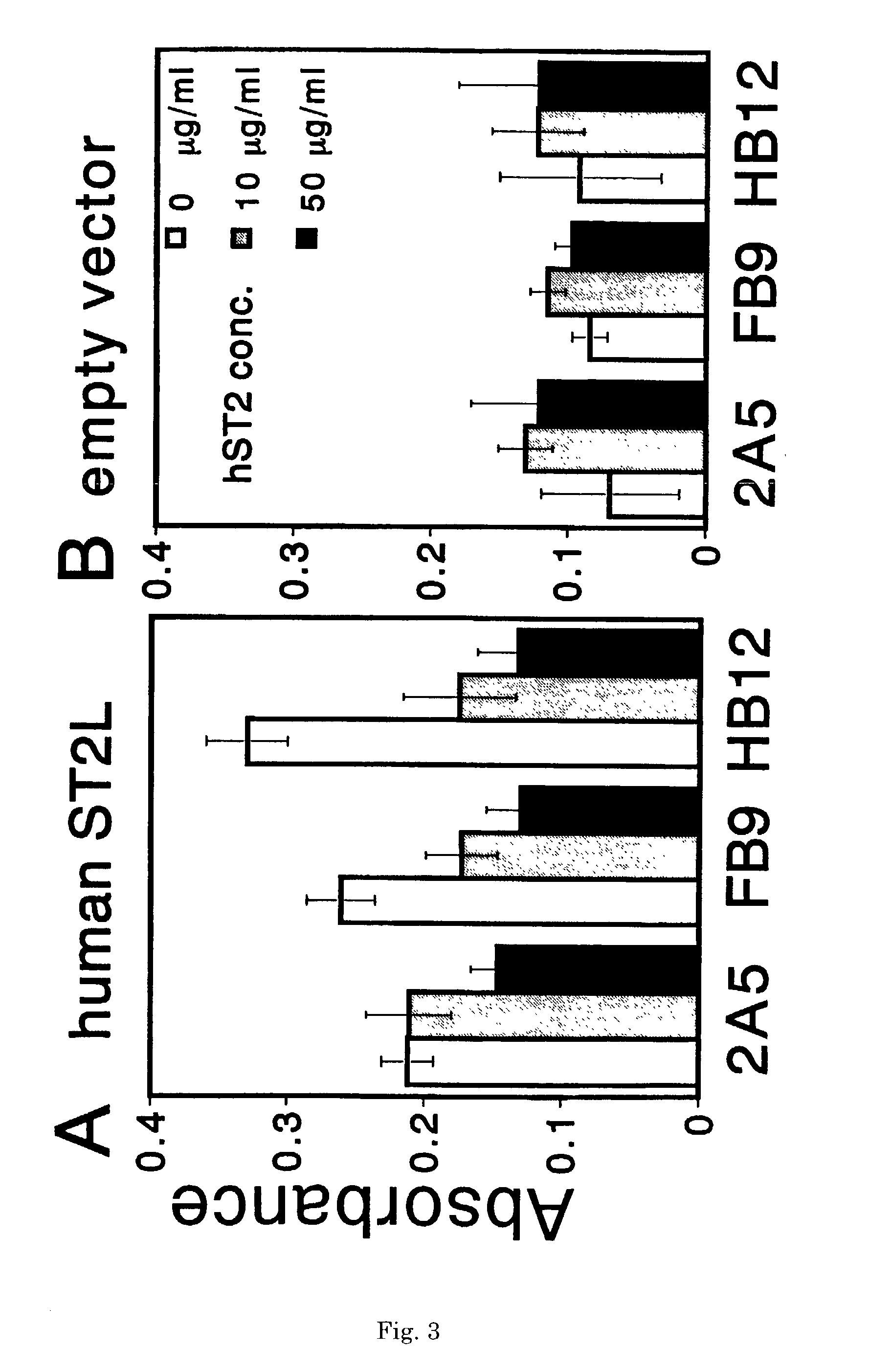
[0119]It was examined whether monoclonal antibodies 2A5, FB9 and HB12 were capable of recognizing a non-denatured hST2 or not by a cell ELISA employing a viable cell which was expressing the hST2L on its cell surface.

[0120]A COS7 cell was transfected with a pEF-BOS plasmid obtained by integrating the hST2LcDNA into the expression vector pEF-BOS, and the transfected cell was incubated in a 96-well plate for 48 hours. Subsequently, each well was made free of the culture medium, and charged with each monoclonal antibody labeled with the HRP, together with a varying concentration of the rhST2. Thereafter, the plate was incubated in the presence of 5% CO2 at 37° C. for 15 minutes. After these reactions, the cell in each well was washed three times with a PBS gently, and the HRP enzymatic activity in each well was assayed.

[0121]The graph of results of the assay are shown in FIG. 3. In the figure gr...


PUM

PropertyMeasurementUnit
pHaaaaaaaaaa
pHaaaaaaaaaa
pHaaaaaaaaaa

Abstract

A method for determining a soluble human ST2 in a sample conveniently at a high sensitivity and an assay kit are provided. By an immunological method comprising a step for bringing a sample into contact with an immobilized antibody formed by binding to an insoluble support a first anti-human ST2 antibody which binds specifically to a non-denatured human ST2, a step for labelling a first reaction product generated in the previous step by reacting said first reaction product with a second anti-human ST2 antibody which binds specifically to a non-denatured human ST2 by recognizing a site different from the site on ST2 where said first anti-human ST2 antibody binds and which is labelled with a label, and a step for determining the amount of the label on said first reaction product which has been labelled, a soluble human ST2 in a sample is determined. In addition, a recombinant ST2 is employed as a standard to prepare a calibration curve, based on which the ST2 in a sample is quantified.

Description

CROSS REFERENCE TO RELATED APPLICATIONS[0001]This application is continuation of international application No. PCT / JP00 / 08723 field Dec. 7, 2000, which claims priority of Japanese application No. 2000-77383 field Mar. 21, 2000.FIELD OF THE INVENTION[0002]The present invention relates to a monoclonal antibody which binds specifically to a human ST2 which is a protein related to diseases in an immune system. The invention also relates to an immunological method and an assay kit for determining a soluble human ST2 using an anti-human ST2 monoclonal antibody as well as an assay kit.BACKGROUND OF THE INVENTION[0003]A helper T cell (Th cell) is known as a cell which regulates immune reactions in a body. Via an interaction resulting from the contact between cells or via a release of a substance called cytokine, the helper T cell acts on and regulates a cell which executes an immune reaction.[0004]A helper T cell can be classified, based on the type of the cytokine it produces, broadly into...

Claims


Application Information

Patent Timeline
Patent Type & Authority Patents(United States)
IPC IPC(8): C07K16/28C12N5/20G01N33/535G01N33/543G01N33/577G01N33/564G01N33/68
CPCC07K16/28G01N33/6893G01N33/564Y10S435/975
Inventor TOMINAGA, SHIN-ICHIARAI, TAKAOKUROIWA, KENJIOSHIKAWA, KATSUHISA
Owner MEDICAL & BIOLOGICAL LAB CO LTD
Learn More