Methods and compositions comprising Renilla GFP
a technology of green fluorescent proteins and renilla, which is applied in the field of methods and compositions using renilla green fluorescent proteins (rgfp) and ptilosarcus green fluorescent proteins, can solve the problems of weak fluorescence, failure to fold properly at higher temperatures, and difficulty in assessing whether any particular peptide has been expressed
- Summary
- Abstract
- Description
- Claims
- Application Information
AI Technical Summary
Benefits of technology
Problems solved by technology
Method used
Image
Examples
example 1
Vector Construction and Expression in Mammalian Cells
[0371]Retroviral constructs were based on a pCGFP vector that carries a composite CMV promoter fused to the transcriptional start site of the MMLV R-U5 region of the LTR, an extended packaging sequence, deletion of the MMLV gag start ATG, and a multiple cloning region encoding EGFP, an Aequoria Victoria GFP variant codon optimized for expression in human cells (Clontech, Palo Alto, Calif.) and a Kozak consensus start, described in Kozak (1986) Cell 44: 283–292. The vector used to express flag tagged EGFP, pEf, is identical to pCGFP but has additional restriction sites in the open reading frame of EGFP (resulting in 8 non-human optimized codons) and a Flag tag fused to the C-terminus of EGFP with the linker EEAAKA.
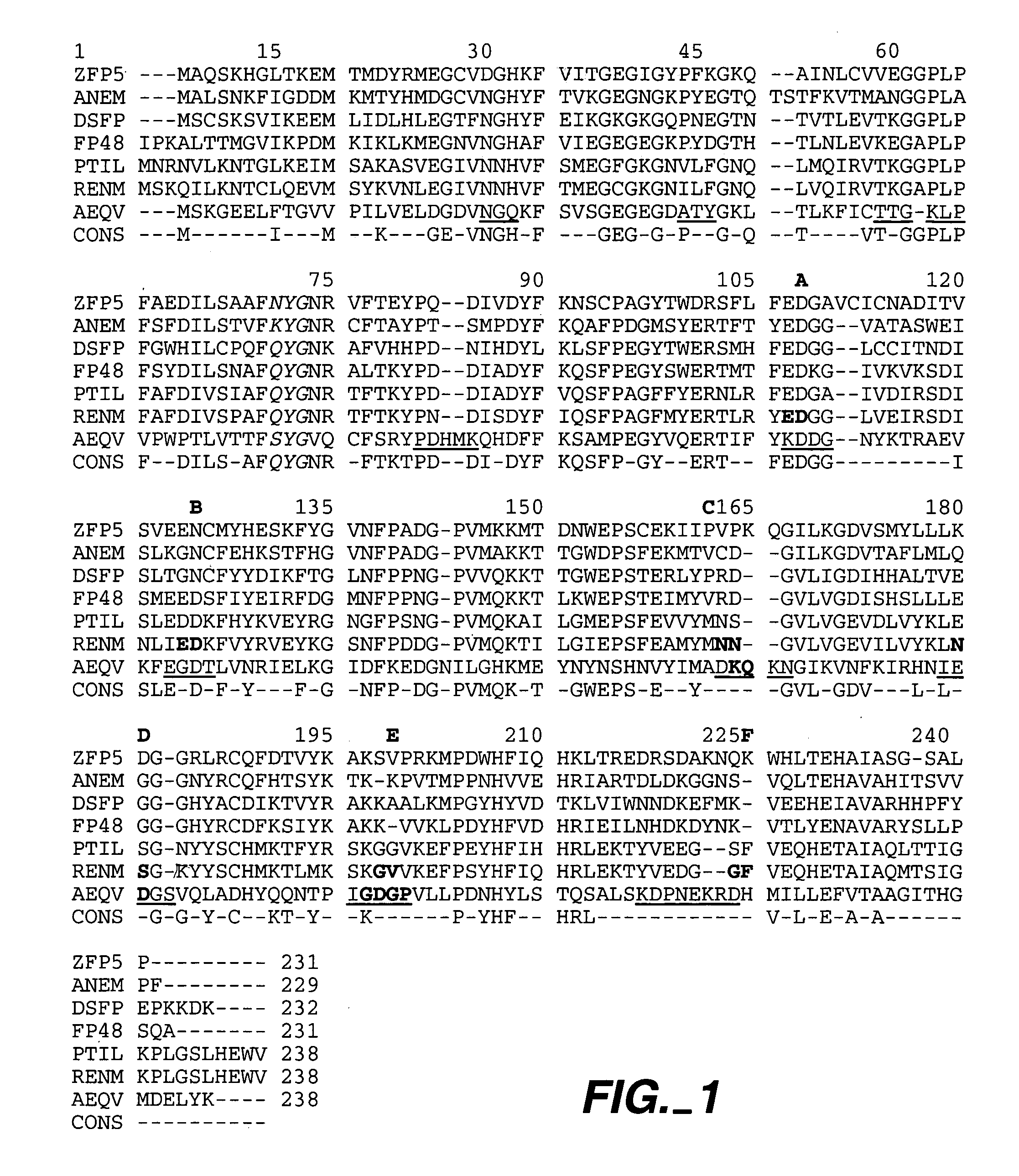
[0372]pR and pP are retroviral expression vectors comprising Renilla muelleri and Ptilosarcus gumeyi GFPs (containing 9 and 11 non-optimized codons, respectively, to introduce restriction sites). Each has a Kozak consensu...
example 2
Expression of Renilla GFP Codon Optimized for Expression in Human Cells
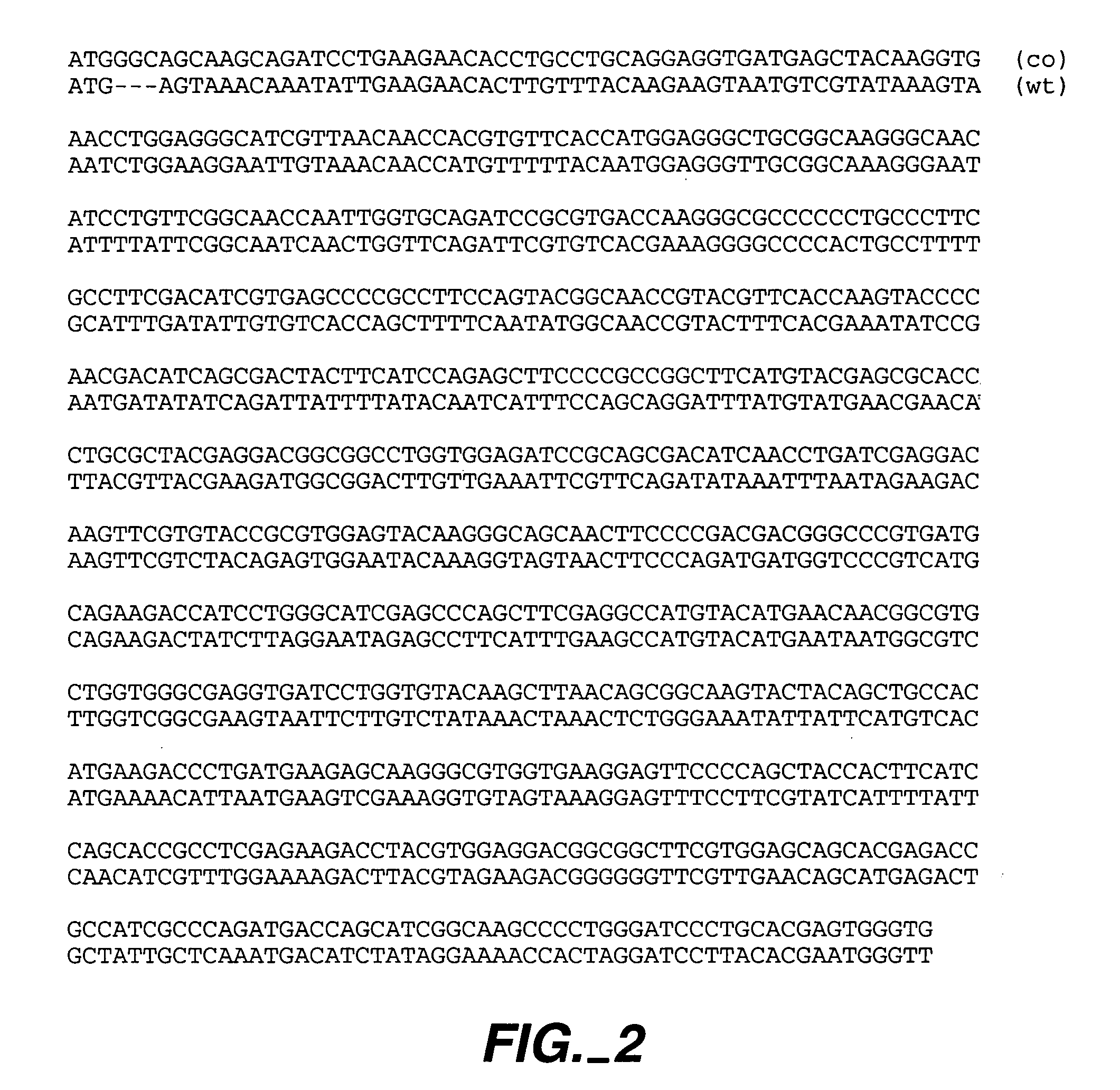
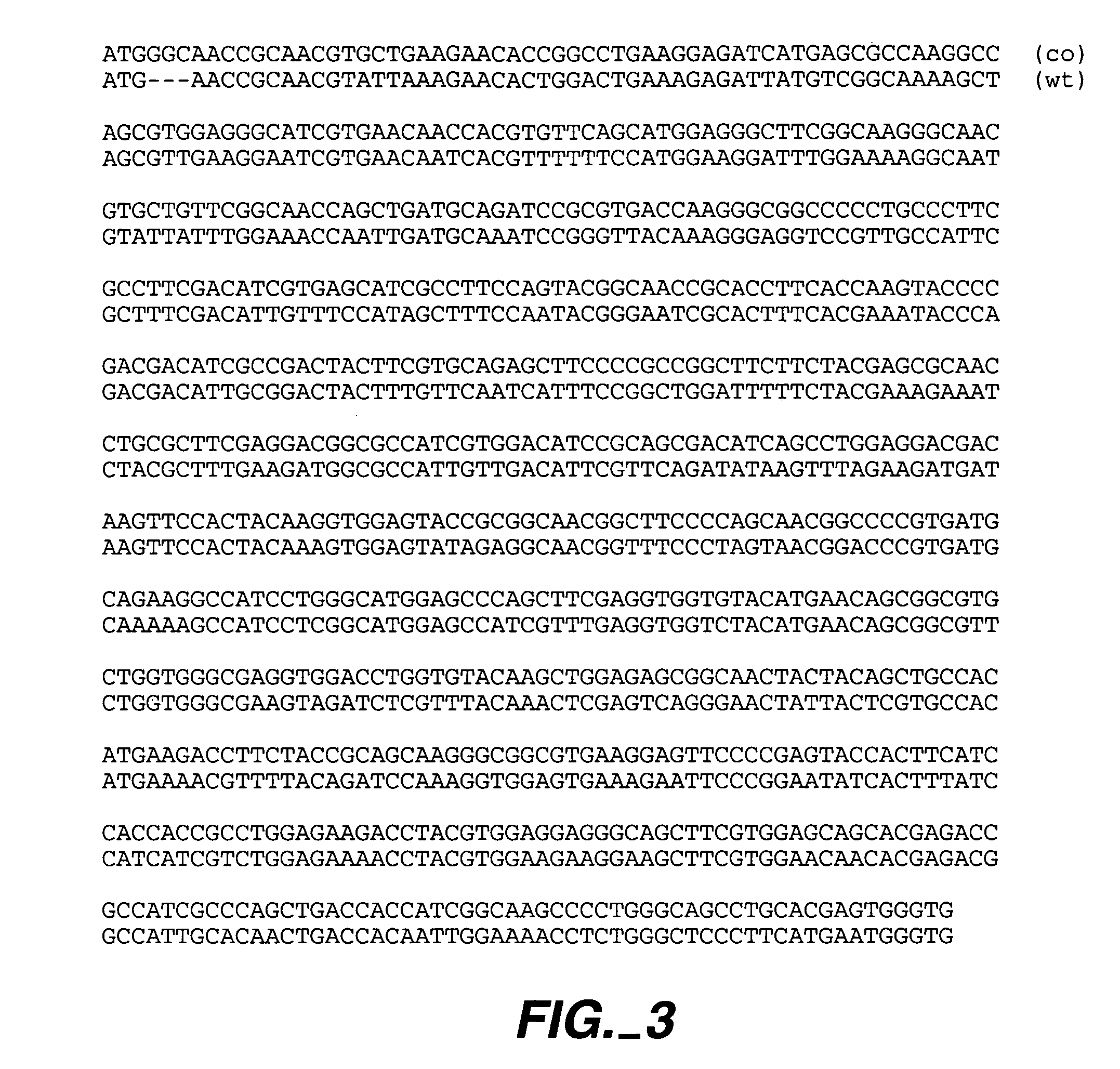
[0386]Renilla muelleri and Ptilosarcus GFP genes were constructed with a glycine following the initial methionine to optimize translations (see Experiment 1). The sequences were codon optimized for efficient expression in human cells. These GFPs were introduced into Jurkat-E cells by retroviral delivery using the protocol of Swift et al., supra. Based on FACS analysis of scatter and propidium iodide staining of cell populations from 13 hours to 8 days post infection, there was no observed toxicity of either Ptilosarcus or Renilla GFP. By 2 days post infection, the accumulation of intracellular GFP slowed to a steady state level. Based on FL1 channel fluorescence, the rate for reaching the steady state level occurred more rapidly for Ptilosarcus and Renilla GFPs than for EGFP. The excitation and emission spectra were 501 and 511 nm; 498 and 509 nm; and 489 and 510 nm for Ptilosarcus GFP, Renilla GFP, and Aequoria ...
example 3
Epitope Tag Insertion for Loop Suitable for Presentation of Peptides
[0389]To test for the location of potential surface loops in Renilla GFP, the peptide sequence GQGGGYPYDVPDYASLGQAGGG (SEQ ID NO: 105) containing the influenza hemagglutinin epitope tag flanked by two flexible linker sequences was inserted into candidate sites corresponding to putative loops of Renilla GFP (see Experiment 1). Following retroviral delivery into human cells, the fluorescence of the modified GFPs were examined. Six different insertion sites, A–F were tested in codon optimized GFP. FIG. 7 shows the fluorescence of the different modified Renilla GFPs retrovirally expressed in Jurkat-E cells and analyzed by FACS 4 days post infection. The geometric mean fluorescence values for the populations indicated by the gates are shown in the upper right corner for each FACS plot. Comparisons of these values are for samples that have populations present within the same dynamic range. All modified Renilla GFPs, excep...
PUM
 Abstract
Description
Claims
Application Information
 - R&D
- Intellectual Property
- Life Sciences
- Materials
- Tech Scout
- Unparalleled Data Quality
- Higher Quality Content
- 60% Fewer Hallucinations
Browse by: Latest US Patents, China's latest patents, Technical Efficacy Thesaurus, Application Domain, Technology Topic, Popular Technical Reports.
© 2025 PatSnap. All rights reserved.Legal|Privacy policy|Modern Slavery Act Transparency Statement|Sitemap|About US| Contact US: help@patsnap.com


