Application of bifluorescence reporting system in tumor stem cell targeted drug screening and method thereof

A cancer stem cell and reporting system technology, which is applied in the application field of dual fluorescent reporter system in the screening of cancer stem cell targeted drugs, can solve the problems of affecting evaluation results, increasing system errors, and unreliable evaluation results, and improving the screening effect. Effect

Inactive Publication Date: 2015-05-06
THE FIRST AFFILIATED HOSPITAL OF THIRD MILITARY MEDICAL UNIVERSITY OF PLA
View PDF2 Cites 9 Cited by
  • Summary
  • Abstract
  • Description
  • Claims
  • Application Information

AI Technical Summary

Problems solved by technology

[0003] At present, there are many limitations in CSCs drug screening, mainly reflected in the difficulty of obtaining CSCs on the one hand, and on the other hand, after CSCs are obtained, some cells will spontaneously differentiate into non-CSCs during the drug screening process. Therefore, if the cell population is used as the object at this time The evaluation results are unreliable, and it is time-consuming and labor-intensive to use antibodies and other methods to mark and distinguish CSCs. Not only cannot it be carried out on a large scale, but it will also increase system errors and affect the evaluation results.

Method used


Image

  • Application of bifluorescence reporting system in tumor stem cell targeted drug screening and method thereof
  • Application of bifluorescence reporting system in tumor stem cell targeted drug screening and method thereof
  • Application of bifluorescence reporting system in tumor stem cell targeted drug screening and method thereof

Examples


Embodiment 1

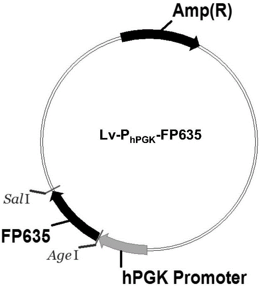
[0034] Example 1Lv-P hPGK -Construction of FP635 vector

[0035] In order to achieve the labeling of all experimental cells, first construct the lentiviral vector Lv-P with the promoter of the housekeeping gene PGK (phosphoglycerate kinase) expressing red fluorescent FP635 hPGK -FP635, see the structure diagram figure 1 . The build process is as follows:

[0036] 1. Construction of the intermediate vector pMD-19-T-pTurboFP635: the primers were designed with pTurboFP635-N plasmid as the template to amplify the coding region of the red fluorescent protein FP635 (SEQ ID No. 1). The primers are as follows:

[0037] 5'upstream primer: 5'-ccaccggtcg ccaccatggt gggtgaggat agcgtgctga tc-3', (SEQ ID No. 2);

[0038] 3'downstream primer: 5'-ttgtcgacgc ggccgcttta cttgtacgtc agctgtgccc cagtttgc-3', (SEQ ID No. 3).

[0039] Introduce AgeI restriction site into the 5'upstream primer, add SalI restriction site to the 3'downstream primer, and amplify by PCR. The amplification system is: pTurboFP63...

Embodiment 2

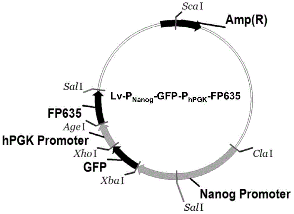
[0049] Example 2Lv-P Nanog -GFP-P hPGK -Construction of FP635 vector

[0050] Since the system described in Example 1 needs to be re-infected twice, which is relatively cumbersome, we further add Lv-P Nanog -GFP and Lv-P hPGK -FP635 The effective elements of the two viral vectors are fused into one vector (see the vector structure figure 2 ) To simplify the operation process. The build process is as follows:

[0051] 1. Construction of pMD-19-T-GFP intermediate vector: Design primers using pT-copGFP plasmid as template to amplify the coding region of green fluorescent protein GFP (SEQ ID No.5) shown in it. The primers are as follows:

[0052] 5'upstream primer: 5'-atcgattcta gacgccacca tgcccgccat g-3', (SEQ ID No. 6);

[0053] 3'downstream primer 5'-ctcgagttat caggcgaagg cgatg-3', (SEQ ID No. 7).

[0054] Introduce ClaI and XbaI restriction sites into the 5'upstream primers, add XhoI restriction sites to the 3'downstream primers, and use PCR for amplification. The amplification sy...

Embodiment 3

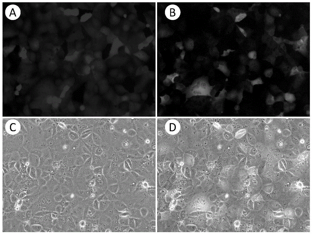
[0059] Example 3 GFP / RFP dual fluorescence reporter system is used for drug screening

[0060] Will be infected with Lv-P alone Nanog -GFP-P hPGK -FP635 virus or simultaneous infection with Lv-P Nanog -GFP and Lv-P hPGK -Huh7 cells of FP635 virus were plated into a 96-well plate, 1×10 per well 3 The tumor cells were cultured until the cell growth confluence reached 40-80%; different drugs and drug solvents were added as controls, and then continued to be cultured in a 37°C cell incubator; 24 hours later, the live cell dye Hoechst 33342 was added to mark the nucleus. Then immediately use the high-content imaging system to detect the expression of green fluorescent and red fluorescent protein in the well plate. After the detection, the cells can be cultured for 24h to 48h, and multiple time points of drug action can be detected separately; use the test results The MetaXpress software attached to the MD high-content imaging system performs analysis. The process is to define the n...


PUM


Abstract

The invention discloses application of a bifluorescence reporting system in tumor stem cell targeted drug screening and a method thereof. Red and green fluorescence proteins are utilized to respectively mark colony tumor cells and tumor stem cells, and the targeting properties of the drug for the tumor cells and tumor stem cells can be respectively evaluated by using a high-content imaging system. No targeted drug screening methods for tumor stem cells exist at present. The tumor stem cell screening method established by the invention is hopeful to be used for screening novel antineoplastic drugs for tumor stem cells, thereby enhancing the screening effect of the antineoplastic drugs.

Description

Technical field [0001] The invention belongs to the field of biotechnology, and specifically relates to the application and method of a dual fluorescence reporter system in tumor stem cell targeted drug screening. Background technique [0002] The market demand for new anti-tumor drugs is great, and the social and economic benefits are great. The domestic and foreign pharmaceutical industry has always regarded anti-tumor drugs as the top priority of new drug research and development. At present, there are at least 860 anti-tumor drugs in the world in clinical trials. This number is twice the sum of heart disease and stroke drugs in clinical trials, and twice the sum of Alzheimer's disease and other neurological diseases. However, the current drug screening is based on the tumor cell population as the object of investigation, and recent studies point to the existence of cancer stem cells (Cancer Stem Cells, CSCs) in tumors is the internal support for the malignant manifestations ...

Claims


Application Information

Patent Timeline
Patent Type & Authority Applications(China)
IPC IPC(8): C12Q1/68C12Q1/02
CPCG01N33/5073G01N33/5011
Inventor 钱程沈俊杰单娟娟
Owner THE FIRST AFFILIATED HOSPITAL OF THIRD MILITARY MEDICAL UNIVERSITY OF PLA
Why Eureka
Try Eureka