Porin B (PorB) as a therapeutic target for prevention and treatment of infection by Chlamydia

a technology of porin and chlamydia, which is applied in the field of prevention and treatment of infectious diseases, can solve problems such as significant health problems, conventional treatments are problematic, and heart disease, and achieve the effects of reducing the risk of infection

Inactive Publication Date: 2006-09-12
RGT UNIV OF CALIFORNIA
View PDF19 Cites 31 Cited by
  • Summary
  • Abstract
  • Description
  • Claims
  • Application Information

AI Technical Summary

Benefits of technology

[0129]To investigate surface exposure of PorB, purified EB were digested with proteases and proteins from EB were assessed for binding by the anti-PorB antibody. Using the dot blot method, EB were treated with various concentrations of trypsin or proteinase K, immobilized on a nitrocellulose membrane and probed with the anti-PorB antibody, as well as to antibodies to MOMP and the anti-pgp3 antibody. A reduction in binding by anti-PorB antibodies was observed for EB-digested proteins suggesting that PorB has surface accessible trypsin and proteinase K cleavage sites, and thus is an outer membrane protein.
[0130]Since PorB is an outer membrane protein with surface exposed regions, antibodies made to PorB were tested for ability neutralize infectivity of C. trachomatis (serovar L2). The anti-PorB sera produced using either the entire protein or an amino-terminal fragment (amino acids 24–71) at a concentration of 100 mg / ml neutralized infectivity by up to 88% and 70%, respectively, further supporting the conclusion that PorB is a surface exposed outer membrane protein (FIG. 3). The control antibody without specificity to outer membrane proteins, anti-pgp3, as well as the pre-immune sera did not neutralize infectivity (FIG. 3). A monoclonal antibody to serovar L2 MOMP (IH5) at a concentration of 50 and 100 mg / ml neutralized infectivity up to 78% (FIG. 3). This neutralization assay confirms that antibodies to PorB can inhibit infectivity by C. trachomatis since this assay is an art-recognized in vitro correlate for the assessment of protective immunity (Byrne et al. (1993) J. Infect. Dis. 168:415–20).
[0131]The pore-forming capabilities of PorB were tested using the liposome reconstitution assay (Nikaido (1983) Methods Enzymol. 97:85–95). The liposome swelling assay for study of porin function is used not only because it is well established, but because this assay gives precise information on the rates of diffusion of solutes through the porin channels (Nikaido & Rosenberg (1983) J. Bacteriol 153:241–252). This assay involves the formation of liposomes incorporated with pore-forming protein and then determination of whether and how fast test solutes can diffuse through the protein channels. This assay was used to test and compare pore-forming activity of the C. trachomatis PorB and MOMP.
[0132]Purification of MOMP using mild detergents causes a loss in porin activity (Bavoil, et al. (1984) Infect. Immun. 44:479–485, Wyllie, et al. (1998) Infect. Immun. 66:5202–5207), therefore, MOMP was expressed in E. coli and outer membrane fractions enriched for MOMP were used. It has been shown in liposome swelling assays that the predominant porin activity of the outer membrane fraction of E. coli expressing MOMP is due to MOMP (Jones et al. (2000) Gene 258:173–181). This was also found to be the case for PorB except purified PorB also functioned in liposome swelling assays (FIG. 4) and was used in all subsequent experiments. To control for potential contaminants that may occur during PorB purification, another predicted outer membrane protein from C. trachomatis serovar D (CT241) was cloned, expressed in E. coli and purified by the same procedure used for PorB. Like PorB, CT241 also contains a predicted leader sequence and ends in phenylalanine and was incorporated into liposomes and tested for pore forming activity. This protein as well as liposomes without protein did not show pore-forming activity with any of the solutes tested.
[0133]The smallest sugars tested in the liposome swelling assay were the monosaccharides arabinose and glucose. These sugars penetrated the PorB- and MOMP-containing liposomes faster than the disaccharide, sucrose, while the tetrasaccharide, stachyose, was too large to enter (FIG. 4). This diffusion selectivity of PorB- or MOMP-containing liposomes with sugars was similar to what has been observed with COMC-containing liposomes (Bavoil, et al. (1984) Infect. Immun. 44:479–485, Wyllie, et al. (1998) Infect. Immun. 66:5202–5207). Larger solutes enter into PorB or MOMP porin slower, suggesting that there is a size restriction of molecules that can enter via these porins. However, the liposomes containing PorB permitted the diffusion of arabinose or glucose at a slower rate than liposomes containing MOMP.
[0134]Since Chlamydia have been proposed to utilize amino acids from host cells (Ossowski et al. (1965) Isr. J. Med. Sci. 1:186–193; Hatch et al. (1982) J. Bacteriol. 150:379–385; Pearce, (1986) Ann. Inst. Pasteur Microbiol. 137A:325–332), diffusion of amino acids through PorB and MOMP were tested using the liposome swelling assay. MOMP liposomes allow for the diffusion of all of the amino acids at different rates based predominantly on size selectivity and alanine and glycine enter through MOMP liposomes slightly faster than arabinose (Jones et al. (2000) Gene 258:173–181). In contrast, PorB liposomes did not efficiently allow for any of 20 amino acids to enter liposomes including the small amino acids such as alanine (FIG. 5). These data indicate that PorB is less efficient than MOMP as a non-specific porin.

Problems solved by technology

Chlamydia pneumoniae usually infects the lungs and causes no more than a mild cold; however, it can travel to the blood vessels and thrive in clots, causing heart disease.
Diseases caused by Chlamydia represent significant health problems worldwide.
These conventional treatments are problematic for several reasons, including patient non-compliance with multi-day, multi-dose regimens and side effects such as gastrointestinal problems.
As with many pathogens, the development of a vaccine to Chlamydia has proven difficult.
A potential problem in making a vaccine to an antigenically variant region is that a vaccine to one region of MOMP may only confer protection to that serovar.
Also, making a subunit vaccine to an antigenic variable region may prove difficult since conformational antigenic determinants may be essential to elicit effective immunization (Fan et al.
Although the use of MOMP as a vaccine still seems promising, these potential problems strongly suggest that other vaccine targets should be explored.

Method used


Image

  • Porin B (PorB) as a therapeutic target for prevention and treatment of infection by <i>Chlamydia </i>
  • Porin B (PorB) as a therapeutic target for prevention and treatment of infection by <i>Chlamydia </i>
  • Porin B (PorB) as a therapeutic target for prevention and treatment of infection by <i>Chlamydia </i>

Examples


example 1

Analysis of PorB Sequence—Comparison to Major Outer Membrane Protein (MOMP)

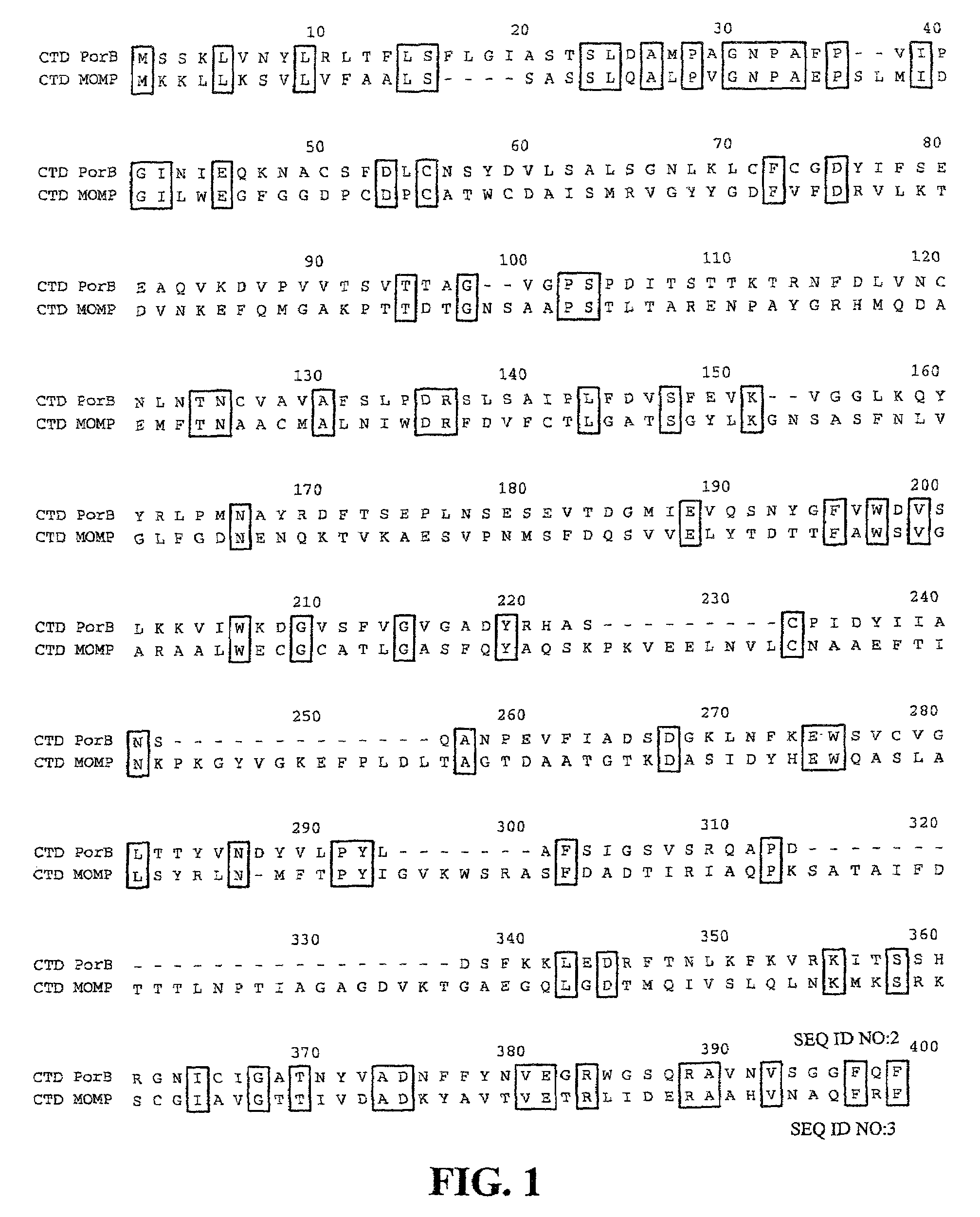
[0116]Genome sequence analysis revealed a number of predicted outer membrane proteins (see Stephens et al. 1998 “Genome sequence of an obligate intracellular pathogen of humans: Chlamydia trachomatis” Science 282:754–759). One such predicted outer membrane protein, encoded by the predicted open reading frame CT713, was selected for analysis, and referred to herein as PorB. The nucleotide and amino acid sequences of PorB (CT713) are available within the complete sequence of the genome at GenBank Accession No. NC—000117, with the amino acid sequence at GenBank Accession No. gi|3329169. The open reading frame corresponding to PorB is the complement of nucleotide residues 3616 to 4638 of GenBank Accession No. AE001342. The nucleotide and amino acid sequences of PorB of C. trachomatis are provided in the Sequence Listing as SEQ ID NOS:1 and 2, respectively. Alignment of the amino acid sequence of PorB with the ami...

example 2

Analysis of PorB Sequence—Comparison of PorB Amino Acid Sequences from Different Serovars

[0119]When compared with other serovars of C. trachomatis, MOMP has four distinct variable segments which correspond to surface exposed regions of the protein. Serovar designations have been related to the differences in these variable segments of MOMP (Stephens et al. (1988) J. Exp. Med. 167:817–831). In order to determine whether this serovar variation is also characteristic for PorB, the sequence of PorB between serovars was compared.

[0120]FIG. 2 provides an alignment of the amino acid sequences of PorB from the C. trachomatis serovars D (CT-D) (SEQ ID NO:2), L2 (CT-L2) (SEQ ID NO:5), and C (CT-C) (SEQ ID NO:6), as well as the amino acid sequence of PorB from C. pneumoniae (CPn) (SEQ ID NO:4). The PorB of C. trachomatis and C. pneumoniae are 59.4% identical. C. trachomatis serovar L2 and C differences are indicated below the amino acid sequence. The cysteines are indicated with an asterisk ab...

example 3

Expression of PorB in E. coli

[0123]PorB was predicted to be in the outer membrane through a variety of protein localization programs such as PSORT (K. Nakai, Human Genome Center, Institute for Medical Science, University of Tokyo, Japan). A leader sequence cleavage site for C. trachomatis PorB was predicted to be at amino acid 26 (FIG. 2). The complete gene including the leader sequence was cloned into E. coli with a HIS tag at the C-terminal end of PorB and expressed. The protein was affinity purified by nickel column chromatography.

[0124]PorB expressed in E. coli was localized to the outer membrane fraction as determined by an immunoblot using an antibody to the C-terminal HIS tag. E. coli porins were also detected in this outer membrane fraction by Coomassie stain. The presence of PorB was primarily localized to the outer membrane suggesting that PorB has the necessary signal(s) to be transported to the outer membrane by E. coli.


PUM

PropertyMeasurementUnit
densityaaaaaaaaaa
pHaaaaaaaaaa
volumeaaaaaaaaaa

Abstract

The present invention features peptides of a PorB polypeptide, which PorB peptides are useful in production of antibodies that bind the full-length PorB polypeptide and as a therapeutic agent. In specific embodiments the invention features a composition comprising one or more PorB peptides (other than a full-length PorB polypeptide), which peptides contain at least one epitope that can elicit Chlamydia-neutralizing antibodies. The invention also features methods for induction of a protective immune response against infection by Chlamydia and Chlamydiophila.

Description

STATEMENT AS TO FEDERALLY SPONSORED RESEARCH[0001]This invention was made, at least in part, with a government grant from the National Institutes of Health (Grant Nos. NIH grants AI40250, AI39258, and AI42156). Thus, the U.S. government may have certain rights in this invention.FIELD OF THE INVENTION[0002]The invention relates generally to the field of treatment, prevention, and diagnosis of infectious disease, particularly to prevention of infectious disease caused by the bacterial pathogen Chlamydia and Chlamydophila (formerly classified as, for example, C. psittacci and C. pneumoniae).BACKGROUND OF THE INVENTION[0003]Chlamydiae are obligate intracellular pathogens that cause a spectrum of diseases including trachoma, the leading cause of preventable blindness worldwide, as well as a variety of sexually transmitted diseases such as lymphogranuloma venereum, urethritis, cervicitis, endometritis, and salpingitis (Thylefors et al. (1995) Bull W H O 73:115–121). For example, Chlamydia...

Claims


Application Information

Patent Timeline
Patent Type & Authority Patents(United States)
IPC IPC(8): A61K39/118A61K38/00A61K39/00A61K39/02C07K1/00C07K14/295G01N33/569
CPCC07K14/295A61K39/118G01N33/56927A61K39/00A61K2039/53Y10S530/806Y10S530/825A61P31/04A61P37/04
Inventor STEPHENS, RICHARD S.KAWA, DIANE
Owner RGT UNIV OF CALIFORNIA
Why Eureka
Try Eureka