Method and apparatus for aneurismal treatment

an aneurismal treatment and anesthetic technology, applied in the field of anesthetic treatment, can solve the problems of weakened arterial walls, prone to fatal aneurysms, and prior art coil insertion methods suffered from several drawbacks, so as to reduce neurological effects, reduce the risk of rupture, and strengthen the aneurysm wall

Active Publication Date: 2007-02-27
MEDTRONIC VASCULAR INC
View PDF6 Cites 27 Cited by
  • Summary
  • Abstract
  • Description
  • Claims
  • Application Information

AI Technical Summary

Benefits of technology

[0021]In contrast to the prior art methods, the present invention is specifically directed to shrinking the aneurysm thereby providing at least a partial cure for the expansion of the arterial wall at the aneurysm site and potentially reducing neurological effects due to a brain aneurysm. This is significant since, shrinking the aneurysm wall strengthens the aneurysm wall and reduces the risk of rupture. Specifically, P=T(1 / R1+1 / R2), where P=transmural pressure across the aneurysm wall, T is the tension in the wall, R1& R2 are the principal radii of curvature. If we assume a spherical aneurysm (R1=R2), then the equation becomes P=2T / R, or put another way, T=PR / 2. This means that as the aneurysm shrinks, the radius decreases and the tension in the wall decreases (the transmural pressure remains essentially the same before and after treatment). This effect is even greater for a cylindrical tube (like an artery) where R2 is infinite. Then 1 / R2 approaches 0, and the equation becomes P=T(1 / R), or T=PR. Now the tension in the wall decreases twice as much for a given decrease in radius (compared to the sphere). Consequently, in contrast to the prior art, the method in accordance with the present invention addresses the underlying disease rather than simply treating the symptom.
[0022]In addition, in embodiments in accordance with the present invention, there are no coils used and therefore no risk of the coil, or ensuing thrombus, migrating from the aneurysm to the parent artery or vessel and causing a thrombo-embolic stroke or of the coil compacting and / or rupturing the aneurysm wall. Consequently, the method according to the invention is safer than prior art methods and can be applied to more types and shapes of aneurysms than prior art methods
[0023]In addition, embodiments in accordance with the present invention can be used on aneurysms of all sizes and shapes and any aneurysm can be treated using the method according to the present invention as soon as it is detected. In addition, embodiments in accordance with the present invention can be used to treat any type of aneurysm including brain aneurysms, abdominal aneurysms and thoracic aortic aneurysms. The fact that the present invention allows for early and immediate treatment provides the method according to the present invention with a significant advantage over the prior art.

Problems solved by technology

The weakened areas of the arterial wall caused by an aneurysm are problematic because they are subject to rupture and if a rupture occurs, the aneurysm can prove fatal.
While at times an effective treatment, the prior art coil insertion method suffered from several drawbacks.
First, there was a risk of the coil, or ensuing thrombus, migrating from the aneurysm to the parent artery or vessel and causing a thrombo-embolic stroke.
To minimize this risk, the prior art coil insertion method was typically limited to the treatment of saccular aneurysms having small necks to ensure that, once inserted, the coil remained within the aneurysm.
In addition, wide-necked and fusiform aneurysms were also difficult to treat effectively with prior art coil insertion methods.
Another disadvantage of prior art coil insertion methods was that the coils could rupture the aneurysm by poking through the aneurysm wall, resulting in life-threatening subarachnoid hemorrhage.
In addition, the effectiveness of the prior art coil insertion method discussed above was limited by the fact that while the insertion of the coil, and the resulting thrombus, could result in protecting the arterial wall at the aneurysm site, the method did not promote shrinkage of the aneurysm, or in any way address the expansion of the arterial wall at the aneurysm site.
If left untreated, the aneurysm could then continue to enlarge, and the risk of rupture remained.
By not shrinking the aneurysm, prior art coil insertion methods often resulted in the coils ending up contributing to the “mass effect”, in which the aneurysm pressed against adjacent structures such as cranial nerves, other vessels, and the brain parenchyma.
In short, the prior art coil insertion method treated the symptom but did nothing to provide a cure, or partial cure, for the expansion of the arterial wall at the aneurysm site.
In addition, since the coils used with the prior art coil insertion method had to be of a minimum size, typically three millimeters or more, to perform their function and, since the coils had to be inserted into the aneurysm site, the prior art method could only be used on relatively large aneurysms, i.e., aneurysms having diameters of three millimeters or more.
Therefore, the prior art coil insertion method typically could not be used on smaller aneurysms, such as those that might be advantageously found by early detection.
Consequently, in the prior art, many aneurysms had to be either treated by more intrusive, and potentially dangerous, methods or, in some cases, the patient would simply have to wait until the aneurysm was large enough to allow treatment using the prior art coil insertion method.
Since, as discussed above, aneurysms can result in a rupture of the arterial wall, and possibly death, waiting for the aneurysm to expand before treating it was not a desirable situation.
However, like the prior art coil insertion methods, these other prior art methods treated the symptom but did nothing to shrink the aneurysm or provide a cure, or partial cure, for the expansion of the arterial wall at the aneurysm site.

Method used


Image

  • Method and apparatus for aneurismal treatment
  • Method and apparatus for aneurismal treatment
  • Method and apparatus for aneurismal treatment

Examples


Embodiment Construction

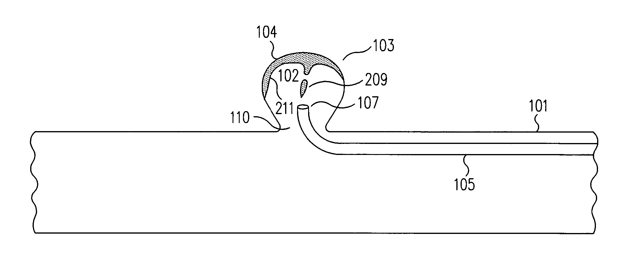
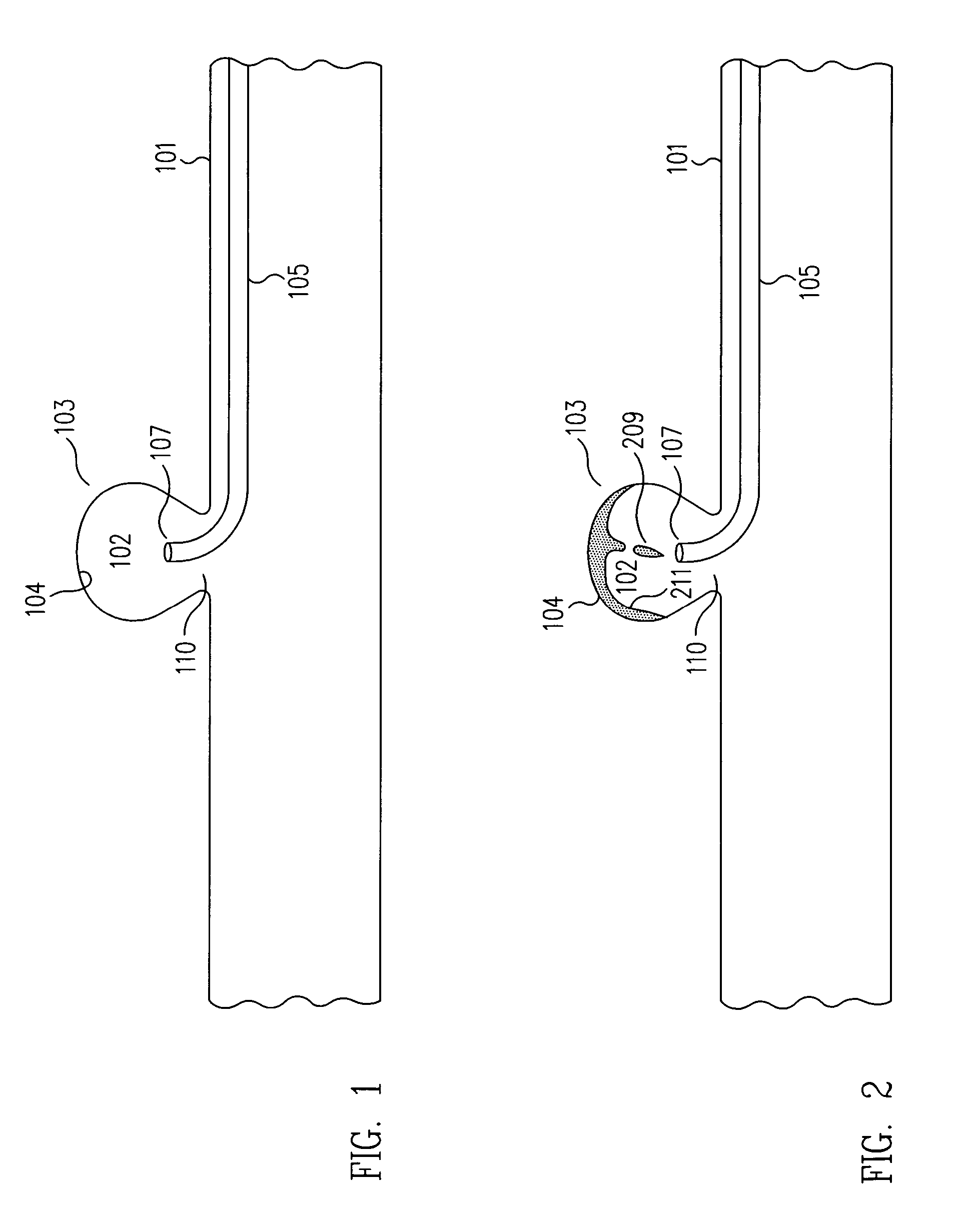
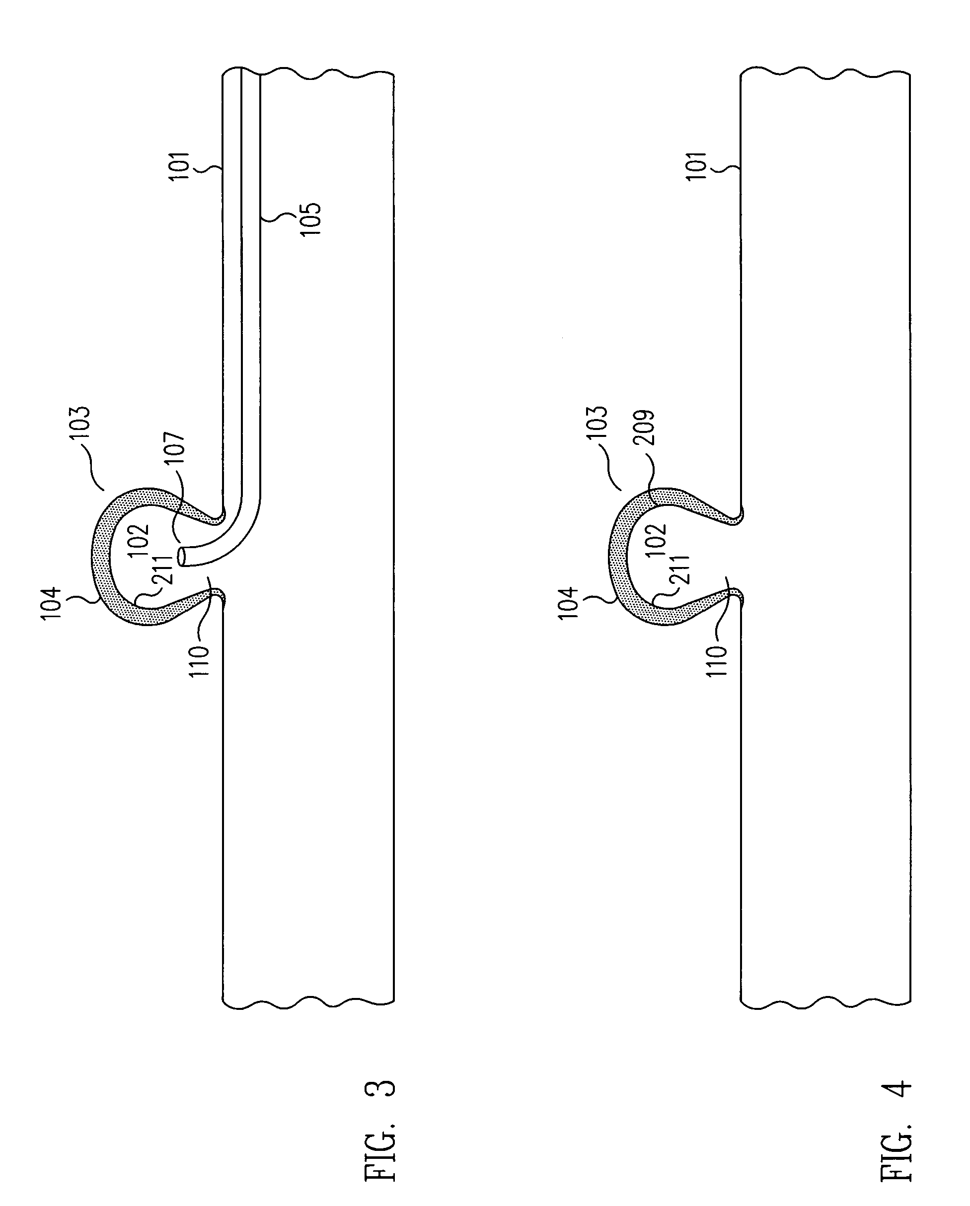
[0042]In embodiments in accordance with the present invention, the inner surface area of an aneurysm (103 in FIGS. 1 to 6 and 803 in FIGS. 8 to 14) is forced to contract thereby shrinking the aneurysm (FIGS. 5, 6, 16 and 17). Consequently, using the method according to the invention, the artery wall is strengthened, the risk of rupture is decreased, and at least a partial cure for the expansion of the arterial wall at the aneurysm site is provided.

[0043]In one example, an irritant (209 in FIGS. 2, 3, 4, 5, and 6) is provided to force the contraction of the aneurysm (103 in FIGS. 1 to 6). A micro-catheter (105 in FIGS. 1, 2 and 3) is inserted into a patient's parent artery or vessel (101 in FIGS. 1 to 6) and positioned at the aneurysm site with the micro-catheter lumen (107 in FIGS. 1, 2 and 3) in the aneurysm. Once the micro-catheter and micro-catheter tip and lumen are properly positioned, an irritant (209 in FIGS. 2, 3, 4, 5, and 6), typically in serum form, is dispensed into the ...


PUM

PropertyMeasurementUnit
diametersaaaaaaaaaa
sizeaaaaaaaaaa
temperatureaaaaaaaaaa

Abstract

An irritant or polymer is dispensed to the inner surface area of an aneurysm to exert a contractile force on the inner surface area of the aneurysm, thereby shrinking the aneurysm. As a result, the artery wall at the aneurysm site is strengthened, the risk of rupture is decreased, and at least a partial cure for the expansion of the arterial wall at the aneurysm site is provided.

Description

BACKGROUND OF THE INVENTION[0001]1. Field of the Invention[0002]The present invention relates to aneurismal treatments. More particularly, the present invention relates to methods for contracting and shrinking aneurysms.[0003]2. Description of the Related Art[0004]Vascular aneurysms are the result of abnormal dilation of a blood vessel, usually resulting from disease and / or genetic predisposition, which weakens the arterial wall and allows it to expand. The weakened areas of the arterial wall caused by an aneurysm are problematic because they are subject to rupture and if a rupture occurs, the aneurysm can prove fatal.[0005]In the prior art, several procedures and methods have been established for the exclusion of aneurysms. One such method involved the insertion of a coil within the aneurysm itself. Using prior art coil insertion methods, the coil precipitated the formation of a thrombus, or clot, within the aneurysm. The thrombus then partially, or completely, occluded the aneurys...

Claims


Application Information

Patent Timeline
Patent Type & Authority Patents(United States)
IPC IPC(8): A61M37/00A61B19/00A61K35/16C02F1/38A61B17/08A61M1/00A61B17/00A61B17/12
CPCA61B17/00491A61B17/12022A61B17/12113A61B17/12186A61B17/12136A61M2025/1052A61B2017/00557A61M2025/0042
Inventor YAMASAKI, SONNYKAEMMERER, WILLIAM F.
Owner MEDTRONIC VASCULAR INC
Why Eureka
Try Eureka