Layered device with capture regions for cellular analysis
- Summary
- Abstract
- Description
- Claims
- Application Information
AI Technical Summary
Benefits of technology
Problems solved by technology
Method used
Image
Examples
example 1
Identification of PSA, Tubulin, Actin, and Cytokeratin in Prostate Tumor
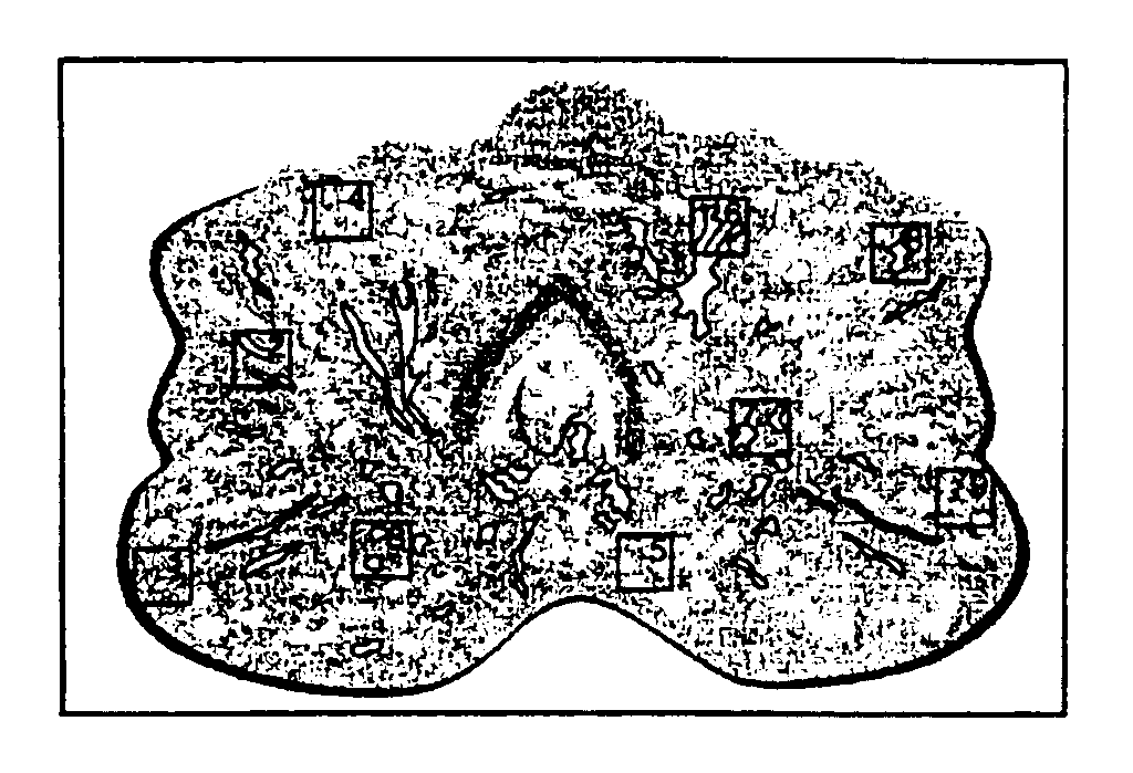
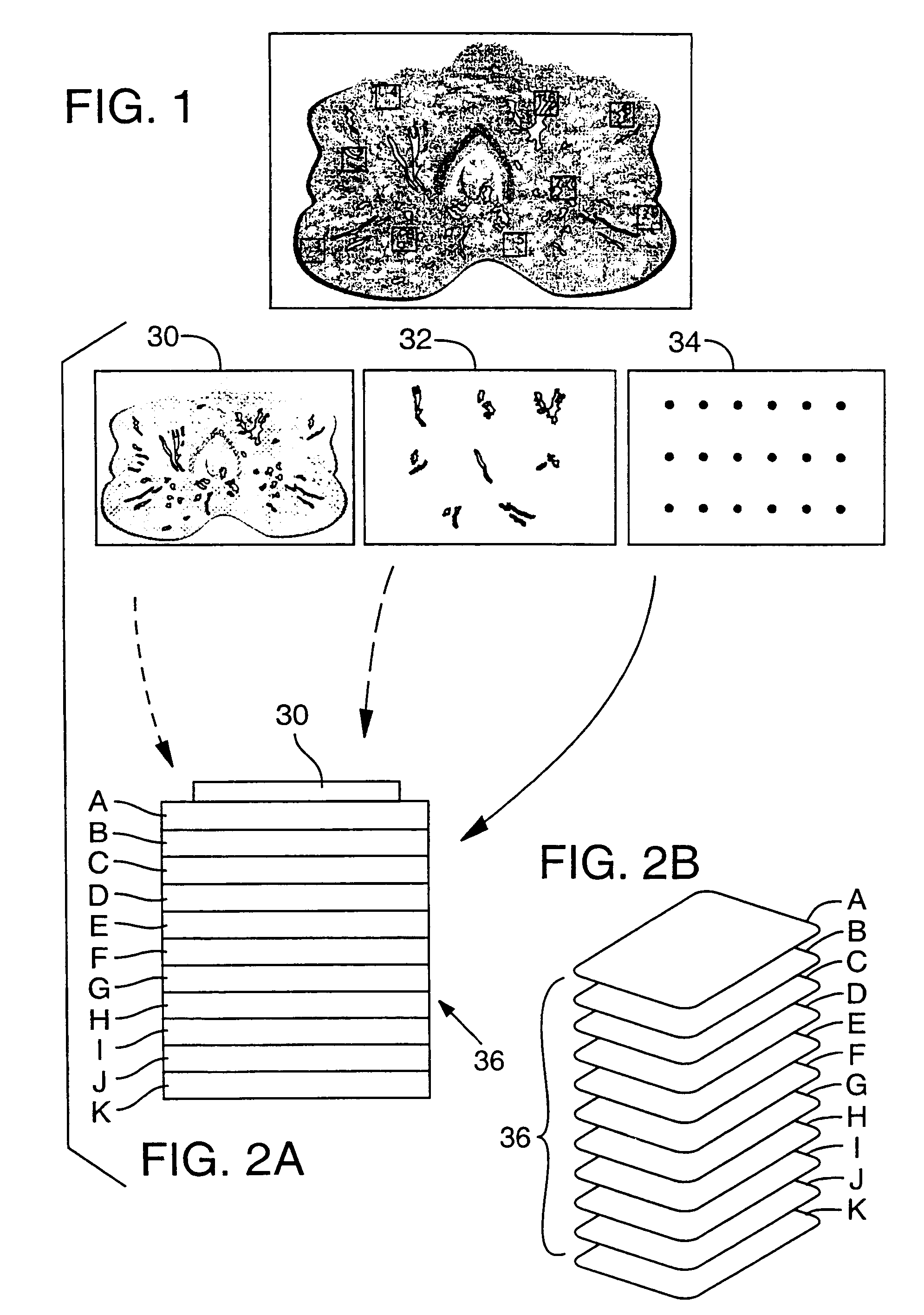
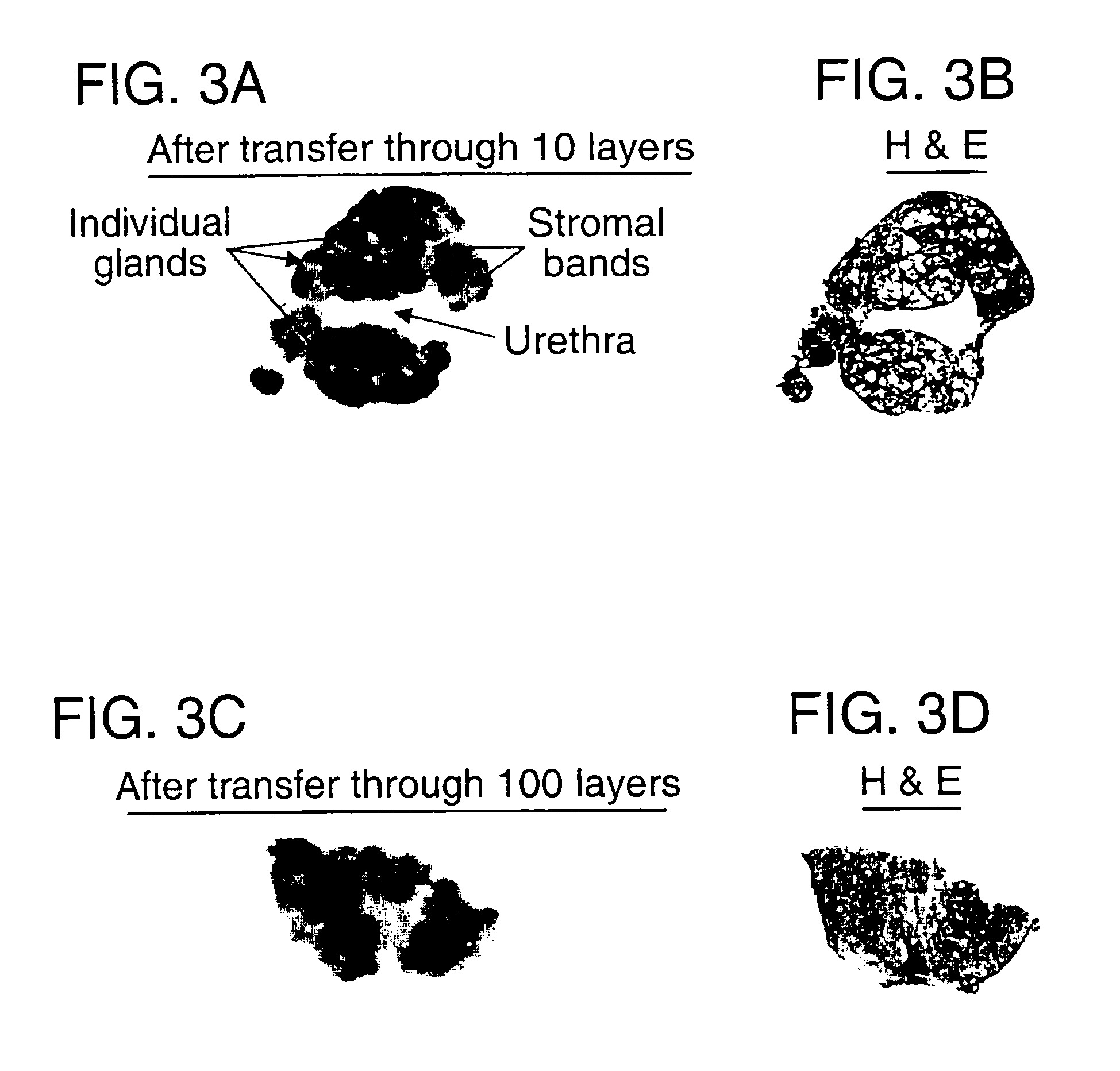
[0056]The LES procedure was performed on prostate tumor sections. The preliminary experiment used cytokeratin as the protein of interest. A whole mount cryostat section of human prostate tissue was prepared by making a thin frozen section of prostate, the section having a thickness of about 10 μm. As shown in FIG. 1, the section includes multiple cell populations of biological interest including normal epithelium, pre-malignant lesions, high and low grade tumor foci, and significant tumor-host interactions such as lymphocytes interacting with cancer cells. This section was placed on an ultrathin 2% agarose gel that had been cast on a glass histology slide. The section was covered with 2% agarose solution. A cover slip was applied on top of the section and the agarose was allowed to polymerize, thus creating a two-layered sample gel with the tissue section in between. The agarose sample gel containing the tissue ...
example 2
Selective Capture of Prostate Specific Antigen (PSA)
[0065]To demonstrate selective molecular capture within substrate layers, cell samples from five separate patients were procured from tissue specimens and solubilized in standard protein extraction buffer. The samples included lysates of normal lung, lung cancer, esophageal cancer, normal prostate, and breast cancer tissue. Each of the cell lysates was placed within a discrete 4 mm diameter spot on the top layer of a capture membrane set. This was accomplished by punching 4 mm diameter holes (“wells”) in a 2 mm thick agarose gel, adding the lysates to 1% liquid agarose, filling the 4 mm wells with the lysate / agarose solution, and allowing them to solidify. The sample gel thus created was placed on the top layer of a capture membrane set. Additionally, purified PSA was used as a positive control sample. In this experiment, the capture membranes consisted of ten nitrocellulose layers, each coupled to a different antibody. Polyclonal ...
example 3
Specificity of PSA Capture
[0067]To show the specificity of the capture process, a single sample of prostate tissue was solubilized and transferred through a set of capture layers as described in Example 2 above, except that polyclonal anti-PSA was placed on membrane 5. After the transfer of the prostate tissue through the layers, each membrane was placed in denaturing buffer to remove captured molecules. The proteins recovered from every membrane were subsequently separated by gel electrophoresis (the proteins recovered from layer 1 were run in Lane 1, the proteins recovered from layer 2 were run in lane 2, and so forth) and analyzed by immunoblot using a monoclonal anti-PSA antibody. FIG. 4B shows the results from each of capture layers one through nine. Lane 5 (representing layer 5, linked to anti-PSA) shows a single, distinct PSA band at Mr=30,000 (30 kDa). The remaining capture membranes are negative for PSA. This result demonstrates that PSA was captured only on the membrane co...
PUM
| Property | Measurement | Unit |
|---|---|---|
| Length | aaaaa | aaaaa |
| Thickness | aaaaa | aaaaa |
| Capillary wave | aaaaa | aaaaa |
Abstract
Description
Claims
Application Information
 - R&D
- Intellectual Property
- Life Sciences
- Materials
- Tech Scout
- Unparalleled Data Quality
- Higher Quality Content
- 60% Fewer Hallucinations
Browse by: Latest US Patents, China's latest patents, Technical Efficacy Thesaurus, Application Domain, Technology Topic, Popular Technical Reports.
© 2025 PatSnap. All rights reserved.Legal|Privacy policy|Modern Slavery Act Transparency Statement|Sitemap|About US| Contact US: help@patsnap.com


