Method and apparatus for biosensor spectral shift detection

a biosensor and spectral shift technology, applied in the field of biosensors, can solve the problems of limited commercial acceptance of label-free biosensor technologies, high cost of production and packaging of biosensors fabricated on semiconductor or glass wafers in batch photolithography/etching/deposition process, arrays pose difficult challenges in terms of package cost and compatibility with sensor exposure to fluids

Inactive Publication Date: 2007-05-15
X BODY
View PDF164 Cites 46 Cited by
  • Summary
  • Abstract
  • Description
  • Claims
  • Application Information

AI Technical Summary

Benefits of technology

[0011]A method and apparatus for performing high-resolution determination of the relative shift of the spectral properties of a biosensor is provided. The shift in the resonance peak of the biosensor is indicative of the amount of material bound to the surface of the biosensor. The apparatus may be used with a colorimetric resonant biosensor having a surface of amine functional groups useful for attaching biological molecules. A preferred biosensor is a Guided Mode Resonant Filter Biosensor (GMRFB).

Problems solved by technology

With the completion of the sequencing of the human genome, one of the next grand challenges of molecular biology will be to understand how the many protein targets encoded by DNA interact with other proteins, small molecule pharmaceutical candidates, and a large host of enzymes and inhibitors.
The widespread commercial acceptance of label-free biosensor technologies has been limited by their ability to provide high detection sensitivity and high detection parallelism in a format that is inexpensive to manufacture and package.
For example, biosensors fabricated upon semiconductor or glass wafers in batch photolithography / etch / deposition processes are costly to produce and package if the sensor area is to be large enough to contain large numbers of parallel assays.
Similarly, the requirement of making electrical connections to individual biosensors in an array poses difficult challenges in terms of package cost and compatibility with exposure of the sensor to fluids.
However, to date, these methods have yet to yield commercially available high-throughput instruments that can perform high sensitivity assays without any type of label in a format that is readily compatible with the microtiter plate-based or microarray-based infrastructure that is most often used for high-throughput biomolecular interaction analysis.

Method used


Image

  • Method and apparatus for biosensor spectral shift detection
  • Method and apparatus for biosensor spectral shift detection
  • Method and apparatus for biosensor spectral shift detection

Examples


Embodiment Construction

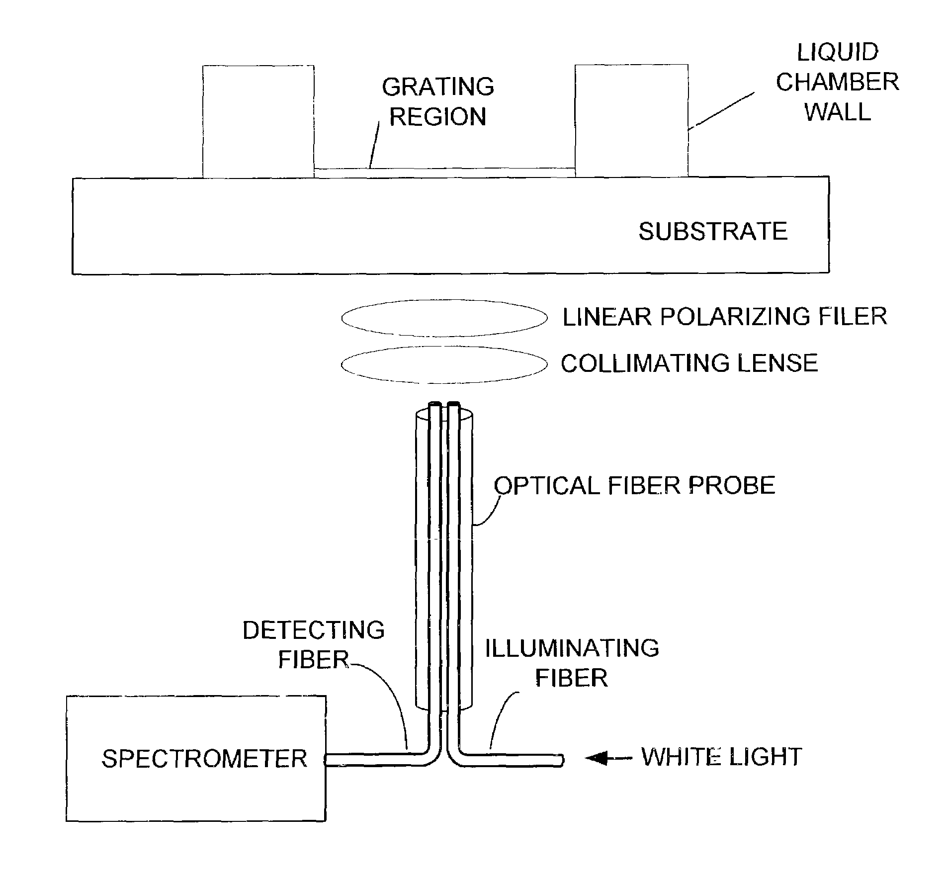
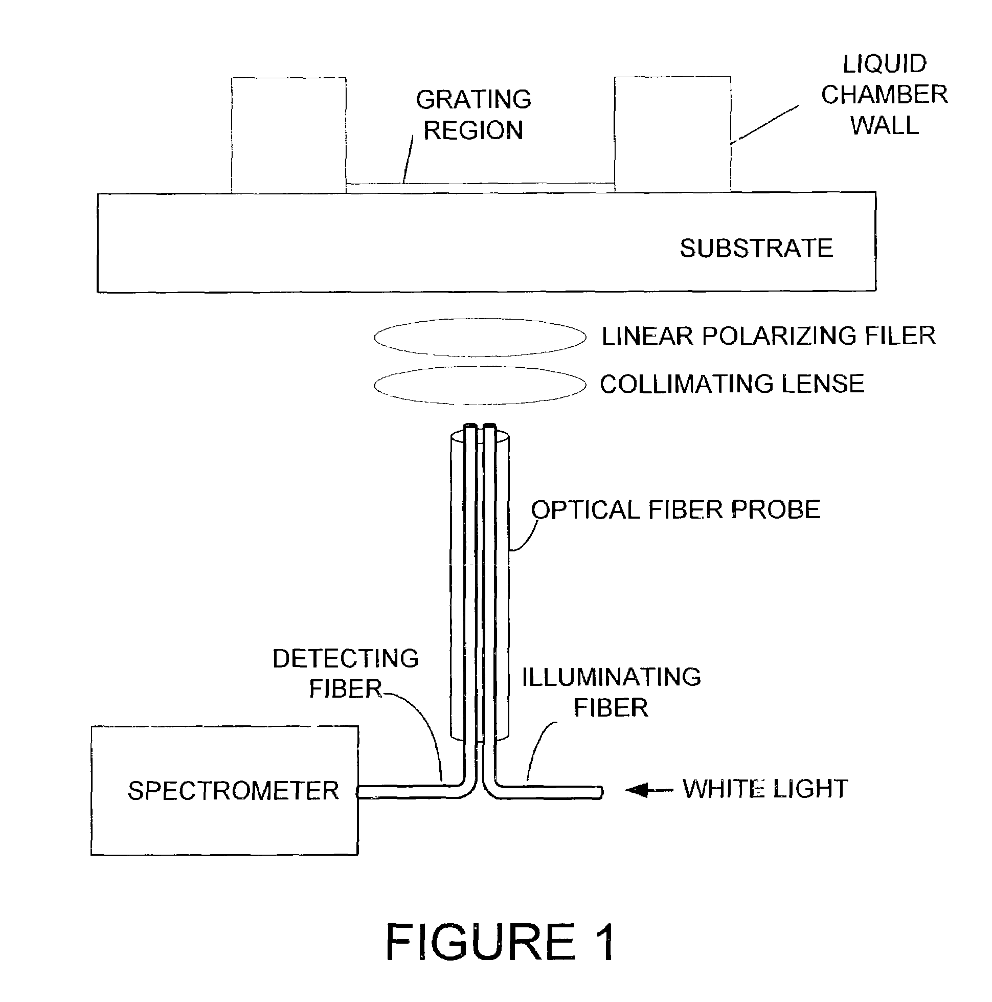
[0027]The preferred calorimetric resonant optical biosensor allows biochemical interactions to be measured on the sensor's surface without the use of fluorescent tags or calorimetric labels. The sensor surface preferably contains an optical structure that, when illuminated with collimated white light, is designed to reflect only a narrow band of wavelengths. The narrow wavelength band is described as a wavelength “peak.”

[0028]One preferred guided mode resonant filter structure is a one-dimensional linear grating surface structure. The sensor can be manufactured by performing sub-micron definition of grating features using photolithography on the sensor. Alternatively, the sensor can be produced inexpensively over large surface areas using sub-micron microreplication of a master sensor surface structure on continuous sheets of plastic film. Alternative structures, including 2-D or 3-D structures, surface relief structures, glass substrates, discrete plastic film, as well as alternati...


PUM

PropertyMeasurementUnit
thicknessaaaaaaaaaa
diameteraaaaaaaaaa
diameteraaaaaaaaaa

Abstract

Performing high-resolution determination of the relative shift of the spectral properties of a biosensor. The shift in the resonance peak of the biosensor is indicative of the amount of material bound to the surface of the biosensor. A preferred biosensor is a Guided Mode Resonant Filter Biosensor (GMRFB). In one aspect of the invention, curve fitting is used to determine the relative location of the spectrum of the unexposed biosensor with respect to those spectra that are altered (e.g., shifted) by the presence of materials bound to the surface of the biosensor. In an alternative embodiment, the cross correlation function is used to detect spectral peak offsets between a reference spectrum and a spectrum measured from an exposed biosensor. In yet another alternative, maximal likelihood estimation techniques are used to determine the spectral shift or offs.

Description

[0001]This application is a Continuation-In-Part of U.S. patent application Ser. No. 10 / 059,060 filed Jan. 28, 2002 now U.S. Pat. No. 7,070,987 and a Continuation-In-Part of U.S. patent application Ser. No. 10 / 058,626 filed Jan. 28, 2002 now U.S. Pat. No. 6,951,715, both of which are Continuations-in-Part of U.S. patent application Ser. No. 09 / 930,352, filed Aug. 15, 2001 now U.S. Pat. No. 7,094,595, which claims the benefit of U.S. provisional application 60 / 244,312 filed Oct. 30, 2000, U.S. provisional application 60 / 283,314 filed Apr. 12, 2001, and U.S. provisional application 60 / 303,028 filed Jul. 3, 2001, all of which are hereby incorporated by reference.BACKGROUND OF THE INVENTION[0002]A. Field of the Invention[0003]The present invention relates to biosensors useful for detecting biological material. More specifically, it is directed to a method and device for determining the amount of shift in an optical biosensor's spectral output.[0004]B. Description of the Related Art[0005...

Claims


Application Information

Patent Timeline
Patent Type & Authority Patents(United States)
IPC IPC(8): G01N21/00B01L3/00G01N21/25G01N21/47G01N33/543G02B5/18
CPCB01L3/5085G01J3/02G01J3/0218G01J3/0224G01J3/1804G01J3/42G01N21/253G01N21/31G01N21/7743G01N33/54373G01N2021/3196G01N2021/7773G02B5/1809Y10S436/805
Inventor PIEN, HOMERKARL, WILLIAM C.PUFF, DEREKLI, PETERCUNNINGHAM
Owner X BODY
Why Eureka
Try Eureka