
Tissue capturing and suturing device and method

a technology of tissue apposition and suture clip, which is applied in the field of new combination of endoscopic tissue apposition device and suture clip delivery device, can solve the problems of invariably long use of such devices, inability to perform the various procedural steps endoscopically, and significant problems with the described gastroplasty procedure, so as to reduce the need for cam surfaces and cam followers, reduce the abrasion of sutures, and reduce the need for friction.

Inactive Publication Date: 2007-05-22
CR BARD INC
View PDF302 Cites 129 Cited by
  • Summary
  • Abstract
  • Description
  • Claims
  • Application Information

AI Technical Summary

Benefits of technology

[0017]The present invention pertains to improvements to endoscopic apposition device and suture clip delivery devices. The improvements may be embodied in a tissue apposition device similar to those disclosed in U.S. Pat. Nos. 5,792,153 or 5,080,663, or a stapling device such as is disclosed in U.S. Pat. No. 5,037,021. The disclosures of the above listed patents are incorporated by reference herein, in their entirety. The prior art endoscopic tissue apposition devices provided a mechanism for capturing only a single fold with one intubation. The present invention provides a tissue apposition device capable of capturing multiple tissue folds and securing the sutures used to capture the folds with one intubation. The elimination of the need for multiple intubations eliminates the need for an overtube with respect to endoscopic procedures performed in the gastro-esophageal tract.
[0021]The tissue apposition device of the present invention offers another advantage over previous designs in that the entire capsule body may be injection molded from a polymer material. A single piece injection molded unit is easier to produce than previous capsule designs, which were machined from metal and comprise several assembled components.
[0023]Also described herein is a suture clip delivery device adapted for navigation through the working channel of an endoscope and through the capsule channel adapted to receive the needle assembly. The delivery device comprises a multi-coaxial catheter with a three or four finger collet jaw affixed at a distal end. The catheter has a distal end from which the collect fingers distally project. The collet fingers are made of a material such as stainless steel or an engineering grade of plastic that allow the collet fingers to flex or spring from a pre-biased first open position to a second closed position and back to the first position. Alternatively, the collet fingers can be designed to flex or spring from a pre-biased first closed position to a second open position and back to the first position. The collet fingers define a cage within which a suture clip assembly is premounted. The cage is defined proximally by a distal end of the collet body and distally by flanges extending radially inwardly from the distal ends of the collet fingers. In one embodiment, the case is sized so that a plurality of suture clip plugs can be preloaded into the cage along with a single suture clip ring. By biasing the collet fingers in an open position, the need for cam surfaces and cam followers is eliminated.
[0024]In one embodiment, a separate control surface provided coaxially about the collet cage is employed to slide the outer sliding sleeve relative to the collet cage and the pusher. With any of the embodiments, the outer sliding sleeve is provided to secure the collet fingers in a closed position when placed in a distally advanced position. The outer sliding sleeve performs the additional function of severing the suture at a point proximal to the suture clip after engagement of the suture upon proximal retraction. One or more suture slots are provided in the distal end of the outer sliding sleeve to provide suture exits. A distal end of the suture slots can be sharpened to sever the suture. Upon proximal retraction of the outer sliding sleeve, the distal end of suture slot engages the suture and severs it when the distal end of the suture slot travels proximally to a cutting edge formed at or near the proximal end of the collet fingers. The design of the delivery device enables the suture clip to be cinched in close proximity to the sutured tissue as well as allow for the severing of the suture tails and release of the suture from the delivery device in one step.
[0025]The suture clips adapted for use with the suture clip delivery device are comprised of a plug and a ring that are configured to allow a suture to inter-wind through the clip in such a manner that the clips move with minimal friction while in an open position. The plug is a headless design that reduces abrasion of the suture as it exits the suture clip ends. In a closed position, the clip captures the suture by frictional engagement. The ring and plug components of the clip can be provided with interlocking features for enhancing the suture capturing effect.

Problems solved by technology

With all the improvements provided by the sewing capsules and suture securing devices, significant problems persist with the described gastroplasty procedure.
One of the significant problems is the time and number of intubations needed to perform the various procedural steps endoscopically.
The use of such devices is invariably long and cumbersome.
Although an overtube provides a barrier between the devices used to perform the procedure and the luminal walls of the tract, the act of inserting the relatively large diameter tube into the tract and the presence of the tube in the tract are perhaps the most significant sources of patient discomfort.

Method used


Image

  • Tissue capturing and suturing device and method
  • Tissue capturing and suturing device and method
  • Tissue capturing and suturing device and method

Examples


Embodiment Construction

[0074]A description of the embodiments of the present invention is best presented in conjunction with an explanation of the operation of a prior art tissue apposition device, which this invention serves to improve. FIGS. 28A–30 depict a prior art endoscopic suturing device disclosed in U.S. Pat. No. 5,792,153. FIG. 28A shows the distal end of a flexible endoscope 90, on which a sewing device 2 is attached. The endoscope is provided with a viewing channel, which is not shown, but which terminates at a lens on the distal face of the endoscope. The endoscope is further provided with a biopsy or working channel 3, and a suction channel 4 the proximal end of which is connected to a source of vacuum (not shown). The suction channel 4 may comprise a separate tube that runs along the exterior of the endoscope, rather than an internal lumen as shown. The sewing device 2 has a tube 5, which communicates with the suction pipe 4 and has a plurality of perforations 6 therein. These perforations ...


PUM


Abstract

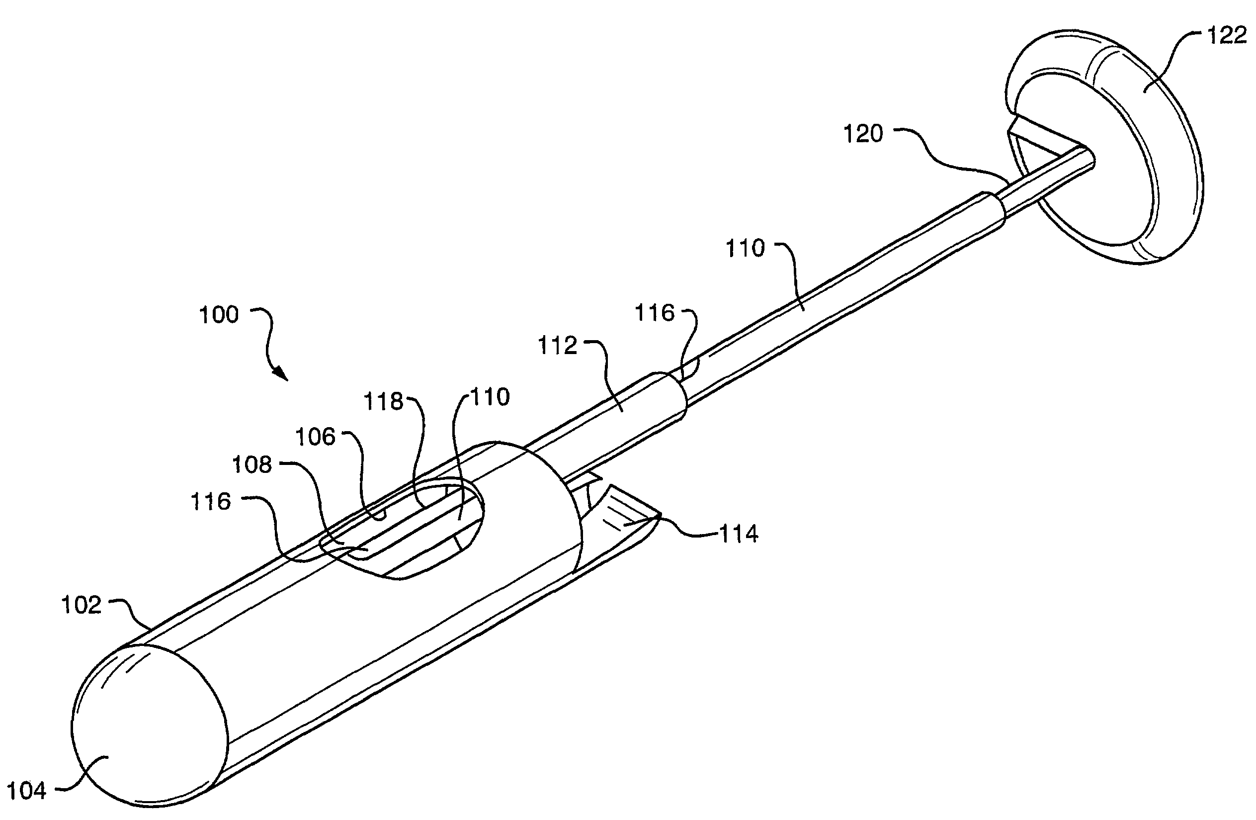
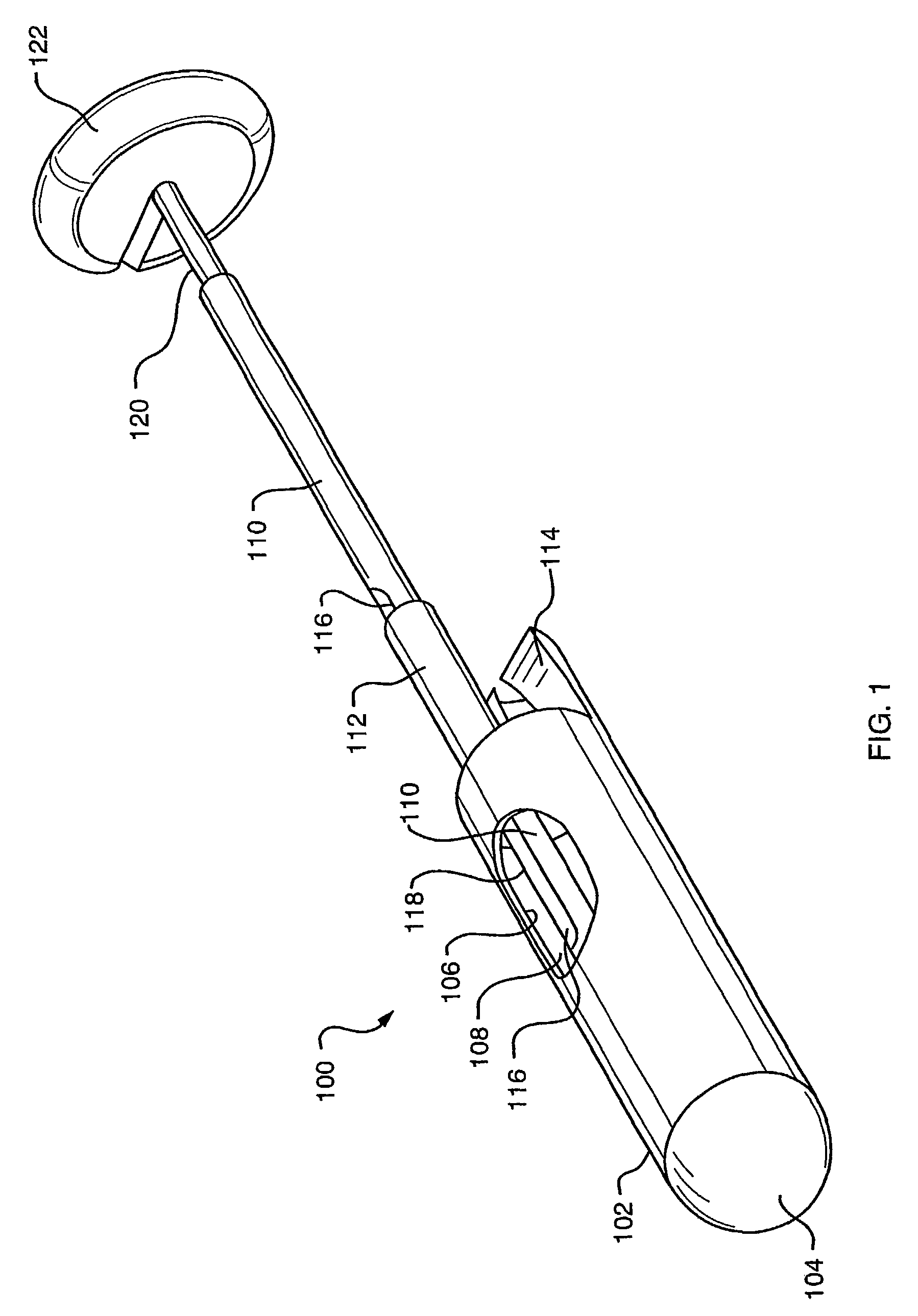
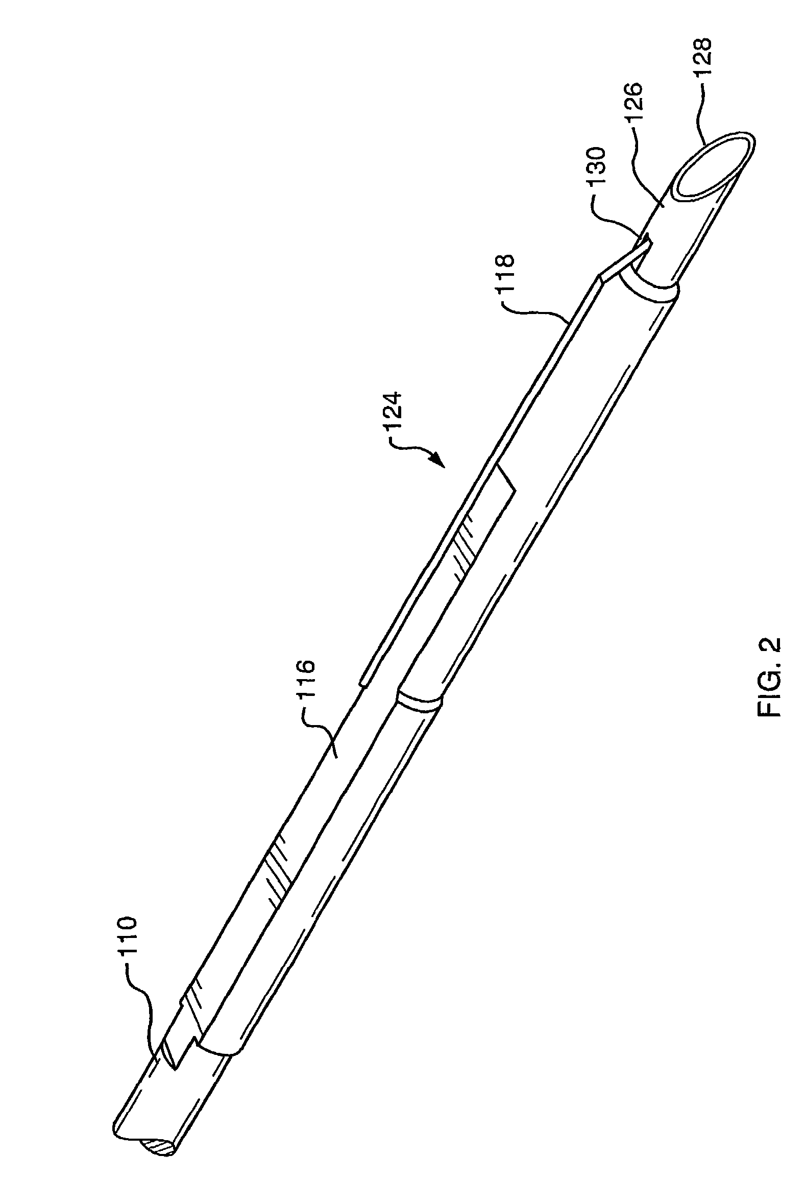
A combination tissue apposition and suture capturing device (100) for performing endoscopic procedures typically in the gastro-esophageal tract. The device (100) is particularly adapted for forming multiple plications used in a gastroplasty procedure devised to cure or ameliorate gastro-esophageal reflux disease. The device includes a tissue sewing capsule (102) attached to the distal end of an endoscope having a needle (120) that is deposited in a capsule (102) distal tip cavity following the suturing of a tissue fold and retrieved to enable the suturing of a subsequent tissue fold without the need for multiple intubations. A suture clip delivery device (200) is also disclosed that is adapted to fit within the capsule (102) to enable suture (118) capture without the need for multiple intubations. The combination device eliminates the need for an overtube and maximizes the speed efficiency of the gastroplasty procedure. A method for using the combination device is also disclosed.

Description

CROSS-REFERENCE TO RELATED APPLICATIONS[0001]A claim of benefit is made to U.S. Provisional Applications Nos. 60 / 205,741 filed 19 May 2000, 60 / 205,444 filed 19 May 2000, 60 / 253,970 filed 29 Nov. 2000 and PCT Application No. PCT / US01 / 07349, the contents of which are incorporated in their entirety herein by reference.FIELD OF THE INVENTION[0002]The present invention relates to a novel combination of endoscopic tissue apposition devices and suture clip delivery devices. Specifically, the invention provides an endoscopic apposition device configured to collect and secure a plurality of tissue portions with a single intubation of the device. The invention is applicable to both flexible and rigid endoscopy.BACKGROUND OF THE INVENTION[0003]Endoscopic apposition devices are devices that can be used in the body of a patient without the need to make an external incision in the patient, the device being controlled externally of the patient by endoscopic means. Apposition devices may comprise a...

Claims


Application Information

Patent Timeline
Patent Type & Authority Patents(United States)
IPC IPC(8): A61B17/04A61B17/10A61B19/00A61B17/06
CPCA61B17/0401A61B17/0469A61B17/0487A61B17/0482A61B2017/0409A61B2017/0417A61B2017/0454A61B2017/0474A61B2017/0475A61B2017/0488A61B2017/06052A61B2017/306
Inventor GAMBALE, RICHARD A.
Owner CR BARD INC
Learn More