
System and method for providing slant-angle collimation for nuclear medical imaging

a technology of nuclear medical imaging and collimation, which is applied in the field of radiation imaging systems and methods, can solve the problems of relatively expensive diagnostic tool subsystems of the gantry system, and achieve the effects of reducing the length of the translational pass, cost saving, and simplifying setup and operation

Active Publication Date: 2007-06-12
SIEMENS MEDICAL SOLUTIONS USA INC
View PDF6 Cites 10 Cited by
  • Summary
  • Abstract
  • Description
  • Claims
  • Application Information

AI Technical Summary

Benefits of technology

The present invention is about a method and arrangement for improving collimation in SPECT scanning to obtain required angular views using only one-dimensional relative linear motion between the camera and the patient. This is important because whole body scanning technologies already use this type of motion, and it reduces the cost and complexity of the support structures required for the instruments and the patient. The invention utilizes a collimator with a slant angle to minimize the length of the translational pass required of the camera and reduce the space requirements for the system. The collimator is positioned between the radiating mass and the radiation detector, and the apertures in the collimator allow gamma rays to strike the detector. The technical effects of the invention include improved radiological imaging quality and simplified and cost-reduced support structures.

Problems solved by technology

One drawback associated with this requirement however, is that such gantry systems are relatively expensive subsystems of the diagnostic tool because they must be capable of providing rapid rotation is of the large and heavy camera heads through very precise orbits about the patient.

Method used


Image

  • System and method for providing slant-angle collimation for nuclear medical imaging
  • System and method for providing slant-angle collimation for nuclear medical imaging
  • System and method for providing slant-angle collimation for nuclear medical imaging

Examples


Embodiment Construction

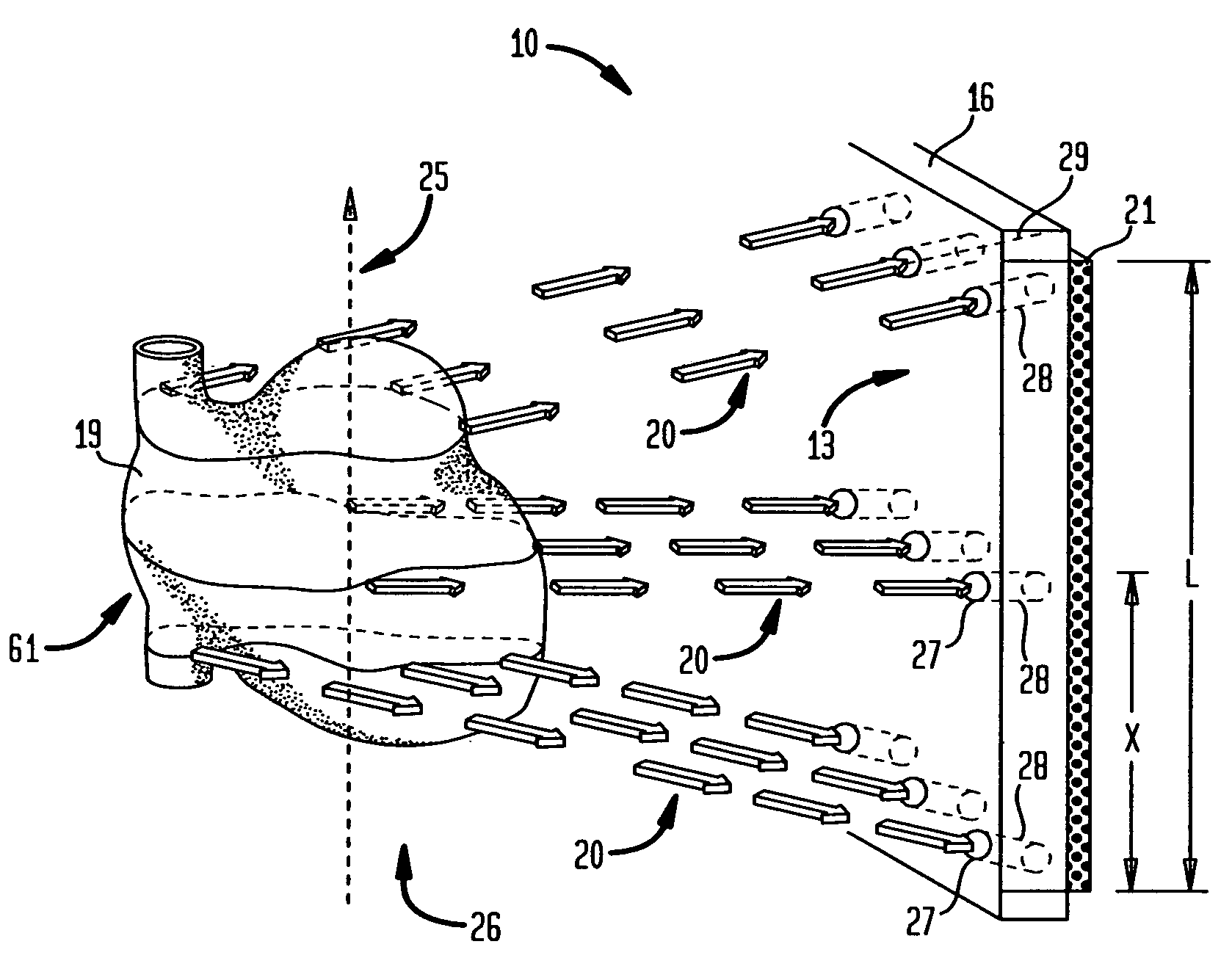
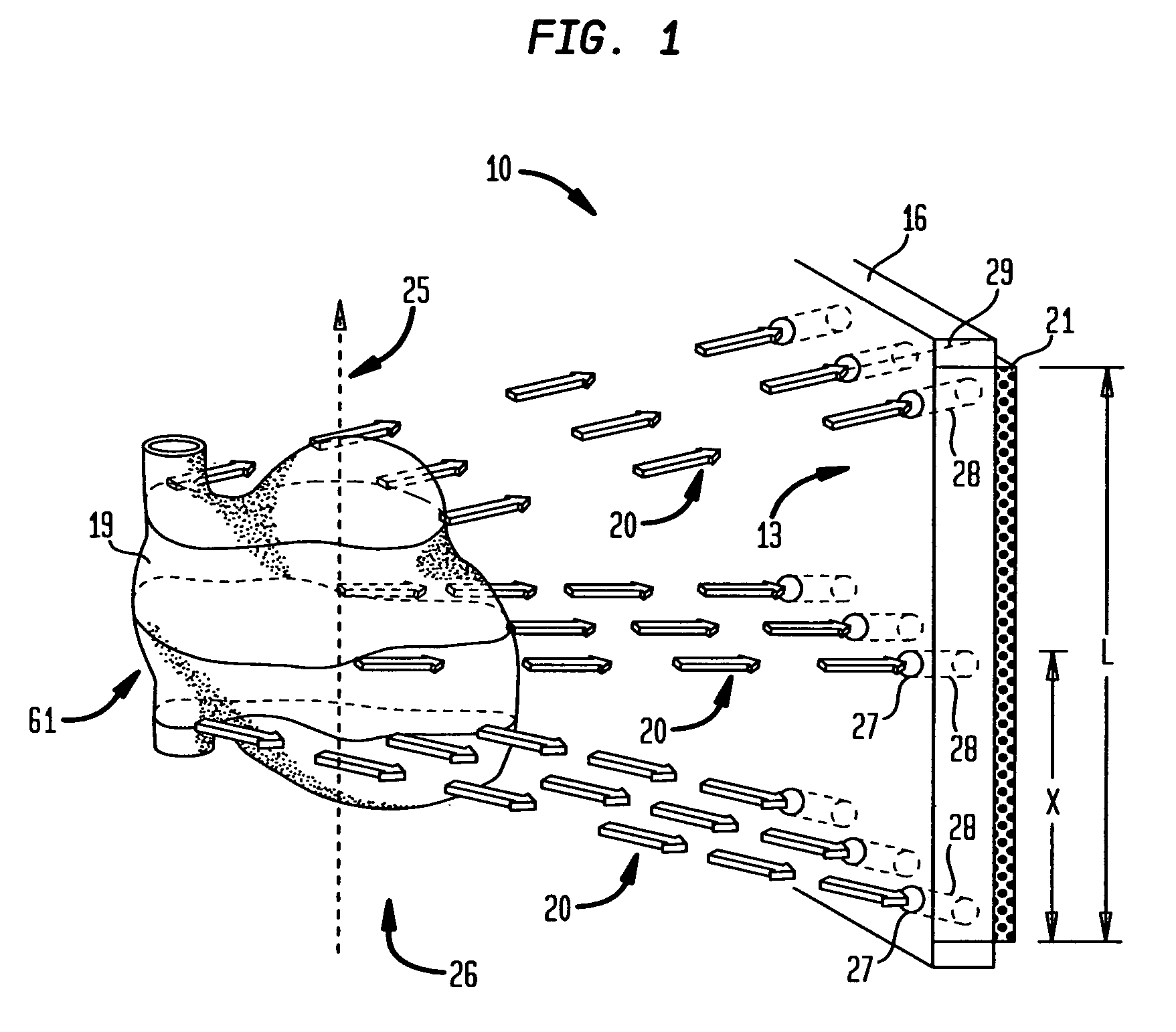
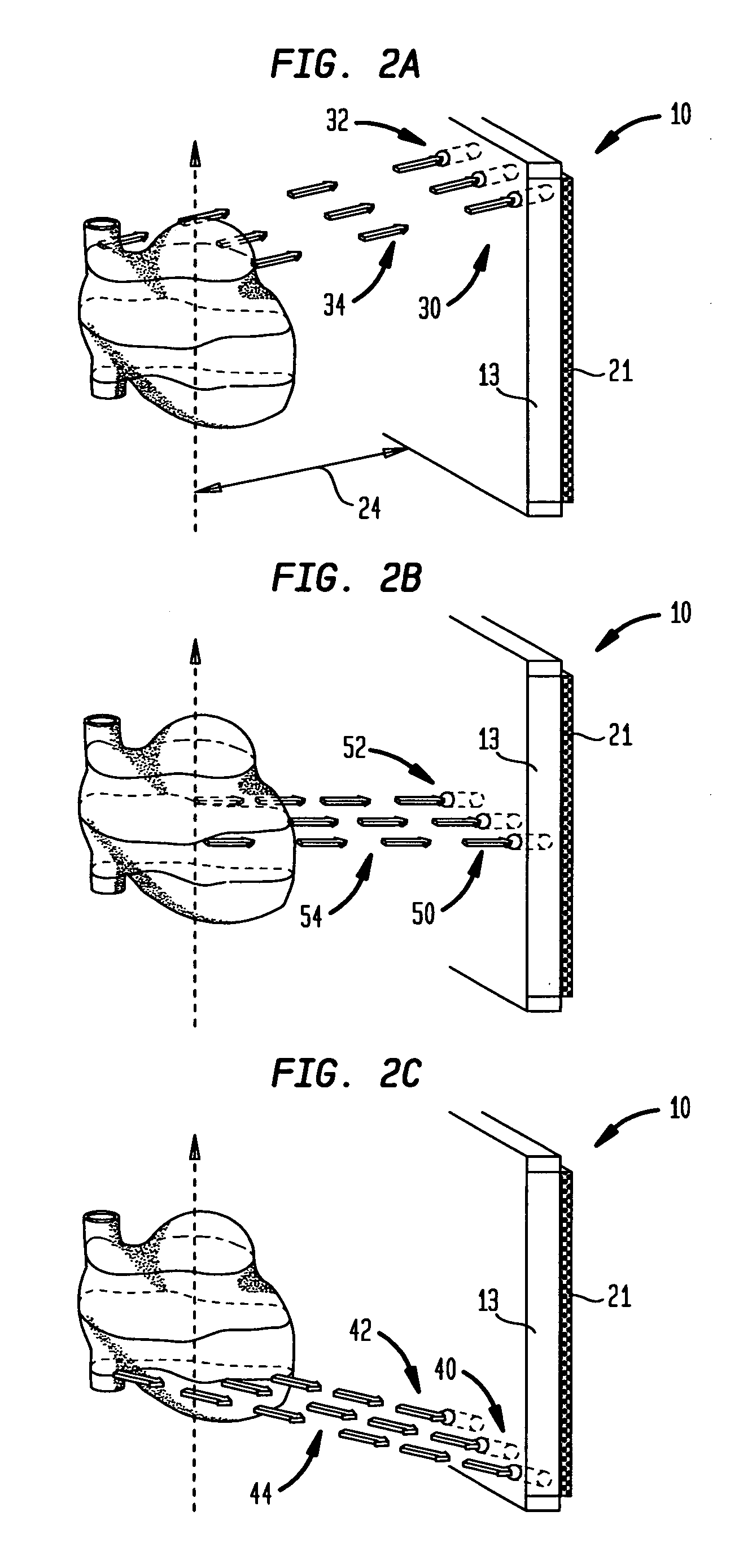
[0021]When considering full angular gamma camera sampling utilizing SPECT parallel projection views, or their equivalents, the image samples should be obtained over one hundred and eighty or three hundred and sixty degrees. According to the present invention, a system (10) is provided that includes a collimator (13) adapted with apertures (27) that serve as openings for slant-angle passageways (28) which accommodate views of a radiating area or object-of-interest (61), such as the heart of a patient (19), from a specific slant-angle.

[0022]Referring to FIG. 1, the specific angle at which the slotted passageway (28) is canted within the collimator (13) is dependent on the relative position (x) of the passageway (28) in the collimator (13). In the instance of FIG. 1, the radiation source is the radiating mass (19) shown as the radioactively infused heart (19) of a patient. For each row of passageways (28) through the collimator (13), the associated collimated (gamma) rays travel along ...


PUM


Abstract

Method and arrangement for implementing a system (10) for providing multi-angular SPECT radiation sampling utilizing slant-angle collimation. The system (10) includes a collimator (13) positioned between a radiating mass (19) within a patient (60) and a radiation detector (21). The collimator (13) is spaced apart from a translational path (25) of the radiating mass (19) at a predefined distance (24). A plurality of apertures (27) extend through the collimator (13) and each forms a passageway (28) for radiation rays (20) emanating from the radiating mass (19) in a direction substantially aligned with a longitudinal axis (29) of the respective passageway (29) and in this manner enables the aligned radiation rays (20) to strike the radiation detector (21). The plurality of passageways (28) include a first group (30) of passageways adjacently aligned in a first row (32) and arranged so that the longitudinal axes (29) of the first group (30) of passageways (32) are substantially contained in a first plane (34) oriented substantially perpendicularly to a central plane (16) of the collimator (13). Each of the parallel longitudinal axes (29) of the first row (32) of passageways are obliquely oriented with respect to the central plane (16) of the collimator (13) with an included angle (36) therebetween. Each of the included angles, when measured clockwise from the central plane (16) or face of the collimator (13) to a respective longitudinal axis, is an acute angle.

Description

BACKGROUND OF THE INVENTION[0001]1. Field of the Invention[0002]The present invention relates to systems and methods used in radiation imaging; and more particularly, to collimators utilized in cooperation with radiation detectors that permit only desired radiation to strike the detector thereby producing a more accurate image-of-interest, typically of an internal portion of a patient when medical and diagnostic applications are considered.[0003]2. Description of the Background Art[0004]In conventional radiation imaging arrangements, collimators are used in a wide variety of equipment in which it is desired to permit only beams of radiation emanating along a particular path to pass a selected point or plane. Collimators are frequently used in radiation imagers to ensure that only radiation beams passing along a direct path from the known radiation source strike the detector thereby minimizing detection of beams of scattered or secondary radiation.[0005]Particularly in radiation imag...

Claims


Application Information

Patent Timeline
Patent Type & Authority Patents(United States)
IPC IPC(8): G21K1/02G21K5/10
CPCG21K1/025
Inventor HAWMAN, ERIC G.
Owner SIEMENS MEDICAL SOLUTIONS USA INC
Learn More