
Sphingomyelin enhancement of tumor therapy

a tumor and sphingomyelin technology, applied in the field of tumor therapy sphingomyelin enhancement, can solve the problems of post-mitotic cell death, inefficiency of dna repair, and not all lesions are repairable, and achieve the effect of enhancing tumor therapy

Inactive Publication Date: 2007-10-30
CENT FOR MOLECULAR BIOLOGY & MEDICINE
View PDF9 Cites 6 Cited by
  • Summary
  • Abstract
  • Description
  • Claims
  • Application Information

AI Technical Summary

Benefits of technology

[0015]In accordance with another aspect of the present invention, there is provided a method of treating rheumatoid arthritis in a mammalian patient, comprising administering to the patient an amount of sphingomyelin effective to increase ceramide production and resultant apoptosis in proliferating synovial fibroblasts.

Problems solved by technology

While mammalian cells are proficient in repairing most DNA double strand breaks, not all such lesions are repairable.
Residual unrepaired DNA lesions can lead to post-mitotic cell death.
Therefore, until recently, inefficiency of DNA repair was thought to play a key role in radiation sensitivity.
However, the mechanism by which such damage induced the apoptotic pathway remained unclear.
While many therapies are successful in initiating the sphingomyelin transduction pathway, the induced apoptotic response may be limited or short-lived.
Tumor tissues typically have higher concentrations of sphingomyelin than normal tissues; however, it is possible that some tumor cells have reduced sphingomyelin synthesis capabilities.
Additionally, altered lipid metabolism in tumor cells can result in changes in the intracellular distribution of sphingomyelin.
Such redistribution within the plasma membrane can lead to misdirected sphingomyelin which is unable to be acted upon by the sphingomyelin hydrolyzing enzymes responsible for generating ceramide in response to cytotoxic treatment.
Consequently, sphingomyelin re-organization within the plasma membrane can impair a tumor cell's ability to generate ceramide-induced apoptosis and lead to reduced sensitivity to certain therapies.

Method used


Image

  • Sphingomyelin enhancement of tumor therapy
  • Sphingomyelin enhancement of tumor therapy
  • Sphingomyelin enhancement of tumor therapy

Examples


example 1

In vivo Evaluation of Sphingomyelin Therapy on GW39 Colonic Tumors

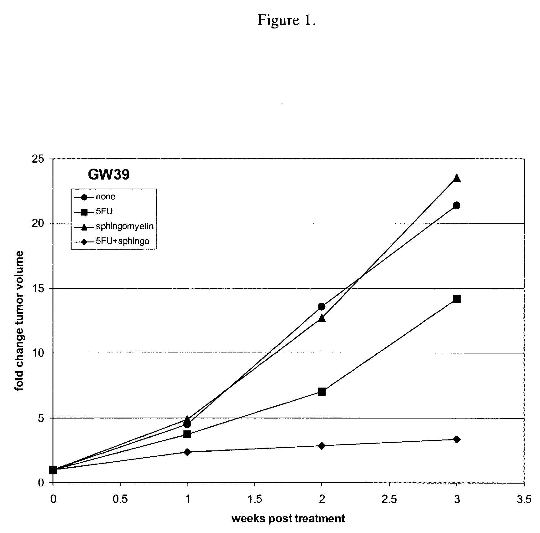
[0045]Sphingomyelin enhancement of chemotherapy was evaluated by measuring its effect on 5-fluorouracil (5FU) treatment of GW39 colonic tumors in mice. Nude mice were implanted subcutaneously with GW39 tumors. After the tumors reached approximately 0.5 cm3, the mice were split into groups of ten and administered one of the following therapies: no treatment (●), 0.45 mg / day of 5-fluorouracil for five days (▪), 10 mg / day of sphingomyelin (SM) for seven days (▴), or 0.45 mg / day of 5-fluorouracil for five days and 10 mg / day of sphingomyelin for seven days (♦). Both the 5-fluorouracil and the sphingomyelin were administered by intravenous injection. The group receiving both 5-fluorouracil and sphingomyelin was administered both therapies for five days and then continued to receive injections of sphingomyelin for 2 days. The tumor volume in each animal was assessed at weekly intervals for three weeks following treatment.

[00...

example 2

In vivo Evaluation of Sphingomyelin Therapy on HT29 Colonic Tumors

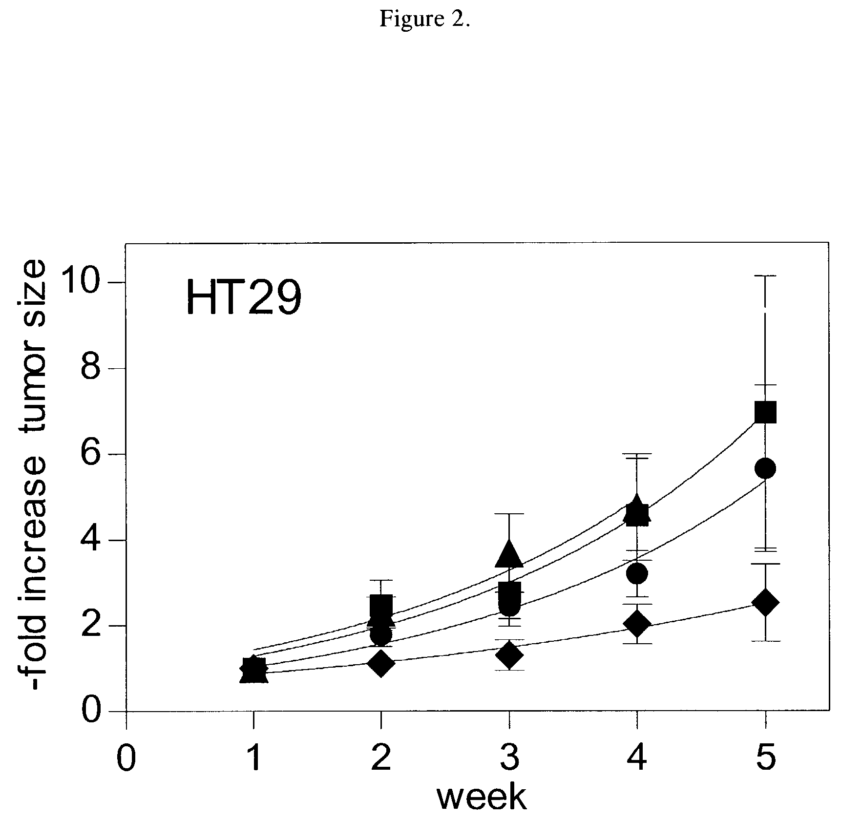
[0047]Sphingomyelin enhancement of chemotherapy was evaluated by measuring its effect on 5-fluorouracil treatment of HT29 colonic tumors in mice. Nude mice were implanted subcutaneously with HT29 tumors. After the tumors reached approximately 0.5 cm3, the mice were split into groups of ten and administered one of the following therapies: no treatment (●), 0.45 mg / day of 5-fluorouracil for five days (▪), 10 mg / day of sphingomyelin for seven days (▴), or 0.45 mg / day of 5-fluorouracil for five days and 10 mg / day of sphingomyelin for seven days (♦). Both the 5-fluorouracil and the sphingo-myelin were administered by intravenous injection. The group receiving both 5-fluorouracil and sphingomyelin was administered both therapies for five days and then continued to receive injections of sphingomyelin for 2 days. The tumor volume in each animal was assessed at weekly intervals for five weeks following treatment, except for th...

example 3

In vitro Evaluation of Sphingomyelin Therapy on Colonic Tumors

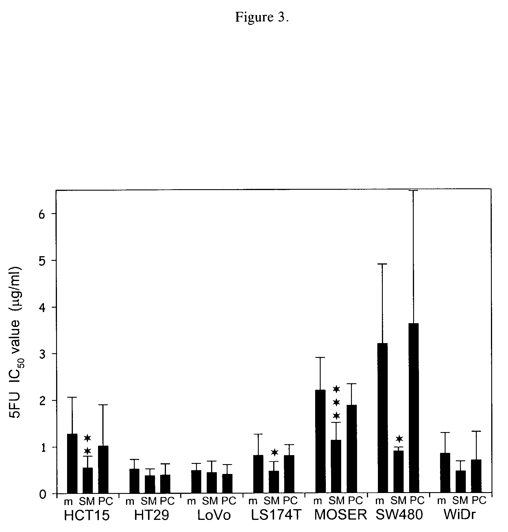
[0049]Sphingomyelin enhancement of chemotherapy was evaluated by measuring its effect on 5-fluorouracil or doxorubicin (DOX) treatment of colonic tumors grown in culture. Cell viability was measured using the dye MTT (3-(4,5-dimethylthiazol-2-yl)-2,5-diphenyl tetrazolium bromide) in a 24-well chamber format. See Mosmann, T., J. Immunol. Methods, 65:55-63 (1983). HCT15, HT29, LoVo, LS174T, MOSER, SW480 and WiDr human colonic tumor cells were maintained in RPMI media supplemented with 10% fetal calf serum. Human umbilical cord venous endothelial cells (HUVEC) from pooled donors (Clonetics / BioWhittaker, San Diego, Calif.) were used as controls. Cells (104 / well) were plated in the presence of varying concentrations of drug and sphingomyelin and grown in a humidified incubator. As an additional control, egg yolk phosphatidylcholine (PC) (Sigma, St. Louis, Mo.) was added to the cells instead of sphingomyelin. Drugs and lipids w...


PUM


Abstract

Cytotoxic tumor therapy in a patient is enhanced by co-administration of sphingomyelin. The invention most likely enhances a tumor cell's ability to undergo ceramide-induced apoptosis by increasing the levels of sphingomyelin in all cellular compartments, thereby providing sufficient substrate for activated sphingomyelinase. A method of treating rheumatoid arthritis also is provided.

Description

[0001]This application is based on U.S. Provisional Application Ser. No. 60 / 126,189, filed on Mar. 25, 1999 and U.S. patent application Ser. No. 09 / 533,799 filed Mar. 24, 2000.BACKGROUND OF THE INVENTION[0002]Traditionally, the efficacy of many cancer therapies was believed to arise from the cytotoxicity derived from chemotherapy- or radiation-induced DNA damage. Such DNA damage was considered to trigger an apoptotic response. See Eastman et al., Cancer Invest., 10: 229-240 (1992); Allan, D. J., Int. J. Radiat. Biol., 62: 145-152 (1992). Apoptosis is conceptualized as an inducible preprogrammed pathway of sequential biochemical events, leading to activation of calcium- and magnesium-dependent endonucleases that cleave the nuclear chromatin at selective internucleosomal linker sites. Signals generated at the membrane of the affected cell activate neighboring cells and infiltrating macrophages to phagocytize the dying cell and its disintegrating nucleus.[0003]An early hypothesis on th...

Claims


Application Information

Patent Timeline
IPC IPC(8): A01N57/26A01N43/04A61K51/00A61K47/42A61K31/513A61K31/688A61K31/704A61K45/06A61P19/02A61P29/00A61P35/00
CPCA61K31/513A61K31/688A61K31/704A61K45/06A61K2300/00A61P19/02A61P29/00A61P35/00
Inventor MODRAK, DAVIDGOLDENBERG, DAVID M.
Owner CENT FOR MOLECULAR BIOLOGY & MEDICINE
Learn More