Method for detection of melanoma

a detection method and technology for melanoma, applied in the field of melanoma patients, can solve the problems of no approved therapy for patients with intermediate risk of relapse, no cure, and high cost of excised lesions for the publi

Active Publication Date: 2007-11-20
DERMTECH INT
View PDF8 Cites 34 Cited by
  • Summary
  • Abstract
  • Description
  • Claims
  • Application Information

AI Technical Summary

Benefits of technology

[0017]In another aspect the present invention provides a kit for detecting malignant melanoma. The kit includes one or more probes or primers that selectively bind to one or more of Interleukin-1 RI (IL-1 RI), endothelin-2, ephrin-A5, Insulin-like Growth Facto

Problems solved by technology

Currently, there is no approved therapy for patients having intermediate risk of relapse.
There is no cure at this time for patients in whom metastasis to distant sites has occurred.
Erroneously excised lesions are costly for the public and cause scars often in cosmetically important body regions.
Furthermore, even after a lesion is taken out differentiation of benign melanocytic lesions from melanomas can be very difficult in a subset of about 10-15%, even for skilled dermatopathologists.
In addition, histological examination of cancer cells does not adequately reflect the complicated series of molecular events

Method used


Image

  • Method for detection of melanoma
  • Method for detection of melanoma
  • Method for detection of melanoma

Examples


example 1

Non-Invasive Recovery of Sub-Stratum Corneum Cells

A. Recovery Using a Rigid Surface

[0125]Skin cells can be recovered non-invasively by scraping the skin with a sterile #15 scalpel. The scalpel is held at an angle approximately 15 degrees from horizontal and repeatedly but gently scraped across an area of skin that is approximately 1×1 cm in size. The epidermal cells are transferred to a sterile tissue culture well by scraping the blade against the interior wall of the well. When the glistening epidermal layer is reached, the scraping is stopped prior to causing any bleeding, to avoid contaminating the scraping(s) with blood. The cells are deposited in a sterile 1 cm petri dish and about 300 ml of lysis buffer is added to the culture well. The lysis buffer is pipetted up and down until the epidermal cells are completely lysed.

[0126]RNA lysis buffer is added within 10 minutes of initiation of the scraping. The sterile tissue culture well is maintained on dry ice. The cells are dissolv...

example 2

Analysis Of Cells Obtained By Tape Stripping

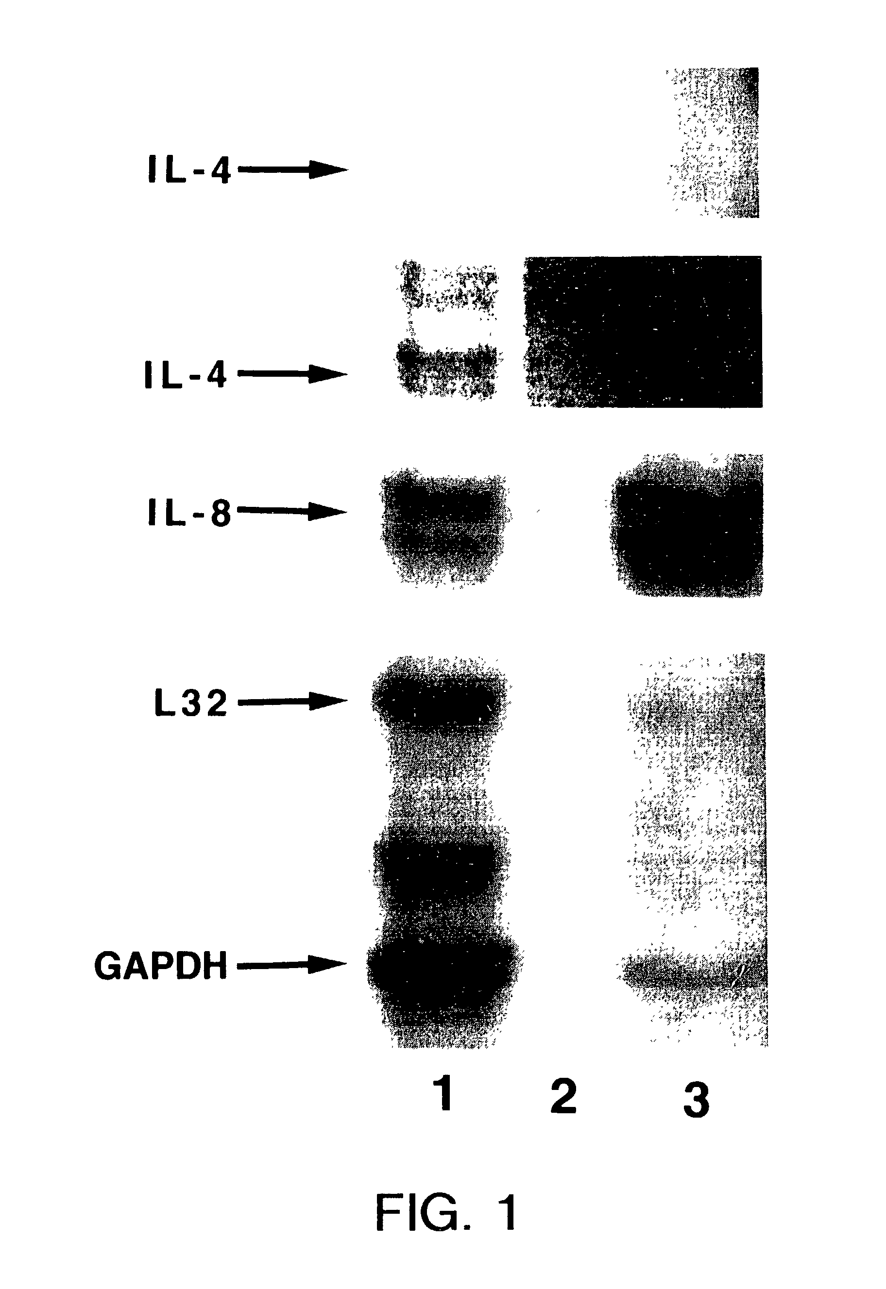
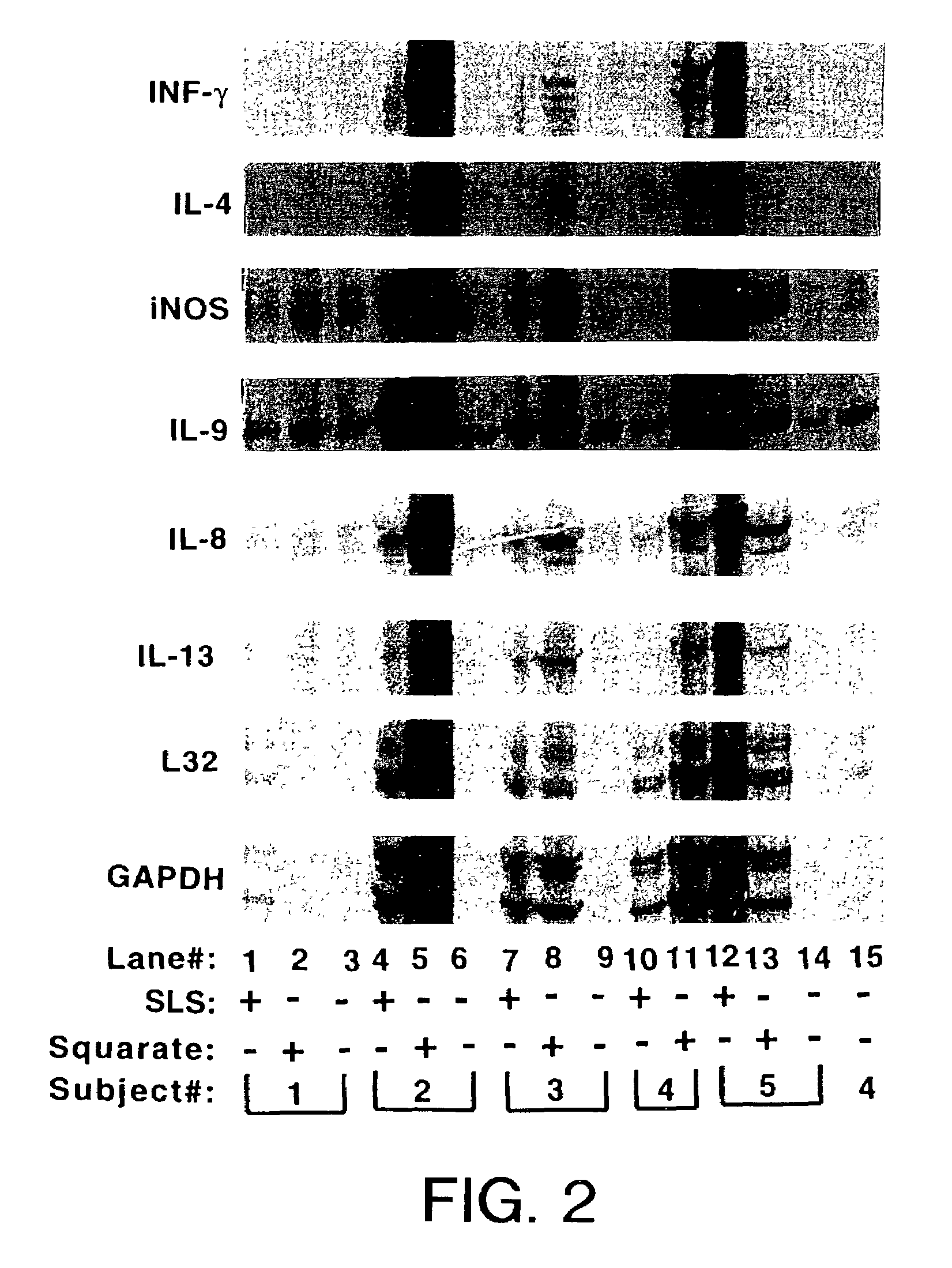
[0130]Irritant contact dermatitis (ICD) was induced by applying 0.5% sodium lauryl sulfate (SLS) in distilled water for 72 hours to the upper arm. After this exposure, the erythema was graded according to standard scoring sales (Fisher's Contact Dermatitis. 4th ed. Rietschel, R. L. and Fowler, J. F. Jr. eds. Williams & Wilkins, Baltimore, 1995, pg. 29). Allergic contact dermatitis (ACD) was induced by applying dibutyl squarate in acetone to the upper arm of the same subject under occlusion for 48 hours. The upper arms of the same individual (subject #1) were tape stripped 12 times and processed as described in Example 2 above.

[0131]FIG. 1, lane 1 shows the RNA isolated from an ACD erythematous area of skin, read clinically as 3+erythema, that was induced by squarate. Lane 3 is the RNA from ICD erythematous skin, clinically scored as 2+erythema, induced after exposure to 0.5% SLS. After exposure of the x-ray film, the band for cytokine IL-4...

example 3

[0138]To further examine the relationship between the cytokines and the degree of inflammation in subject numbers 3-5, the IL-4, IL-8 and IL-13 RNA levels were normalized to the corresponding housekeeping gene levels (Table 3). Among the three subjects analyzed, a correlation exists between the RNA levels and the severity of the reactions. Table 2 shows that the samples from the strongest skin reactions were also the ones that demonstrated the largest relative amount of IL-8 in the ACD reaction. For example, subject #4 with a 2+reaction at the ACD site and only a slight (low+1) reaction at the ICD site showed an approximate two fold difference in the IL-8 / GAPDH ratios when comparing the ICD and ACD reactions using the RPA method described above. In addition, one would predict an ACD reaction if, on the gel, there is a band for IL-4 and a value for IL-4 / GAPDH of about 0.001 or higher. Also, an ACD reaction can be confirmed where there is an IL-13 band with an IL-13 / GAPDH value of abo...


PUM

PropertyMeasurementUnit
Levelaaaaaaaaaa

Abstract

The present invention provides non-invasive methods for detecting, monitoring, staging, and diagnosing malignant melanoma in a skin sample of a subject. The methods include analyzing expression in skin sample of one or more melanoma skin markers. The melanoma skin markers include IL-1 RI, endothelin-2, ephrin-A5, IGF Binding Protein 7, HLA-A0202 heavy chain, Activin A (βA subunit), TNF RII, SPC4, and CNTF Rα. The skin sample can include nucleic acids, and can be a human skin sample from a lesion suspected of being melanoma.

Description

CROSS REFERENCE TO RELATED APPLICATIONS[0001]This application claims priority under 35 U.S.C. § 119(e) to Provisional Application Ser. No. 60 / 302,348, filed Jun. 28, 2001, which is incorporated herein by reference in its entirety.FIELD OF THE INVENTION[0002]This invention relates to methods related to melanoma patient for detecting expression of genes in a skin sample of the epidermis related to malignant melanoma.BACKGROUND OF THE INVENTION[0003]Malignant melanoma ranks second among adult cancers (behind adult leukemia) in potential years of life lost. Each year, over 47,000 new cases are diagnosed, and the incidence of cutaneous melanoma appears to be rising rapidly. Treatment of malignant melanoma involves surgical excision of the primary lesion, and vigilant monitoring to detect recurrence. Currently, there is no approved therapy for patients having intermediate risk of relapse. High-dose interferon, which can have serious side effects, is approved for treatment of patients havi...

Claims


Application Information

Patent Timeline
IPC IPC(8): C12Q1/68C12P19/34C12P21/04C12Q1/6886G01N33/574
CPCC12Q1/6886G01N33/5743Y10S435/912C12Q2600/158
Inventor VOGT, THOMAS
Owner DERMTECH INT
Why Eureka
Try Eureka