Method for detecting carcinomas in a solubilized cervical body sample

a solubilized and cervical body technology, applied in the field of early diagnosis of carcinoma, can solve the problems of high inter- and intra-observer variance of experienced examiners, unsatisfactory false positive and false negative results of screening tests, and difficulty in preserving and preparing samples,

Active Publication Date: 2007-12-11
VENTANA MEDICAL SYST INC
View PDF14 Cites 18 Cited by
  • Summary
  • Abstract
  • Description
  • Claims
  • Application Information

AI Technical Summary

Problems solved by technology

This is inter alia due to the fact that up to 30% of the results of the Pap test are false-negative.
Thus, for examination of swabs cytologists and pathologists have to be especially trained, and even experienced examiners have a high inter- and intra-observer variance in the assessment of a diagnosis based on cytological specimens.
As a result, the rate of false positive and false negative results in the screening tests remains unsatisfying high.
Yet the problem with the preservation and preparation of the samples may not be overcome by just additionally using molecular markers.
One further complication when performing cytological or histological examinations for screening purposes and especially when applying methods for the detection of molecular markers originates from strict precautions in preserving the samples from causing artifacts or improper results.
This is in part due to the instability of the cell-based morphological information and in part to the instability of the molecular markers to be detected during the tests.
If the samples are not prepared, transported or stored in an appropriate manner, the cell-based information, or even the molecular information may be lost, or may be altered.
So the diagnosis may be impossible, or may be prone to artifacts.
For example, the interpretation of biopsies or cytological preparations is frequently made difficult or impossible by damaged (physically or bio- / chemically) cells.
Firstly, the methods are highly dependent on individual perception of the examiners.
Secondly, the morphological information is quite sensitive to decay processes and thus to production of artifacts after preparation of the samples.
Both aspects contribute to improper reproducibility of the results.

Method used


Image

  • Method for detecting carcinomas in a solubilized cervical body sample
  • Method for detecting carcinomas in a solubilized cervical body sample
  • Method for detecting carcinomas in a solubilized cervical body sample

Examples


example 1

Detection of the Overexpression of p16 in Biopsies of the Cervix Uteri

[0086](A) Paraffin sections having a thickness of 3 to 5 μm were produced from 20 biopsies of the cervix uteri, which comprised all degrees of the dysplastic progression from normal tissue (n=2) via CIN I (n=4), II (n=4), III (n=5) lesions to the invasive carcinoma (n=5). They were deparaffinized in xylene for 2×10 min. and rehydrogenated using ethanol. The antigens were demasked in 10 mM citrate buffer (pH 6.0) in an autoclave at 110° C. for 10 min. Thereafter, the endogenous peroxidases were inactivated using 0.25% H2O2 in PBS. Following the blocking of unspecific binding sites with horse serum (Vectastain ABC detection kit, Vector Laboratories, Burlingame, Calif.) at room temperature for 20 minutes, the sections were incubated with a p16-specific monoclonal antibody (Neomarkers, Fremont, Calif.) in the presence of 3% fetal calf serum at room temperature for 45 min. For the detection of the p16-antibody binding,...

example 2

Detection of the Overexpression of Cell Cycle Regulatory Proteins in HPV-transformed Cells

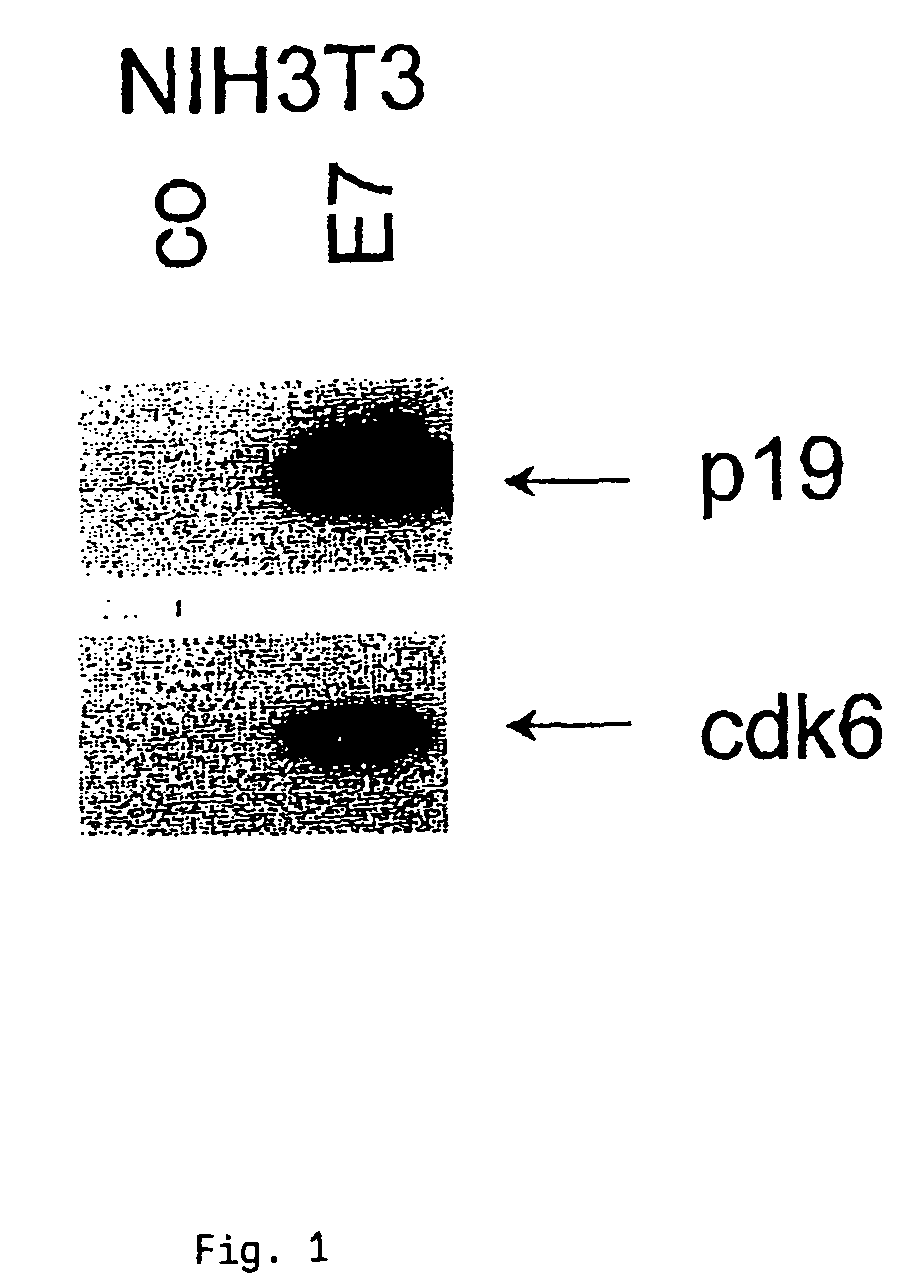
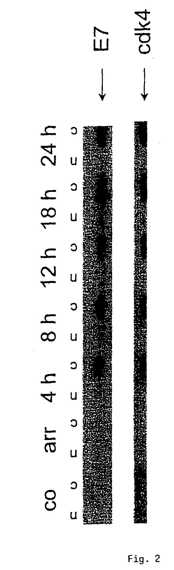
[0094](A) Cervical carcinoma cells CaSki which had been transformed with HPV16 were cultured in the absence of serum for 72 h. Following the addition of serum, cell extracts were collected at various times, subjected to SDS-PAGE and transferred to PVDF membranes (Du Pont). The expression of cdk4 was determined using polyclonal antiserum (1:1000) from Santa Cruz. Furthermore, the expression of HPV16-E7 protein was determined with a monoclonal antibody against HPV16-E7 (1:50) from Triton. The individual immune responses were detected via peroxidase-linked second antibodies and a chemiluminescence detection system (NEN, Du Pont).[0095]FIG. 1 shows that cdk4 was overexpressed.[0096](B) NIH3T3 cells were transformed with HPV16 so as to obtain an expression of HPV16-E7 protein. Cell extracts of the transformed cells were obtained and treated as described in (A). For detecting the expression of cdk6 a...

example 3

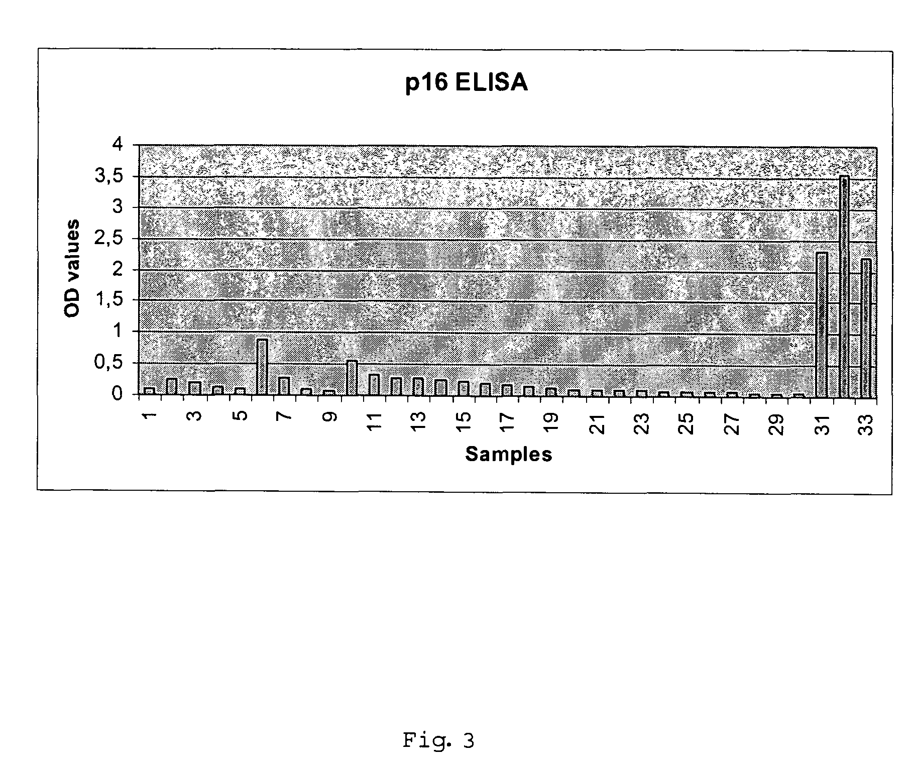
Detection of Cervical Intraepithelial Neoplasia in an ELISA Test Format

[0098]33 cervical swabs provided in a lysis buffer were subjected to ELISA based detection of overexpression of cyclin dependent kinase inhibitor p16 in solutions prepared from the cells contained in the swabs. The ELISA testing was performed as follows:

(A) Cell Lysis

[0099]Cervical swab brushes were given into 15 ml vessels, containing 2 ml of mtm lysis buffer (2% TRITON® X-100, 0.4% SDS, 0.6 mM PMSF in PBS). Cervical cells present in the brush were lysed for at least 20 h. The lysates of the cervical swab samples were then transferred in 2 ml tubes and were centrifuged at 4° C. (15 min at 28.000×g (16.600 rpm HighspeedCentrifuge JEC Multi RF)); Supernatant was transferred to a fresh tube. The supernatant may be stored at −20° C.

(B) Performing the ELISA

[0100]Coating of ELISA-plates[0101]Stock-solution of p16 specific antibody clone mtm E6H4 was diluted in PBS to give ready-to-use coating solution.[0102]50 μl of t...


PUM

PropertyMeasurementUnit
pHaaaaaaaaaa
concentrationsaaaaaaaaaa

Abstract

The present invention relates to a method for the early diagnosis of carcinomas and their preliminary stages, which comprises determining the overexpression of a cell cycle regulatory protein in a solubilized body sample. The present invention is particularly directed to a method for detecting cervical carcinomas, cervical intraepithelial neoplasias, or cervical carcinomas in-situ from a solubilized cervical body sample of a human subject, by solubilizing the cervical body sample in a lysis buffer, and determining the overexpression of cyclin dependent kinase inhibitor p16 in the solubilized cervical sample. The invention also concerns a test kit usable for this purpose as well as an in-vitro diagnostic device.

Description

[0001]This application is a continuation-in-part of U.S. application Ser. No. 10 / 633,484, filed Jul. 31, 2003, which claims the benefit to a foreign application EP 02017313.4, filed Aug. 1, 2002.TECHNICAL FIELD[0002]The present invention relates to a method for the early diagnosis of carcinomas as well as their preliminary stages, particularly carcinomas of the upper respiratory tract or the anogenital tract, from a solubilized body sample.BACKGROUND OF THE INVENTION[0003]Preventive programs have been offered for the most differing carcinomas since the middle of the fifties. Regarding cervical carcinomas, they are based mainly on the morphological and cytological examination of cytosmears of the cervix uteri, what is called the Pap test, which is made on the basis of gynecological routine examinations at regular intervals in women from the 20th year on. By means of the morphology of the cells, the smears are divided into various intensity degrees of dysplastic cellular changes. Acco...

Claims


Application Information

Patent Timeline
Patent Type & Authority Patents(United States)
IPC IPC(8): G01N33/574G01N33/50
CPCG01N33/5076G01N33/5091G01N33/574G01N33/57411G01N33/57423G01N33/57488G01N33/57407G01N2333/4739
Inventor DOEBERITZ, MAGNUS VON KNEBELRIDDER, RUDIGERHERKERT, MATTHIASREICHERT, ANJA
Owner VENTANA MEDICAL SYST INC
Why Eureka
Try Eureka