Method for distinguishing follicular thyroid adenoma (FTA) from follicular thyroid carcinoma (FTC)

a technology of follicular thyroid and adenoma, applied in the field of distinguishing follicular thyroid adenoma from follicular thyroid carcinoma, can solve the problems of fna clinical problem, ftc clinical problem remains, and candidate markers have not proved to have practical value for fna

Active Publication Date: 2008-01-15
DUKE UNIV
View PDF3 Cites 52 Cited by
  • Summary
  • Abstract
  • Description
  • Claims
  • Application Information

AI Technical Summary

Problems solved by technology

A common problem in clinical practice, however, is evaluation and management of thyroid tumors with a follicular pattern.
Nonetheless, these candidate markers have not proved to have practical value for FNA pre-operative diagnosis of FTC (13-15).
Although many of the gene or gene patterns expressed in thyroid tumors have been described, the clinical problem of distinguishing FTC from FTA remains.
In particular, in many areas of the world where health care systems are over-burdened, limited resources for surgery could be directed more rapidly towards those with the highest risk of having carcinoma.

Method used


Image

  • Method for distinguishing follicular thyroid adenoma (FTA) from follicular thyroid carcinoma (FTC)
  • Method for distinguishing follicular thyroid adenoma (FTA) from follicular thyroid carcinoma (FTC)
  • Method for distinguishing follicular thyroid adenoma (FTA) from follicular thyroid carcinoma (FTC)

Examples


example 1

Identification of Diagnostic Markers by SAGE Analysis to Distinguish FTA from FTC

[0027]To directly address the problem of finding diagnostic markers that would distinguish FTA from FTC, gene expression was quantified in FTA tissues, FTC tissues, and normal thyroid tissues using serial analysis of gene expression (SAGE) (17). SAGE counts cDNA transcript tags in large numbers. Thus, SAGE analysis makes it possible to identify a restricted set of genes that are highly expressed in one tissue and not detectable in another. Transcript counts from FTC, FTA, and normal thyroid libraries were generated and compared.

[0028]SAGE Libraries.

[0029]One follicular thyroid adenoma, one follicular thyroid carcinoma, and one normal thyroid were chosen for SAGE (17). SAGE libraries were constructed using a microSAGE procedure (19) and were sequenced through the SAGE portion of the Cancer Genome Anatomic Project (20). Tags were extracted from automated sequence text files; and duplicate ditags, linker s...

example 2

RT-PCR Analysis of Genes Identified by SAGE Analysis to Confirm Expression Level

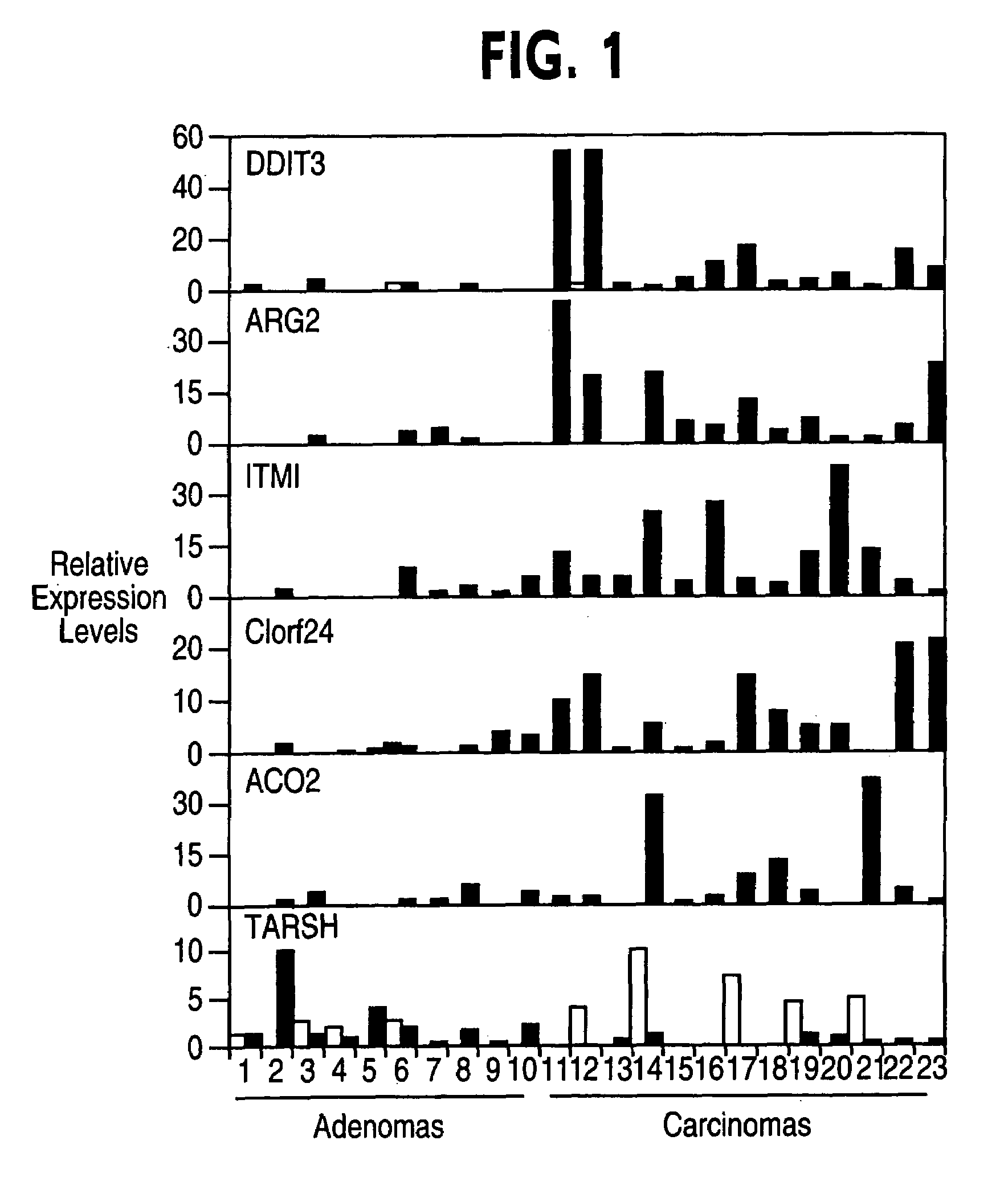
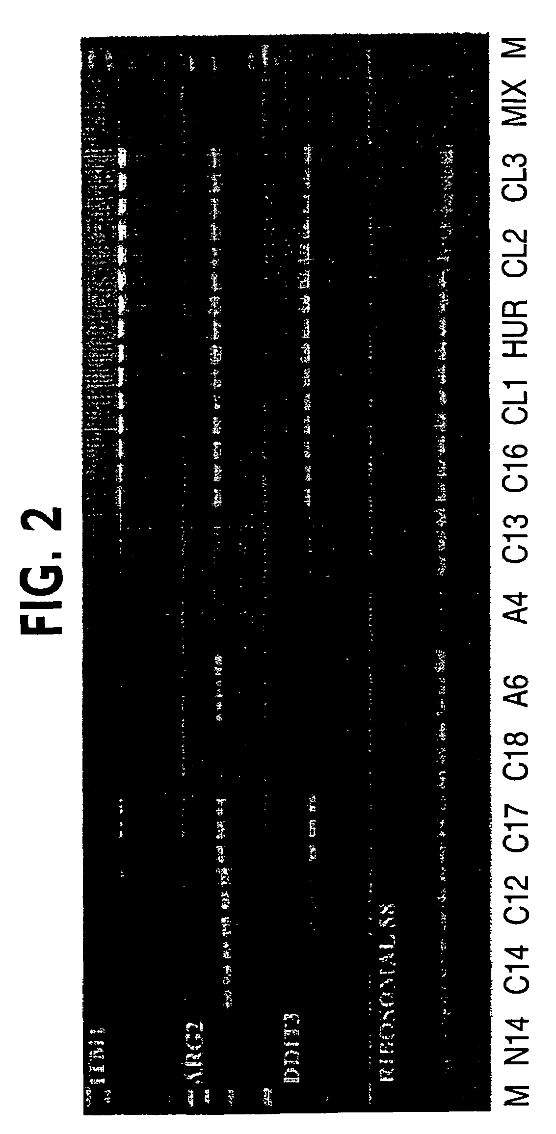
[0038]To validate the differential gene expression profile predicted by SAGE, seventeen genes with the highest fold-induction were tested and analyzed for gene expression by quantitative real-time RT-PCR.

[0039]Tissue Samples.

[0040]For RT-PCR analysis, twenty-three primary tumors were obtained from patients initially diagnosed with follicular thyroid tumor. The tumors were frozen immediately after surgical biopsy. All samples were obtained from patients followed at Hospital São Paulo, Universidade Federal de São Paulo, and Hospital Helópolis, São Paulo, Brazil. The study was approved by the Ethics and Research Committees of the Universidade Federal de São Paulo and Hospital Heliópolis and was in agreement with the 1975 Helsinki statement, revised in 1983. A signed letter of informed consent was obtained from each patient. All patients received post-surgical radioiodine ablation and suppressive thyroxine t...

example 3

Analysis of Gene Expression by Immunohistochemical Staining

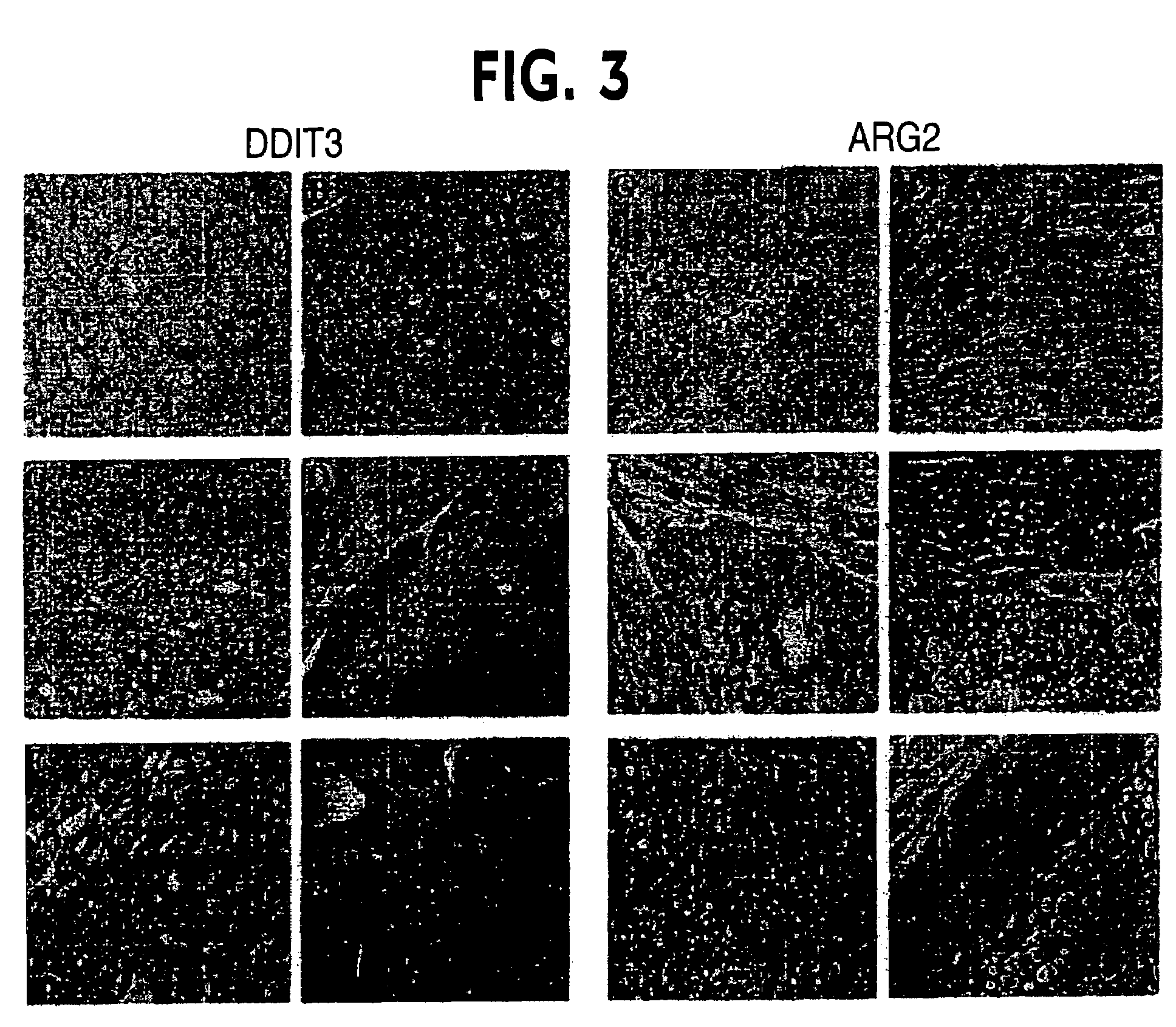
[0053]The expression levels of two genes (DDIT3 and ARG2) were confirmed by immunohistochemistry. For the immunohistochemical study pathologic materials were retrieved from specimens diagnosed with FTC (n=27) and FTA (n=32) at Hospital São Paulo, Federal University of São Paulo in an eight-year period from 1996-2003. Hematoxylin and eosin-stained sections were reviewed by an experienced pathologist.

[0054]Immunohistochemical Analysis.

[0055]Immunohistochemical staining was performed on paraffin-embedded tissue sections (3 μm) placed on 0.1% poly-lysine-coated slides (Sigma), deparaffinized with xylene and rehydrated through a series of graded alcohols. The endogenous alkaline phosphatase activity was blocked by 3% hydrogen peroxide. After pressure-cooking retrieval (10 mmol / L citrate buffer, pH 7.4 for 2 minutes), the sections were blocked in 1× PBS / 0.1% BSA for 1 hour at room temperature and incubated with the first antibody ...


PUM

PropertyMeasurementUnit
temperatureaaaaaaaaaa

Abstract

Follicular thyroid adenoma (FTA) is distinguished from follicular thyroid carcinoma (FTC) by comparing amount of an expression product of at least one gene selected from the group consisting of DDIT3, ARG2, ITM1, C1orf24, TARSH, and ACO1 in a test follicular thyroid specimen to a normal control thyroid specimen. The test follicular thyroid specimen is identified as FTA if the amount of expression product of TARSH is equal to or greater in the test follicular thyroid specimen than in the normal control thyroid specimen. The test follicular thyroid specimen is identified as FTC if the amount of expression product of DDIT3, ARG2, ITM1, C1orf24, or ACO1 is greater in the test follicular thyroid specimen than in the normal control thyroid specimen.

Description

[0001]This application claims the benefit of provisional application Ser. No. 60 / 560,243, filed Apr. 8, 2004, the contents of which are expressly incorporated herein.[0002]The U.S. Government retains certain rights to this invention due to funding by the National Institutes of Health contract number NIH 98X-S146A.FIELD OF THE INVENTION[0003]The invention relates to the field of distinguishing thyroid diseases, and more particularly to the field of distinguishing follicular thyroid adenoma from follicular thyroid carcinoma.BACKGROUND OF THE INVENTION[0004]The incidence of thyroid cancer is increasing, with a global estimate of one-half million new cases this year. Thyroid carcinoma is usually first suspected by a physician when a solitary nodule is palpated on physical exam. Thyroid nodules, however, can be the result of a wide spectrum of causes, and a major concern is to accurately differentiate between benign and malignant nodules.[0005]Cytology of a fine-needle aspiration (FNA) b...

Claims


Application Information

Patent Timeline
Patent Type & Authority Patents(United States)
IPC IPC(8): C12Q1/00G01N33/567G01N33/574
CPCC12Q1/6886G01N33/57407C12Q2600/112C12Q2600/158
Inventor RIGGINS, GREGORY J.CERRUTI, JANETE
Owner DUKE UNIV
Why Eureka
Try Eureka