
Methods for screening compounds for proarrhythmic risk and antiarrhythmic efficacy

a technology of proarrhythmic risk and antiarrhythmic efficacy, applied in the field of safety pharmacology, can solve problems such as qt prolongation, recurrent fainting, and sudden death in humans

Inactive Publication Date: 2008-07-08
MAIN LINE HEALTH HEART CENT
View PDF6 Cites 4 Cited by
  • Summary
  • Abstract
  • Description
  • Claims
  • Application Information

AI Technical Summary

Benefits of technology

"The present invention provides methods for screening compounds to see if they can cause or prevent cardiac arrhythmias. The methods involve measuring the recovery of a specific protein called ICa,L in tissue that expresses the L-type calcium channel. This recovery is compared to the time it takes for the tissue to repolarize after a beat. By measuring this ratio, researchers can determine if a compound affects the function of the L-type calcium channel in a way that could lead to a risk of cardiac arrhythmia. This information can be used to develop safer drugs that target the L-type calcium channel."

Problems solved by technology

Delayed ventricular repolarization that manifests as QT interval prolongation on the ECG is associated with the development of an atypical form of polymorphic ventricular tachycardia termed torsade de pointes (TdP) that can result in recurrent fainting and sudden death in humans.
An increasing number of medications are found to prolong ventricular repolarization, leading to QT prolongation.
However, suboptimal dosing of these drugs in humans in order to avoid TdP greatly attenuates their efficacy in the inhibition of cardiac arrhythmias.
This screening method, however, suffers from several major drawbacks.
The drawbacks include the fact that this method is not well suited for high throughput screening, and that TdP risk is not proportional to the potency of a compound to inhibit hERG current.
In other words, this method has a fairly high potential to produce false negatives and positives.
A false negative may result if the hERG channel is not the K+ channel target of the compound.
This screening method, however, is not a very sensitive assay, and thus is generally done in conjunction with another screen such as hERG inhibition.
This screening method also suffers from an additional drawback in that it has a high potential to cause false positive or negative results.
Thus, the lack of sensitivity of this assay may result in a compound's true propensity to cause TdP to go unrecognized in preclinical screening.
This method suffers from a significant drawback as well.
The drawback is that the electrical stability of the perfused heart preparations is of relatively short duration, on the magnitude of less than two hours.
The instability of the preparations may thus result in false negative or false positive results.
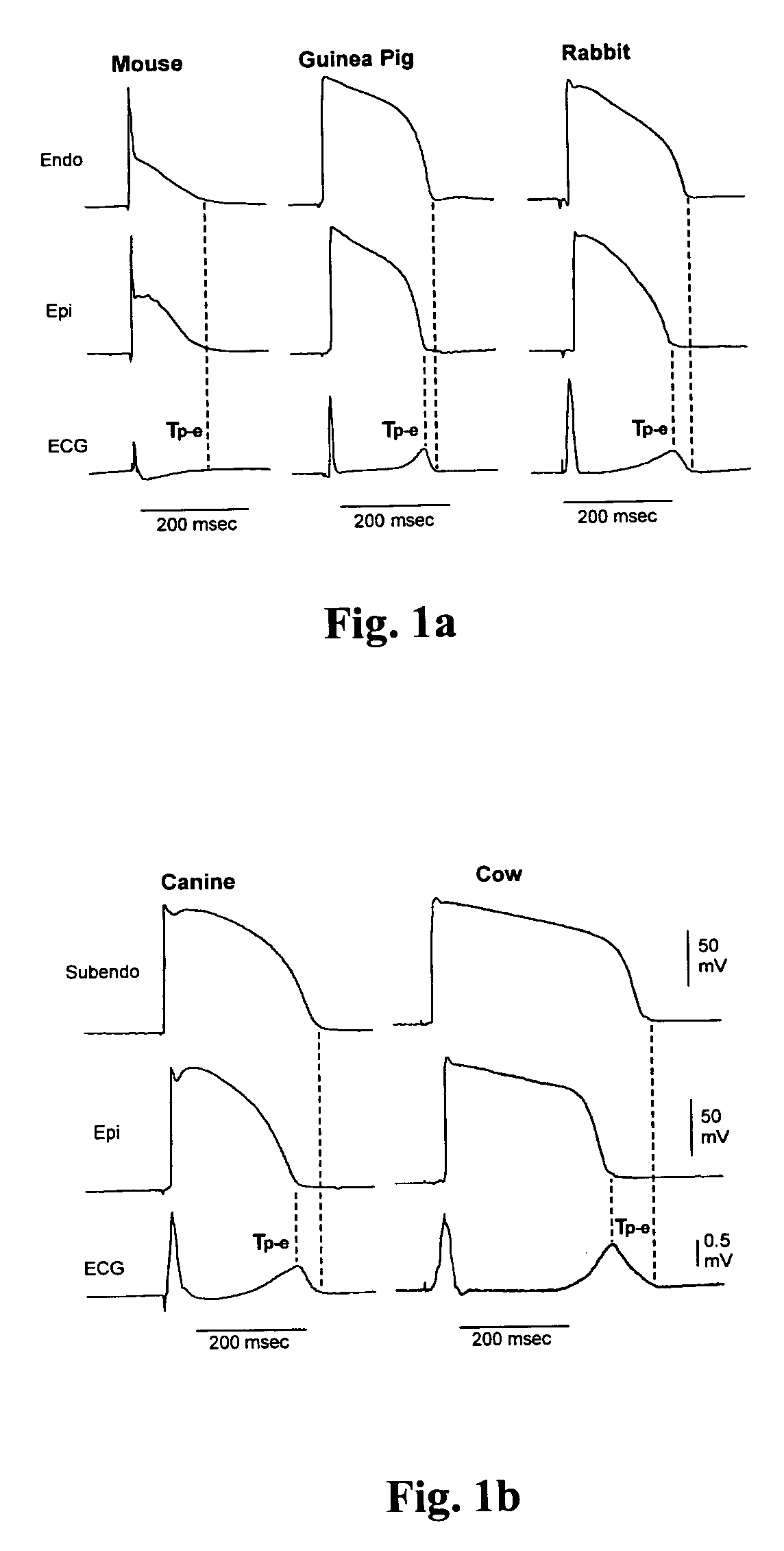
For example, the physiological QT interval in the cow is approximately 400 ms, but the same length of QT would be likely associated with a high risk of TdP in the rabbit.

Method used


Image

  • Methods for screening compounds for proarrhythmic risk and antiarrhythmic efficacy
  • Methods for screening compounds for proarrhythmic risk and antiarrhythmic efficacy
  • Methods for screening compounds for proarrhythmic risk and antiarrhythmic efficacy

Examples


example 1

Comparison of Recovery Time Constant (τ) of ICa,L in Representative Mammals

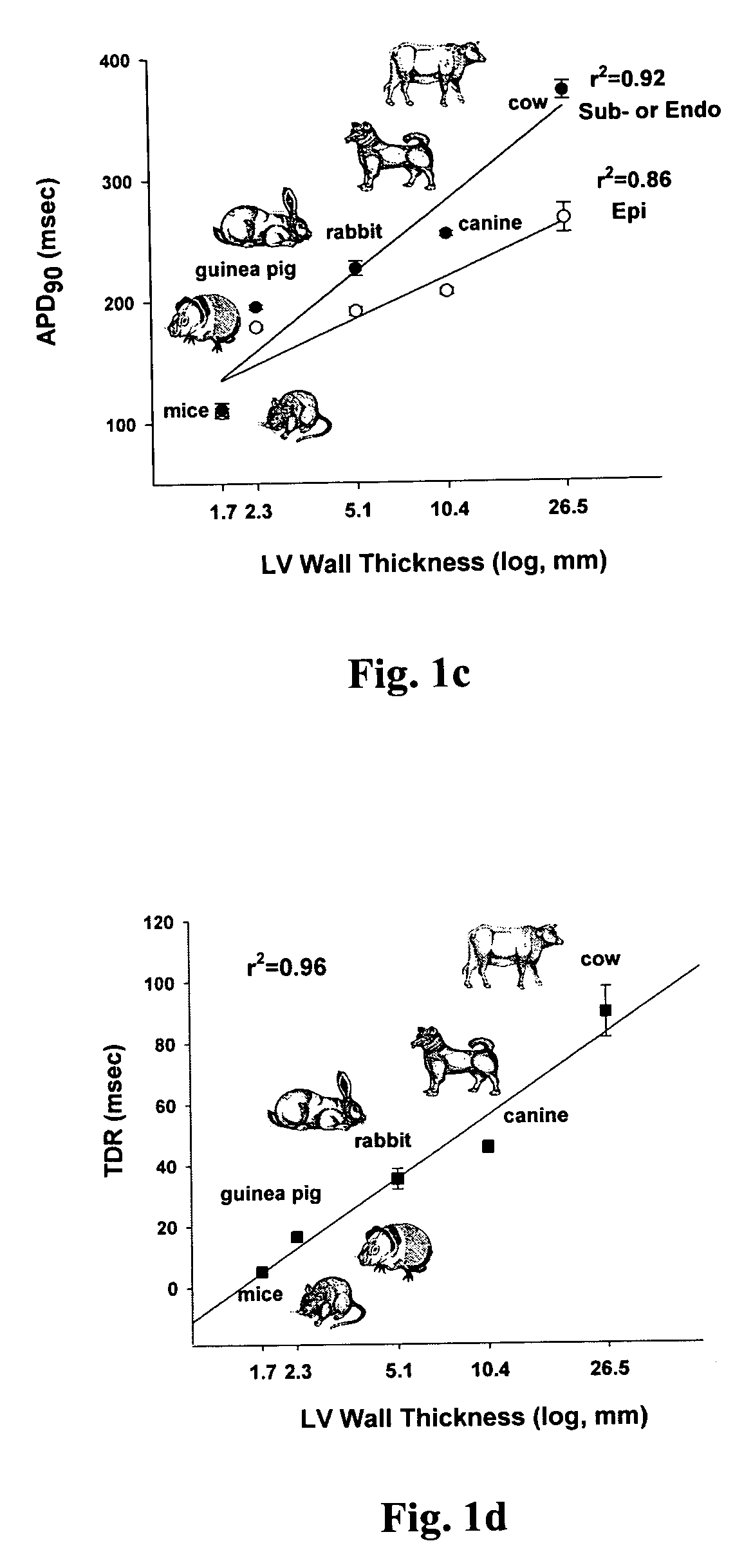
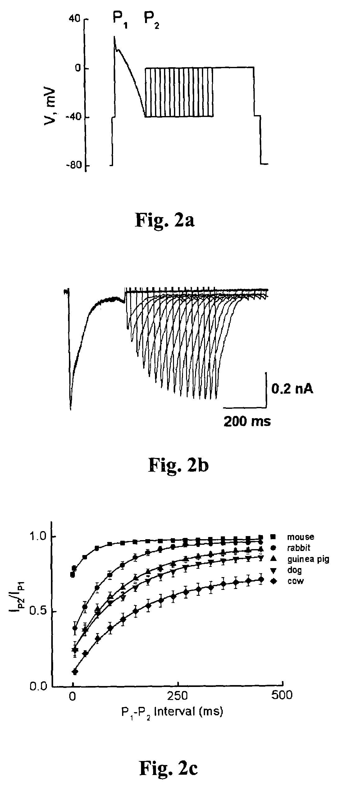
[0058]In preliminary experiments, the time course of L-type calcium current (ICa,L) recovery from inactivation in mice, guinea pigs, rabbits, dogs and cows was examined. As shown in FIG. 2, the recovery kinetics of ICa,L at −40 mV were different among these species, and there was a linear relationship between the time constant (τ) of ICa,L recovery and the corresponding ventricular repolarization time (r2=0.90, pa). The data show that although the left ventricular repolarization time was markedly different among these species (FIG. 1), the ratio of τ of ICa,L recovery to the longest ventricular repolarization time remained fairly consistent among them (FIG. 3b). This indicated that an adequate and constant ratio of the time constant τ of ICa,L recovery to ventricular APD serves as a stabilizer of cardiac cell membrane potential during repolarization phase 2.

[0059]Without intending to be limited to any particu...

example 2

Rabbit Left Ventricular Wedge Preparation and Electrophysiological Recordings

Rabbit Ventricular Wedge Preparation:

[0060]Female rabbits weighting 2.5-3 kg were anticoagulated with heparin and anesthetized with pentobarbital (30-35 mg / kg, i.v.). The chest was opened via a left thoracotomy, and the heart was excised and placed in a cardioplegic solution consisting of cold (4° C.) normal Tyrode's solution. Transmural wedges with dimensions of approximately 1.5 cm wide and 2-3 cm long were dissected from the left ventricle. The wedge tissue was cannulated via the circumflex or left main artery and perfused with cardioplegic solution. The preparation was then placed in a small tissue bath and arterially perfused with Tyrode's solution containing 4 mM K+ buffered with 95% O2 and 5% CO2 (T: 35.7±0.1° C., perfusion pressure: 35-45 mmHg). The ventricular wedge was allowed to equilibrate in the tissue bath until electrically stable, approximately one hour. The preparations were stimulated at b...

example 3

Screening of Compounds in Ventricular Wedge Preparations

[0062]The following compounds were evaluated for their dose-dependent effect on the QT interval in the ventricular wedge preparations: quinidine, sotalol, bepridil, haloperidol, moxifloxacin, flecainide, citalopram, loratadine, fluxetine and verapamil. Each of these ten compounds was tested in 4 doses ranging from their free therapeutic plasma Cmax to the concentration closely to or greater than 30-fold over the Cmax. After the wedge preparations were perfused with normal Tyrode's solution for one hour in the chamber, control transmembrane action potentials and a transmural ECG were recorded. The preparations were then switched to Tyrode's solution containing a test compound with each dose for 20 minutes.


PUM


Abstract

Methods for screening compounds for their potential to induce or inhibit a cardiac arrhythmia are disclosed. The methods comprise determining the ratio of the time constant (τ) of ICa,L recovery in tissue expressing the L-type calcium channel or any subunit or combination thereof of the L-type calcium channel treated with a test compound to the ventricular repolarization time of cardiac tissue treated with a test compound. The methods further comprise determining an arrhythmic risk score for a specified dose of a test compound.

Description

FIELD OF THE INVENTION[0001]The present invention relates generally to the field of safety pharmacology. In particular, the invention features novel methods for screening compounds for their potential to induce a cardiac arrhythmia or to inhibit a cardiac arrhythmia in a subject, as well as methods to generate a risk score for distinguishing among different compounds' capacity to cause torsade de pointes.BACKGROUND OF THE INVENTION[0002]Various publications, including patents, published applications, technical articles and scholarly articles are cited throughout the specification. Each of these cited publications is incorporated by reference herein, in its entirety, for all that it teaches.[0003]Ventricular repolarization time is determined by transmembrane action potential duration (APD) of the ventricular myocardium or the QT interval on the body surface electrocardiogram (ECG). Delayed ventricular repolarization that manifests as QT interval prolongation on the ECG is associated ...

Claims


Application Information

Patent Timeline
Patent Type & Authority Patents(United States)
IPC IPC(8): A61K49/00
CPCG01N33/5088G01N33/6872G01N2800/326G01N2500/04
Inventor YAN, GAN-XIN
Owner MAIN LINE HEALTH HEART CENT
Learn More