Removal of embedding media from biological samples and cell conditioning on automated staining instruments

a technology of cell conditioning and biological samples, applied in the field of biological samples and cell conditioning on automated staining instruments, can solve the problems of only achieving adequate results, affecting the accuracy of analysis, and deteriorating tissue samples, so as to increase the intensity of staining, accurate interpretation of data, and increase the amount of signal obtained

Inactive Publication Date: 2008-08-12
THE CLEVELAND CLINIC FOUND +1
View PDF36 Cites 31 Cited by
  • Summary
  • Abstract
  • Description
  • Claims
  • Application Information

AI Technical Summary

Benefits of technology

[0005]The diagnosis of disease based on the interpretation of tissue or cell samples taken from a diseased organism has expanded dramatically over the past few years. In addition to traditional histological staining techniques and immunohistochemical assays, in situ techniques such as in situ hybridization and in situ polymerase chain reaction are now used to help diagnose disease states in humans. Thus, there are a variety of techniques that can assess not only cell morphology, but also the presence of specific macromolecules within cells and tissues. Each of these techniques requires that sample cells or tissues undergo preparatory procedures that may include fixing the sample with chemicals such as an aldehyde (such as formaldehyde, glutaraldehyde), formalin substitutes, alcohol (such as ethanol, methanol, isopropanol) or embedding the sample in inert materials such as paraffin, celloidin, agars, polymers, resins, cryogenic media or a variety of plastic embedding media (such as epoxy resins and acrylics). Other sample tissue or cell preparations require physical manipulation such as freezing (frozen tissue section) or aspiration through a fine needle (fine needle aspiration (FNA)). Regardless of the tissue or cell sample or its method of preparation or preservation, the goal of the technologist is to obtain accurate, readable and reproducible results that permit the accurate interpretation of the data. One way to provide accurate, readable and reproducible data is to prepare the tissue or cells in a fashion that optimizes the results of the test regardless of the technique employed. In the case of immunohistochemistry and in situ techniques this means increasing the amount of signal obtained from the specific probe (e.g., antibody, DNA, RNA, etc.). In the case of histochemical staining it may mean increasing the intensity of the stain or increasing staining contrast.

Problems solved by technology

Without preservation, tissue samples rapidly deteriorate such that their use in diagnostics is compromised shortly after removal from their host.
Some antigens have particular pH requirements such that adequate results will only be achieved in a narrow pH range.
Although the chemical components of the antigen retrieval solution, including metal ions, may play a role as possible co-factors in the microwave heating procedure, thus far, no single chemical has been identified that is both essential and best for antigen retrieval.
These organic solvents are very volatile causing a variety of problems including requiring special processing (e.g., deparaffinization is performed in ventilated hoods) and requires special waste disposal.
The use of these organic solvents increases the cost of analysis and exposure risk associated with each tissue sample tested and has serious negative effects for the environment.
Presently, there is no available technique for removing inert media from sample tissue by directly heating the slide in an automated fashion.
Neither is it currently possible to remove inert media from sample tissue while conditioning the sample tissue or cell in a one-step automated staining process.

Method used


Image

  • Removal of embedding media from biological samples and cell conditioning on automated staining instruments
  • Removal of embedding media from biological samples and cell conditioning on automated staining instruments
  • Removal of embedding media from biological samples and cell conditioning on automated staining instruments

Examples


example 1

Automated “Exposing” and “Cell Conditioning” with Biological Samples Stained with H&E

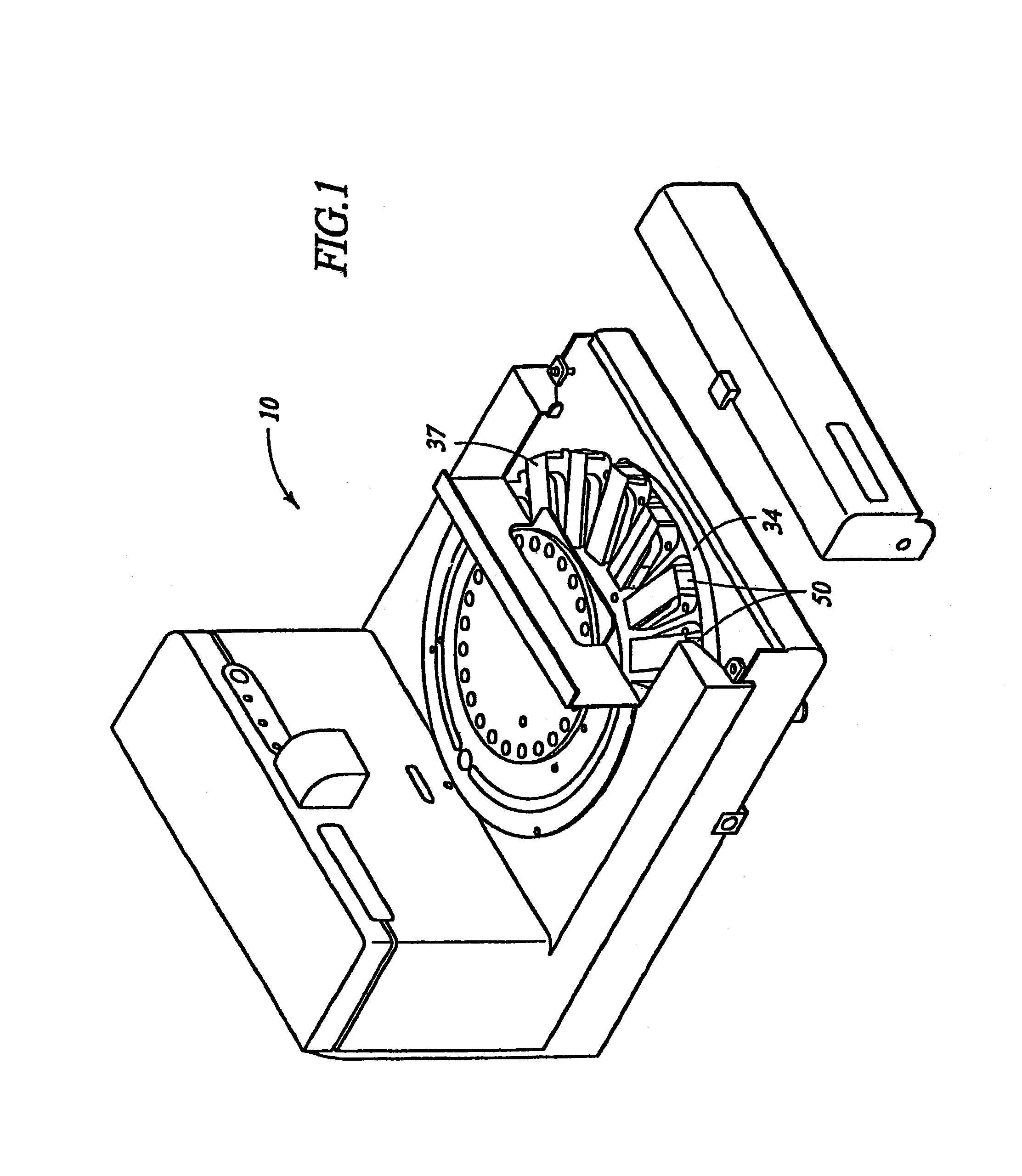
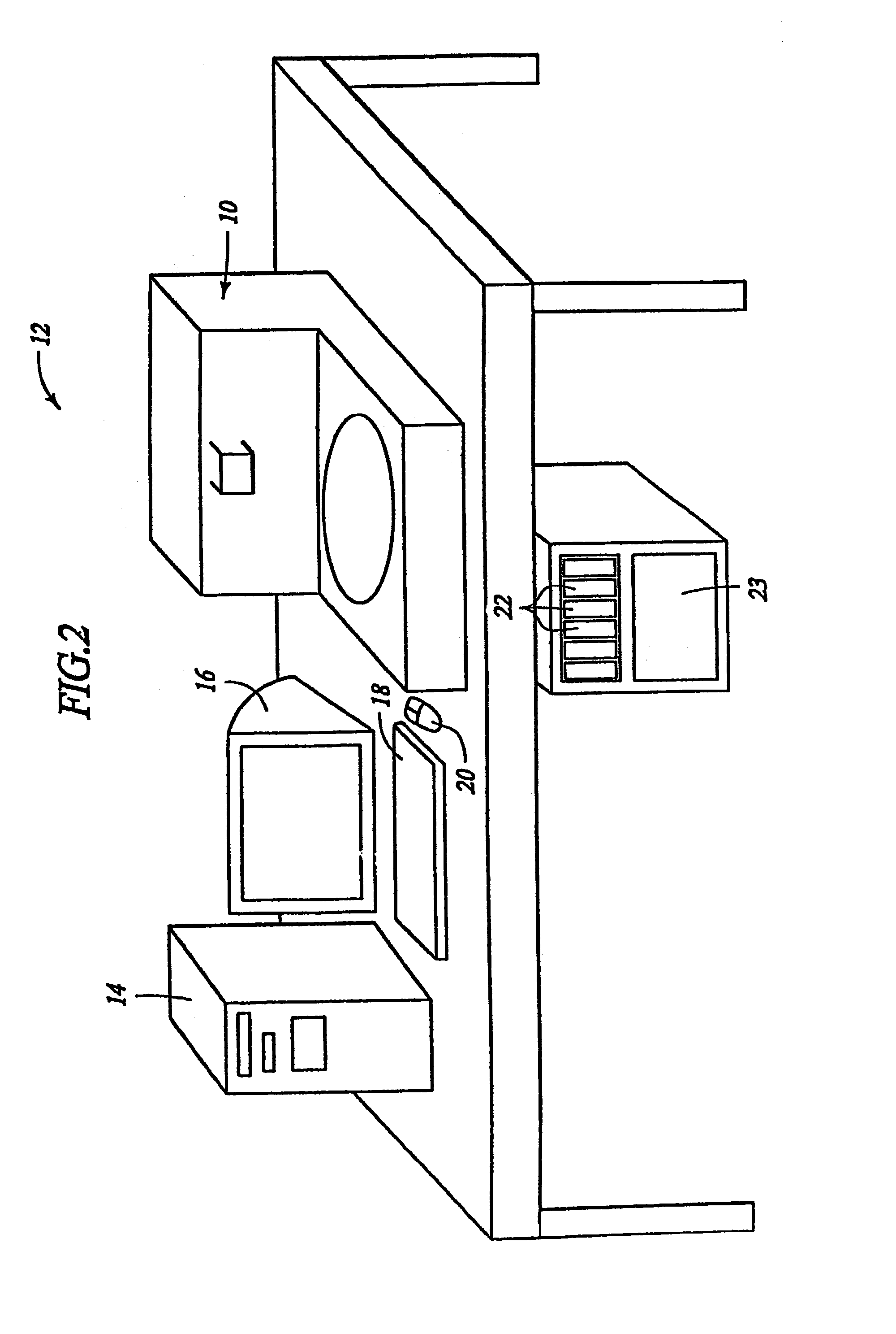
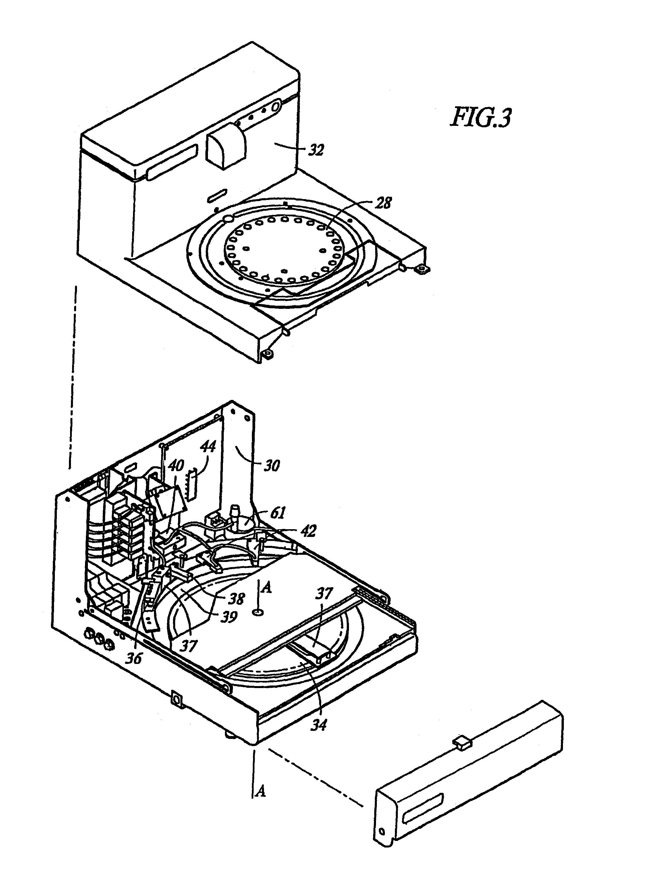
[0088]Biological samples, including breast, stomach, brain, tonsil and kidney, that had been embedded in paraffin were exposed according to the following procedure: slides containing the above referenced biological sample were placed on an automated instrument (Ventana Medical Systems, Inc., Tucson, Ariz.) and subjected to the exposing protocol described below. Generally, the slides containing paraffin embedded biological samples were dry heated to 65° C. for six (6) minutes then rinsed with any of the following: 1) 1× citrate buffer, 2) de-ionized water, 3) 10 mM phosphate buffer (pH=6.3), or 4) 10 mM Tris-HCl buffer (pH=7.4) each containing 0.1% Triton X-100.

Exposing Protocol 1

[0089]1. Incubate for 2 minutes[0090]2. Rinse slide[0091]3. Adjust slide volume and apply LIQUID COVERSLIPLIQUID COVERSLIP™[0092]4. Incubate for 6 minutes[0093]5. Rinse slide[0094]6. Adjust slide volume and apply LCS™[0095]7...

example 2

Automated “Exposing” of Biological Samples with Simultaneous “Cell Conditioning”

[0115]Biological samples of kidney and tonsil that had been formalin fixed and embedded in paraffin were exposed according to the protocol described in Example 1. After automated exposing, the biological sample was subjected to the automated DAB paraffin protocol used for immunohistochemical staining. The protocol for DAB staining is described below:

DAB Protocol

[0116]1. Incubate for 2 minutes[0117]2. Rinse slide[0118]3. Adjust slide volume and apply LCS™[0119]4. Rinse slide[0120]5. Adjust slide volume and apply LCS™[0121]6. Rinse slide[0122]7. Adjust slide volume and apply LCS™[0123]8. Apply one drop of inhibitor[0124]9. Incubate for 4 minutes[0125]10. Adjust slide volume and apply LCS™[0126]11. Apply one drop of primary antibody[0127]12. Incubate for 32 minutes[0128]13. Adjust slide volume and apply LCS™[0129]14. Apply one drop of Biotinylated Ig[0130]15. Incubate for 8 minutes[0131]16. Rinse slide[0132...

example 3

Two Step Automated “Exposing” and “Cell Conditioning”

[0147]Biological samples of tonsil and breast that had been preserved in paraffin and treated with formaldehyde were treated by the following protocol:

Exposing and Cell Conditioning Protocol

[0148]1. Incubate for 2 minutes[0149]2. Increase thermofoil temperature to 65.0° C.[0150]3. Incubate for 6 minutes[0151]4. Rinse slide and apply LCS™[0152]5. Incubate for 6 minutes[0153]6. Rinse slide and apply LCS™[0154]7. Increase thermofoil temperature to 100.0° C.[0155]8. Adjust slide volume and apply LCS™[0156]9. Rinse slide[0157]10. Adjust slide volume and apply LCS™[0158]11. Incubate for 4 minutes[0159]12. Adjust slide volume and apply LCS™[0160]13. Incubate for 4 minutes[0161]14. Adjust slide volume and apply LCS™[0162]15. Incubate for 4 minutes[0163]16. Adjust slide volume and apply LCS™[0164]17. Incubate for 4 minutes[0165]18. Adjust slide volume and apply LCS™[0166]19. Incubate for 4 minutes[0167]20. Adjust slide volume and apply LCS...


PUM

PropertyMeasurementUnit
temperatureaaaaaaaaaa
temperaturesaaaaaaaaaa
melting pointaaaaaaaaaa

Abstract

The present invention provides reagents for use in an automated environment for removing or etching embedding media by exposing a biological sample to be stained in histochemical or cytochemical procedures without the dependence on organic solvents. The reagents comprise components optimized to facilitate removal or etching of the embedding media from the biological sample. The present invention also provides reagents for use in an automated environment for cell conditioning biological samples wherein the cells are predisposed for access by reagent molecules for histochemical and cytochemical staining procedures. The reagents comprise components optimized to facilitate molecular access to cells and cell constituents within the biological sample.

Description

REFERENCE TO RELATED APPLICATIONS[0001]This application claims priority to U.S. Provisional Patent Application No. 60 / 367,015, filed on Mar. 23, 2002. This application is further a continuation-in part of application Ser. No. 09 / 721,096, filed on Nov. 22, 2000 now U.S. Pat. No. 6,855,559, which claims the benefit of priority from International Application No. PCT / US99 / 20353, filed Sep. 3, 1999, which was published on Mar. 16, 2000 as WO 00 / 14507. International Application No. PCT / US99 / 20353, in turn, claims the benefit of priority from U.S. Provisional Patent Application No. 60 / 099,018, filed Sep. 3, 1998; U.S. patent application Ser. No. 09 / 259,240, filed Feb. 26, 1999; and International Application No. PCT / US99 / 04181, filed Feb. 26, 1999, which was published on Sep. 2, 1999 as WO 99 / 44030. All of the patent applications mentioned herein are hereby incorporated by reference.BACKGROUND OF THE INVENTION[0002]1. Field of the Invention[0003]The present invention relates to a method and...

Claims


Application Information

Patent Timeline
Patent Type & Authority Patents(United States)
IPC IPC(8): A01N1/00G01N33/00G01N1/30G01N1/36
CPCG01N1/30G01N1/36Y10T436/21Y10T436/25125Y10T436/25375Y10T436/25Y10T436/2525
Inventor HOPKINS, KERIPUSCH, ALEXISWOLF, CATHERINEHARTMAN, ANTHONYPETTAY, JAMESCHRISTENSEN, KIMBERLYMACREA, ETHELSEBASTIAO, NOEMI
Owner THE CLEVELAND CLINIC FOUND
Why Eureka
Try Eureka