

Methods for identifying stem cells based on nuclear morphotypes
a nuclear morphotype and stem cell technology, applied in the field of methods for identifying stem cells based on nuclear morphotypes, can solve the problems of not being able to identify or isolate such cells, and achieve the effect of improving the morphotype and morphotype of the stem cell
- Summary
- Abstract
- Description
- Claims
- Application Information
AI Technical Summary
Benefits of technology
Problems solved by technology
Method used
Image

Examples
example 1
Experimental Procedures
Sources of Cells and Tissues
[0080]All adult tissue and tumor specimens were obtained as surgical discards at the Massachusetts General Hospital through the Department of Pathology and the MGH Center for Cancer Research. Each tissue section was immediately placed in fresh ice-cold fixative and transported to the MIT cytogenetics laboratory for further analyses. Use of the anonymous discarded sections had been approved by the Institutional Review Boards of both MGH and MIT. The fetal gut sections analyzed were not obtained for this research but were drawn from the archival slide collection of the Chernobyl Scientific Expedition charged with the task of discovering signs of genetic radiation damage in developing fetuses and children after the meltdown of the nuclear reactor at Chernobyl, Ukraine in 1985. Two normal adult colons that were discarded after surgery not related to cancer (five ˜2 to 10 mm diameter polyps of two FAPC colons, four colon tumors, one panc...
example 2
Detection of Bell-Shaped Nuclei
Embryonic Hindgut
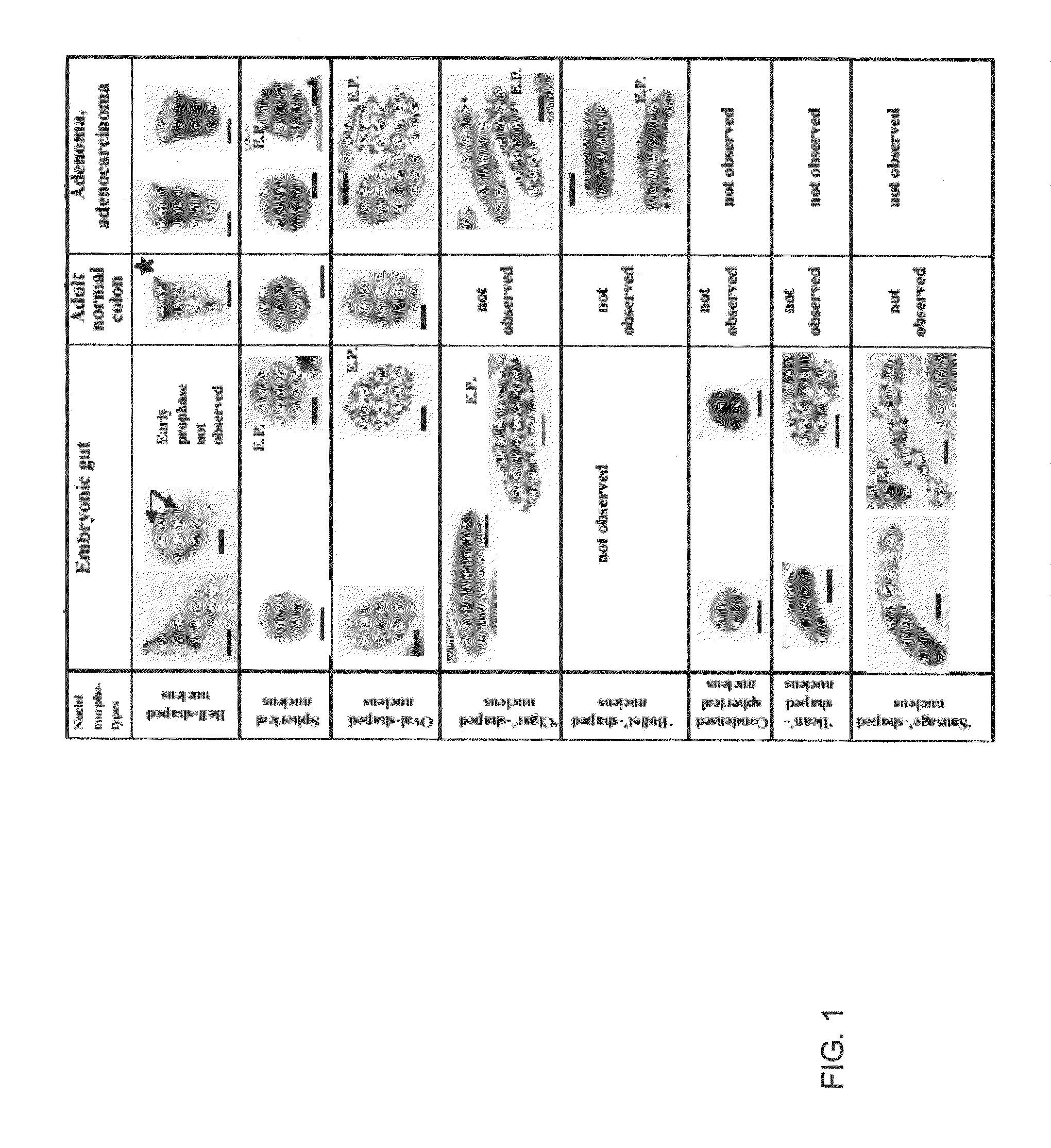
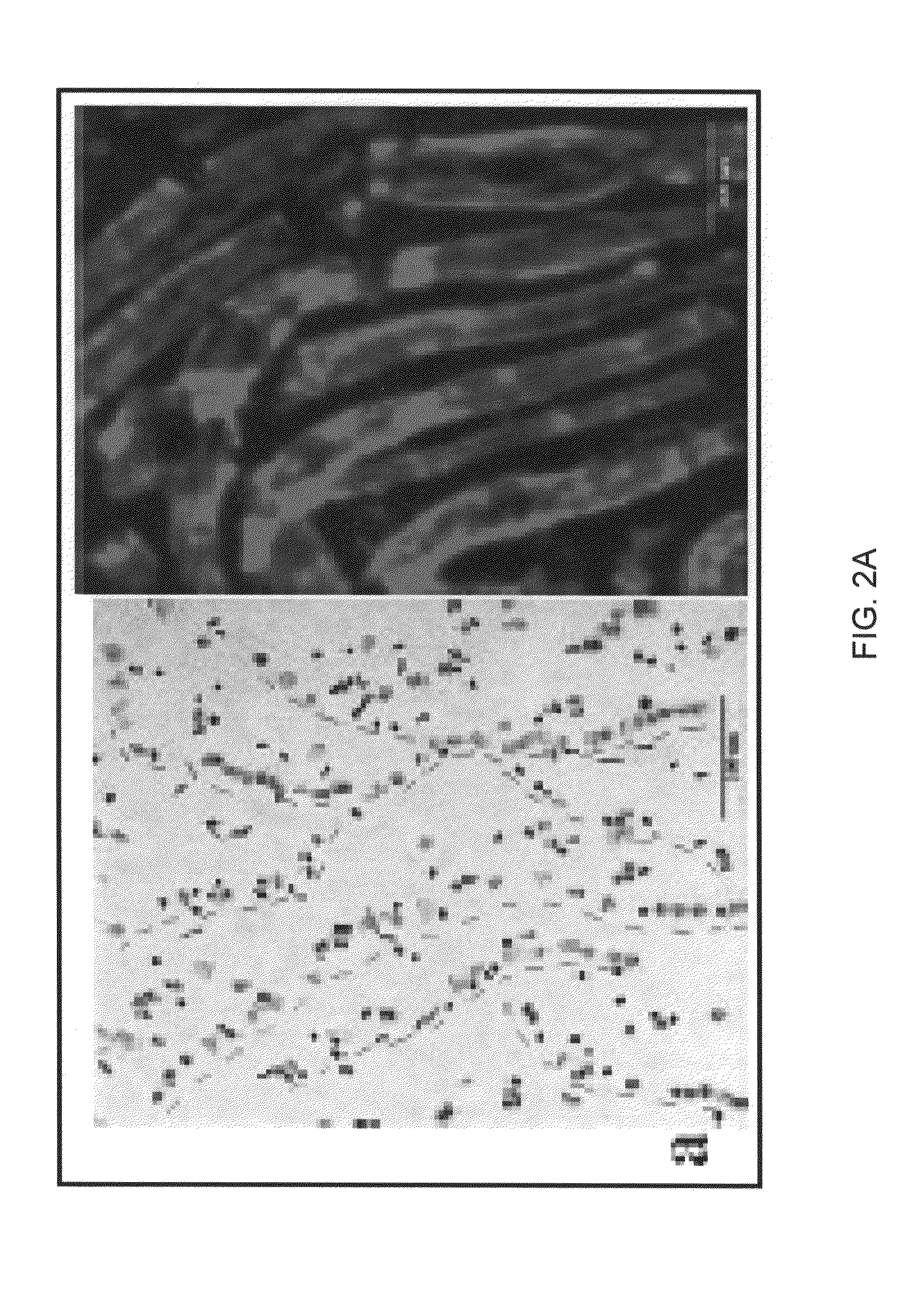
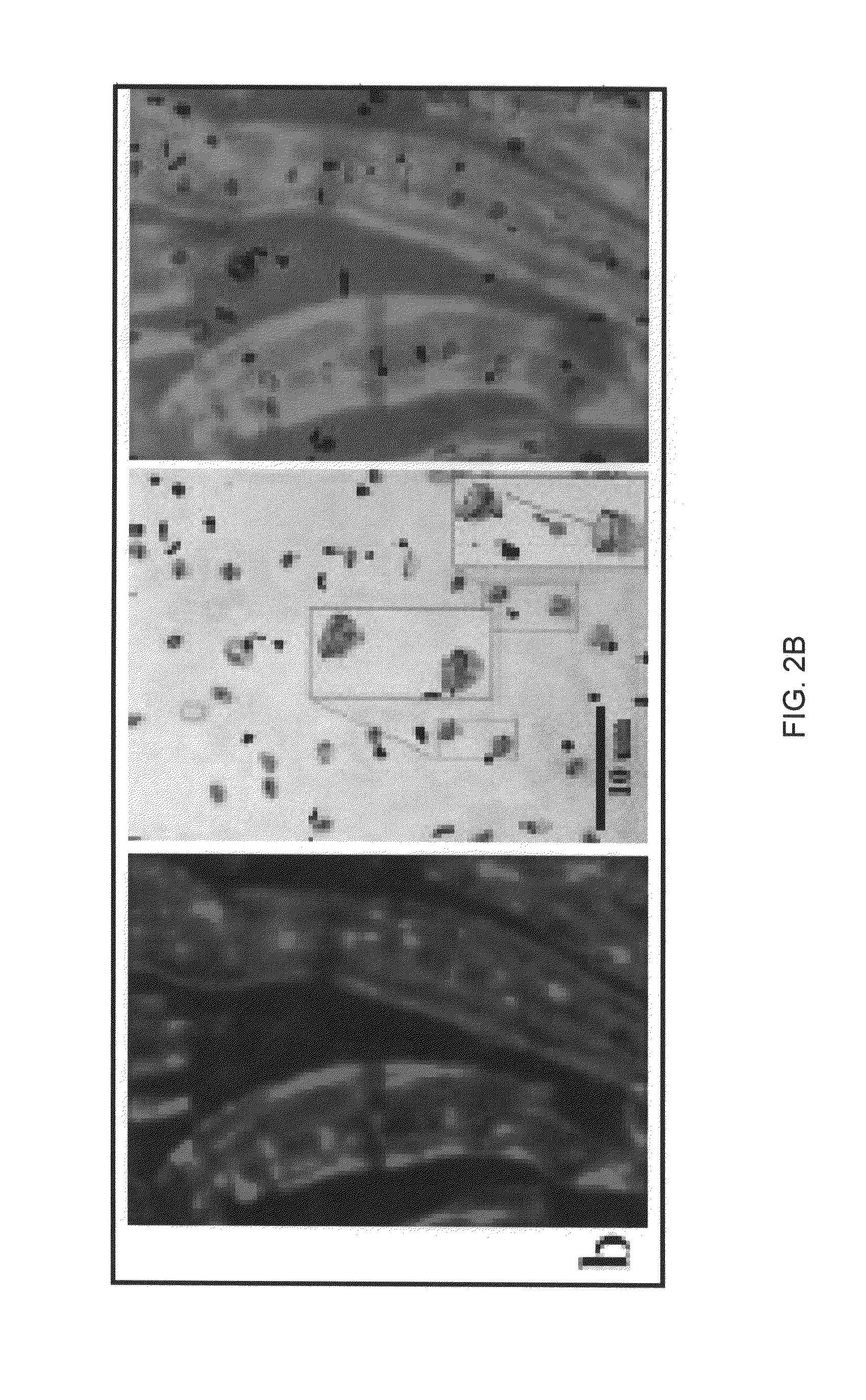
[0088]Observations described herein are from two independent embryo gut sections are shown in FIGS. 1, 2A-2C and 3A-3F. Three observations were made. First, there was the array of seven distinct nuclear morphotypes summarized in FIG. 1. Secondly there was the orderly linear head to toe arrangement of bell-shaped nuclei organized in long (˜20 to 50 micron diameter) tubes as shown in FIGS. 2A-2C. Thirdly, there were the extraordinary forms of symmetrical and asymmetrical amitoses involving bell-shaped nuclei as shown in FIGS. 3A-3F.
[0089]Phase contrast images (left frame, FIG. 2B) and stained nuclear images (middle frame, FIG. 2B) of the identical hindgut section when overlaid (right frame, FIG. 2C) showed that the a linearly oriented nuclei were contained in a previously unreported tube-like structure which is itself about 20-50 microns in diameter. 280× magnification of the nuclei (middle frame, FIG. 2B) shows that these non-spherical ...
example 3
[0102]Post-embryonic organizing stem cells of the fetal colon were specifically identified by an opened-mouth, bell-shaped nuclear morphotype. These peculiar nuclei undergo both symmetric and asymmetric nuclear fission without general chromosome condensation. Nuclear fissions drive net growth and differentiation throughout fetal, neonatal and juvenile life before a final metamorphosis into adult maintenance stem cells. These bell-shaped nuclear morphotypes are rarely found in adult colonic crypt bases but reappear prominently in preneoplasia and neoplasia and appear to drive net growth and differentiation in colon adenomas, adenocarcinomas and metastases.
[0103]Combining these observations with inferences derived from analyses of historical age-specific colorectal cancer rates, present day age-specific colonic adenoma prevalence and direct measurements of genetic change in human tissues suggests a default hypothesis for late-onset carcinogenesis in the colon and perhaps other sites: ...
PUM
| Property | Measurement | Unit |
|---|---|---|
| diameter | aaaaa | aaaaa |
| size | aaaaa | aaaaa |
| size | aaaaa | aaaaa |
Abstract
Description
Claims
Application Information