Fluorescent indicator proteins
a fluorescent indicator and protein technology, applied in the field of protein biosensors, can solve the problem of high degree of technology required for genetic manipulation, and achieve the effect of excellent signal intensity and increased signal intensity
- Summary
- Abstract
- Description
- Claims
- Application Information
AI Technical Summary
Benefits of technology
Problems solved by technology
Method used
Image
Examples
example 1
Construction of Recombinant Vectors for Expression of MBP Fusion Proteins
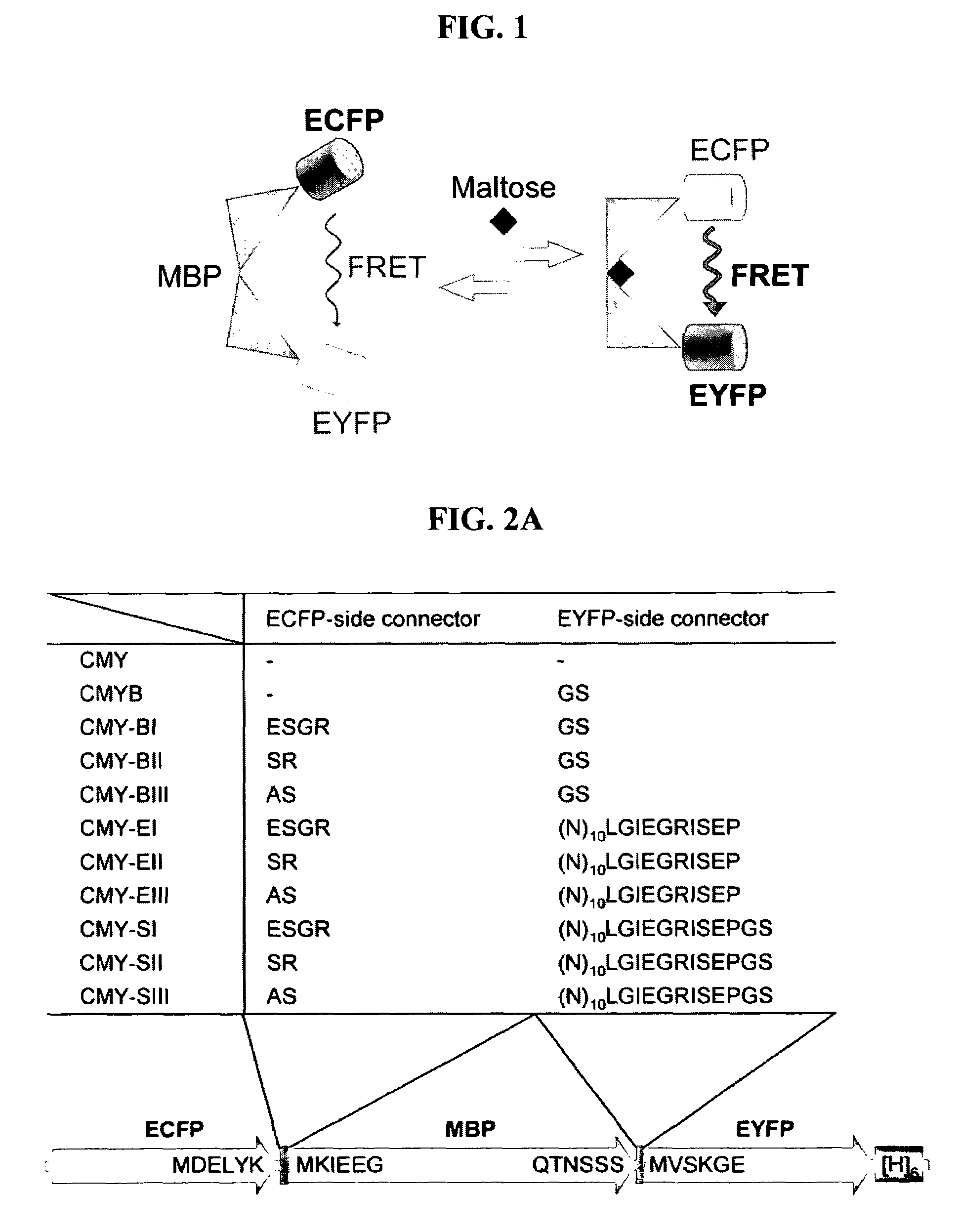
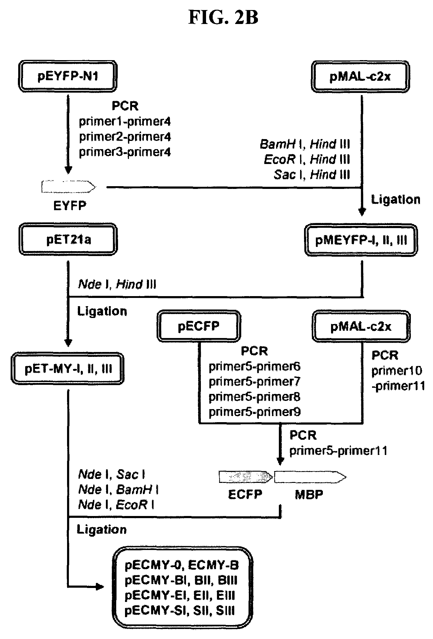
[0066]As shown in FIG. 2A, the fluorescent indicator proteins (MBP fusion proteins) for detection of maltose were constructed of three protein domains (ECFP, MBP, EYFP) and two composite linkers, which composed of connecting peptides (cc, yc) and unstructured terminal regions of flanking proteins: ECFP-cc-MBP-yc-EYFP. In order to construct vectors for the expression of the inventive fluorescence indicators, genes were amplified with primers of SEQ ID NOs: 1 to 11 and were used to construct the expression vectors.
[0067]First, to obtain the gene of EYFP, PCR was performed using a pEYFP-N1 vector (Clontech, USA) as a template with primers of SEQ ID NOs: 1 to 4, which were introduced with a SacI, EcoRI or BamHI restriction enzyme recognition site at the 5′ end and a HindIII restriction enzyme site at the 3′ end. The amplified EYFP gene was digested with SacI and EcoRI, or with BamHI and HindIII, and then inserted i...
example 2
Expression and Purification of MBP Fusion Proteins (Fluorescent Indicator Proteins)
[0070]The MBP fusion protein expression vectors constructed in Example 1 were transformed into E. Coli JM109 (DE3) (Promega, USA), and the transformed recombinant strains were inoculated into LB media (1% bacto-trypton, 0.5% yeast extract, 1% NaCl) containing 50 μg / ml of ampicillin and 0.5 mM IPTG (isopropyl β-d-thiogalactoside), and were cultured with shaking at 28° C. for 24 hours to induce expression. The expressed E. coli were collected by centrifugation and suspended in 50 mM sodium phosphate buffer (pH 7.5). The bacterial cells were disrupted by ultrasonification, centrifuged and then used in a subsequent purification process.
[0071]The MBP fusion proteins were purified with a Histrap™ HP1 column (Amersham Bioscience, Sweden) connected to FPLC (Fast Performance Liquid Chromatography, Amersham Biosciences, Sweden), using His-6 tag expressed at the C-terminus of EYFP. Buffer used in the purificatio...
example 3
Analysis of Fluorescence of MBP Fusion Proteins
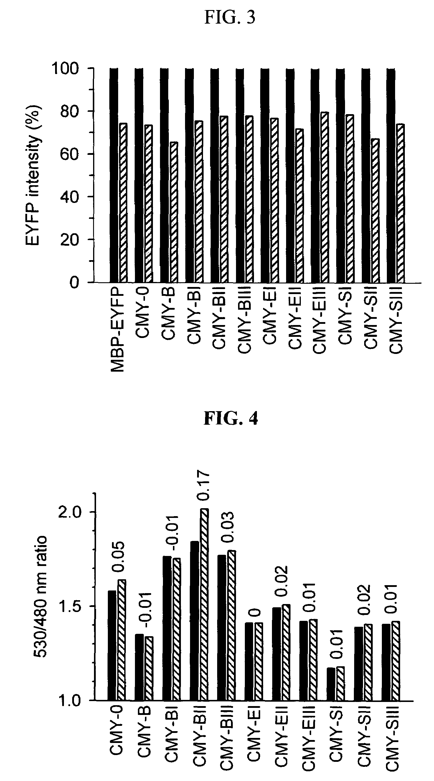
[0072]The fluorescence analysis of the MBP fusion proteins expressed and purified in Example 2 was performed using fluorescence spectrophotometer Cary Eclipse (Varian, Australia) at 25° C., and the MBP fusion proteins were diluted in PBS buffer (pH 7.4) to the same concentrations of 0.2 μM. To define the FRET efficiency in the fluorescence analysis, the emission intensity ratio between the emission intensity of ECFP at 480 nm, measured at an excitation wavelength of 436 nm, and the emission intensity of EYFP at 530 nm, caused by FRET, was substituted into Equation 3 below for convenience:
Ratio=[530 nm / 480 nm] [Equation 3]
[0073]Also, an index of the signal intensity of fluorescence indicators was defined as the change of ratio (ratio) between the FRET efficiency (ratio10 mM) in the presence of 10 mM maltose and the FRET efficiency (ratioapo) in the absence of maltose.
[0074]As shown in FIG. 4, the FRET efficiencies of MBP fusion proteins...
PUM
 Abstract
Description
Claims
Application Information
 - R&D
- Intellectual Property
- Life Sciences
- Materials
- Tech Scout
- Unparalleled Data Quality
- Higher Quality Content
- 60% Fewer Hallucinations
Browse by: Latest US Patents, China's latest patents, Technical Efficacy Thesaurus, Application Domain, Technology Topic, Popular Technical Reports.
© 2025 PatSnap. All rights reserved.Legal|Privacy policy|Modern Slavery Act Transparency Statement|Sitemap|About US| Contact US: help@patsnap.com


