Methods of collecting and transporting vaginal discharge for detection of infectious organisms and to facilitate cervical cancer screening

a technology of vaginal discharge and detection of infectious organisms, applied in the field of collection, storage and transportation of vaginal discharge, can solve the problem of complete loss of the function of the corresponding gen

Inactive Publication Date: 2009-03-10
TONG SUN WING
View PDF8 Cites 0 Cited by
  • Summary
  • Abstract
  • Description
  • Claims
  • Application Information

AI Technical Summary

Benefits of technology

[0027]“Apoptosis” refers to physiological cell death, or programmed cell death, a process that shapes an organism during development, as well as being a means to prevent viral replication or the propagation of irreparable genetic damage to progeny cells. It is an active process requiring gene transcription and the expenditure of energy.

Problems solved by technology

When LOH occurs, inactivation of the remaining allele by mutational inactivation results in complete loss of function of the corresponding gene.

Method used


Image

  • Methods of collecting and transporting vaginal discharge for detection of infectious organisms and to facilitate cervical cancer screening

Examples


examples

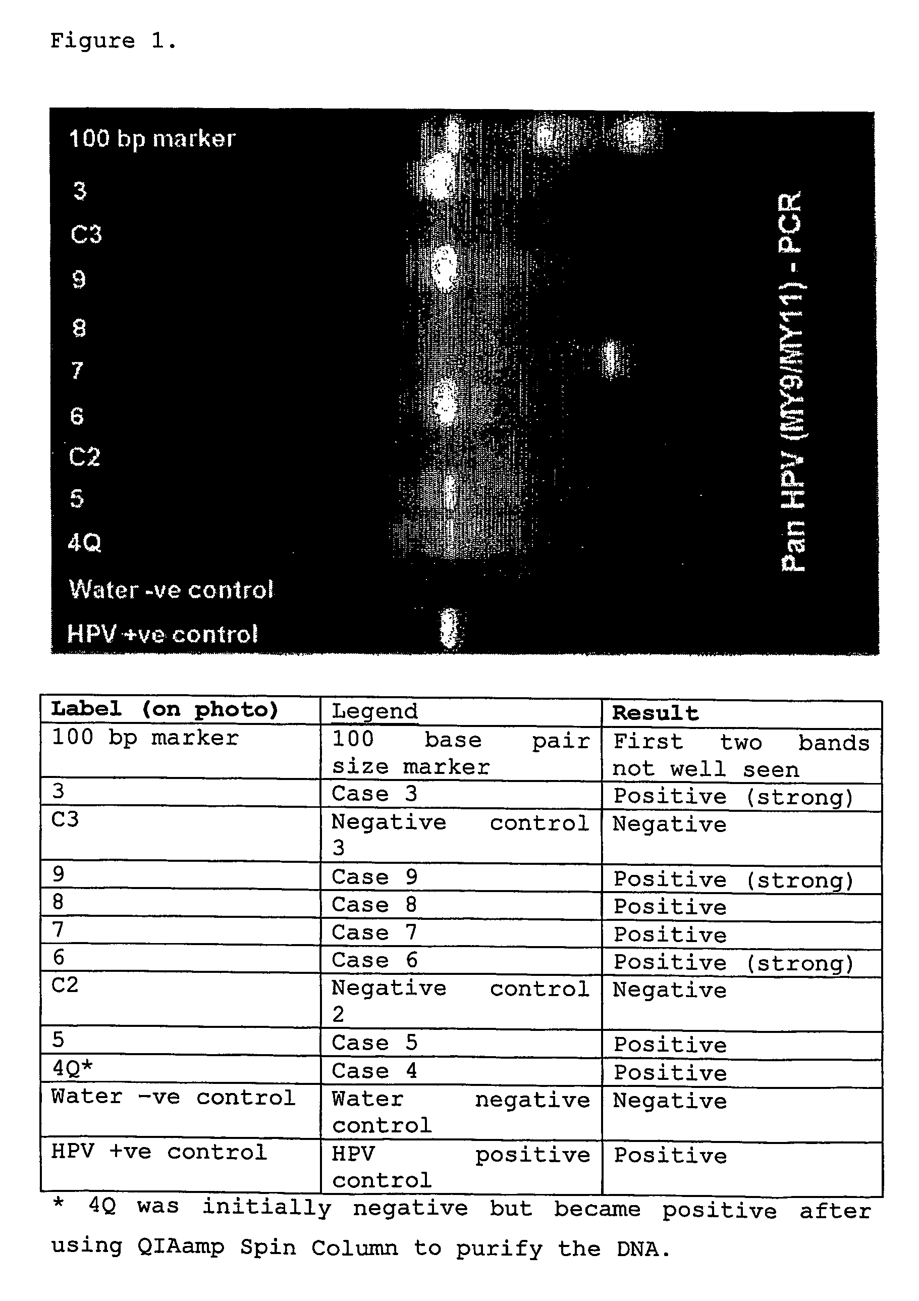
1. Detection of HPV in vaginal discharge.

[0072]We were able to detect HPV in air-dried vaginal discharge from 45 samples (100%) obtained from women with histological confirmation of cervical HPV infection or cervical neoplasia.

[0073]We extracted DNA from the specimens using the following protocol:[0074]1. Add 600 ul Cell Lysis Solution and 3 ul Proteinase K (20 mg / ml). Vortex to mix and incubate at 56° C. overnight.[0075]2. Cool sample to room temperature (RT). Remove the specimen using a sterile long 10 ul pipette tip fitted to an autopipette and expel all lysate by pressing it against the inside of the tube.[0076]3. Add 200 ul Protein Precipitation Solution to the cell lysate. Vortex vigorously at high speed for 20 sec. to mix.[0077]4. Centrifuge at 14,000 rpm for 5 min. The precipitated proteins will form a tight pellet. If the protein pellet is not tight, repeat the centrifugation.[0078]5. Pipette the supernatant containing the DNA (leaving behind the precipitated protein pellet...


PUM

PropertyMeasurementUnit
thicknessaaaaaaaaaa
nucleic acid amplificationaaaaaaaaaa
areaaaaaaaaaaa

Abstract

The present invention provides a method of testing for the presence of infectious disease agents or host genetic markers comprising applying a device comprising an absorbent and porous material onto the introitus of a female patient; encouraging air drying of at least a portion of the collected vaginal discharge while the device is proximate to the introitus; and determining the presence of infectious disease agents or host genetic markers in the at least partially dried vaginal discharge.

Description

BACKGROUND OF THE INVENTION[0001]1. Field of the Invention[0002]The present invention relates to methods of collection of clinical samples of biological fluids or body discharge. In particular, the invention relates to methods of collection, storage and transportation of vaginal discharge on a simple device, so as to maximize the specimen quantity and quality and to enable the detection of infectious disease agents or host genetic markers by nucleic acid amplification or other technologies.BACKGROUNDUterine Cervical Cancer[0003]The cervix, or neck of the uterus, is the lower part of the uterus that opens into the upper vagina. It is also the thoroughfare for sperms entering the womb as well as being part of the birth canal. The part of the cervix exposed to the vaginal lumen is usually lined by squamous epithelium, whereas the lining of the lumen of the cervix is glandular. The junction between the two epithelia (transformation zone) is susceptible to infection by HPV (human papillo...

Claims


Application Information

Patent Timeline
Patent Type & Authority Patents(United States)
IPC IPC(8): C12Q1/70A61B10/00A61F5/455C12Q1/68G01NG01N33/52G01N33/569
CPCC12Q1/6886C12Q1/708G01N33/528A61B2010/0074G01N2333/025
Inventor TONG, SUN-WINGCHAN, OLIVIA WAI-HINGCHOW, TAT-CHONGYU, VIVIAN
Owner TONG SUN WING
Why Eureka
Try Eureka