Remote interpretation of medical images

a remote interpretation and medical image technology, applied in the field of can solve the problems of inaccurate results of image analysis operations and unworkable remote viewing, and achieve the effect of facilitating timely diagnosis of tissue samples and facilitating remote interpretation of medical images

Inactive Publication Date: 2009-03-17
CARL ZEISS MICROSCOPY GMBH
View PDF31 Cites 23 Cited by
  • Summary
  • Abstract
  • Description
  • Claims
  • Application Information

AI Technical Summary

Benefits of technology

[0006]In general, the invention facilitates the remote interpretation of medical images. In order to facilitate the timely diagnosis of a tissue sample, it is desirable that a medical professional, such as a pathologist, be able to remotely view and interpret a medical image. The immense size of a medical image for a single tissue sample typically makes remote viewing unworkable due to bandwidth constraints. Compression algorithms can produce an image suitable for transmission, but the data lost during compression can lead to inaccurate results from the image analysis operations.

Problems solved by technology

The immense size of a medical image for a single tissue sample typically makes remote viewing unworkable due to bandwidth constraints.
Compression algorithms can produce an image suitable for transmission, but the data lost during compression can lead to inaccurate results from the image analysis operations.

Method used


Image

  • Remote interpretation of medical images
  • Remote interpretation of medical images
  • Remote interpretation of medical images

Examples


Embodiment Construction

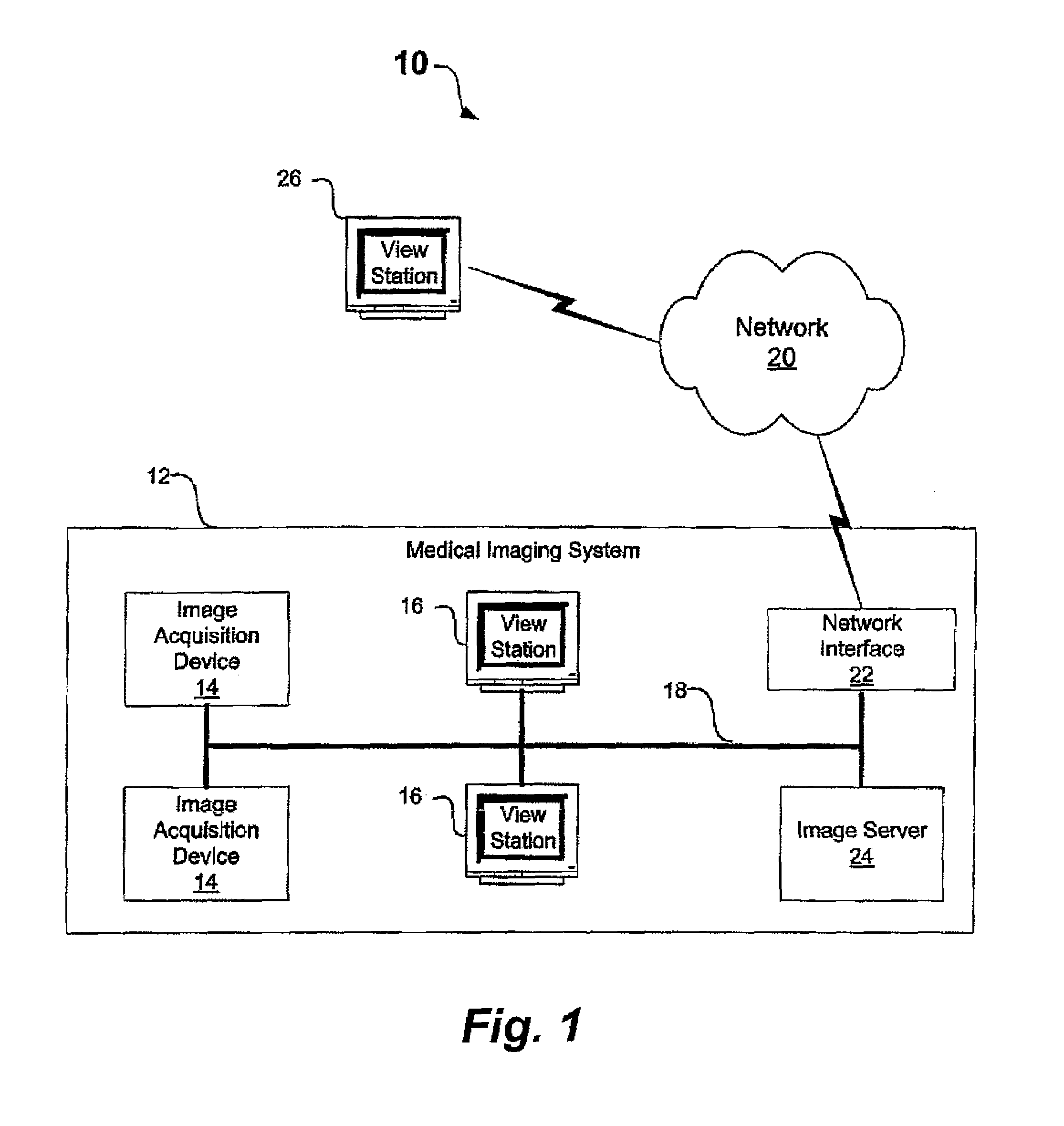
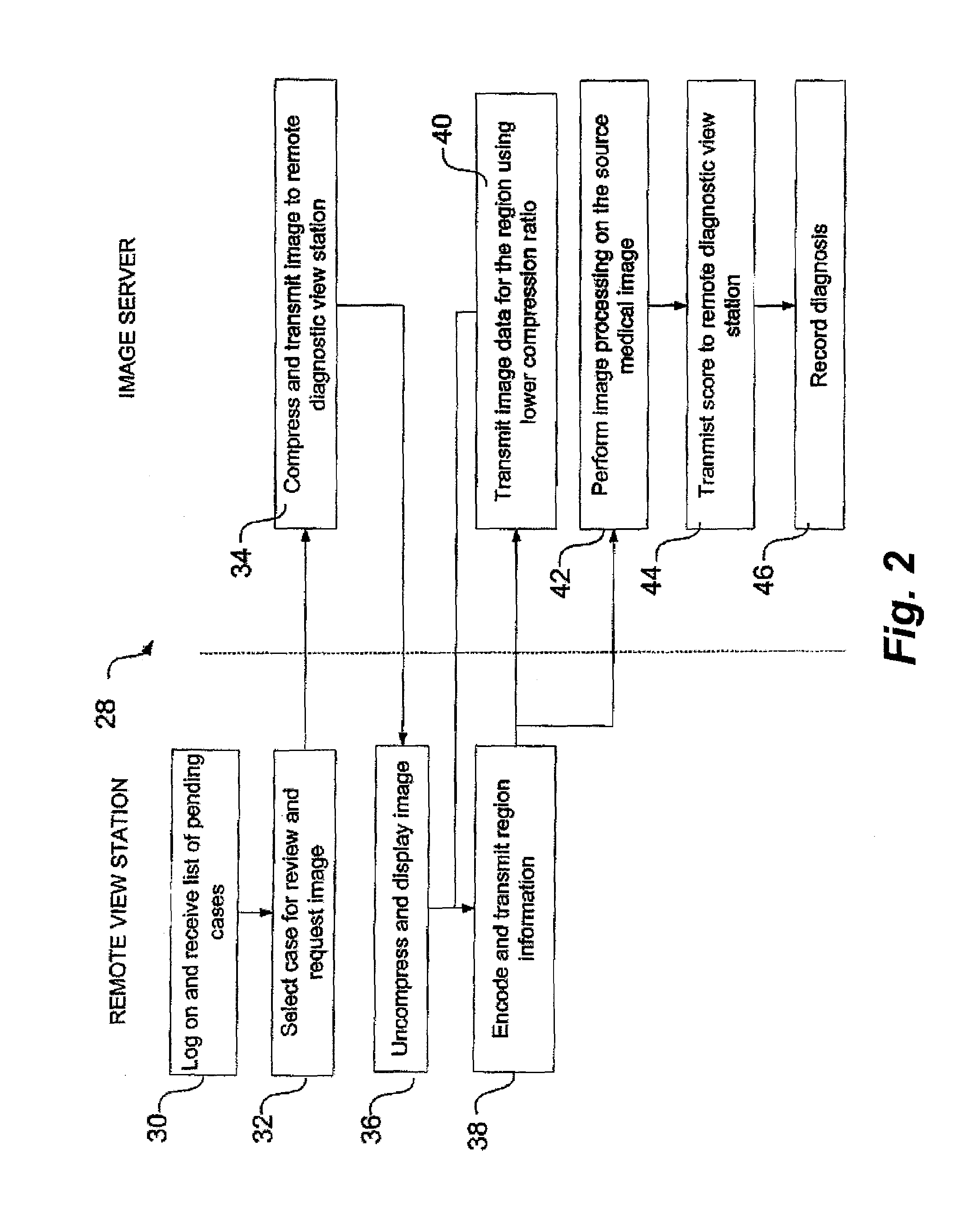
[0013]FIG. 1 is a block diagram illustrating a system 10 that facilitates remotely viewing and interpreting medical images. System 10 includes remote view station 26 this is communicatively coupled to medical imaging system (MIS) 12 by network 20, which represents any packet-switched network such as a local area network or the Internet. MIS 12 includes image acquisition devices 14 that represent any medical imaging device that generates digital medical images, such as an electronic camera used in conjunction with an automated microscope. Other image acquisition devices include computed tomography (CT), nuclear medicine, magnetic resonance imaging (MRI), ultrasound and X-ray devices. Image server 24 stores the images that are generated by image acquisition device 14 and, upon request, communicates the images to view stations 16 for display. Using view stations 16, a medical professional, such as a pathologist, can perform a variety of image processing techniques on selected regions t...


PUM


Abstract

A remote view station is communicatively coupled to an image server and receives a compressed version of source medical images. The remote view station uncompresses and displays the received medical image. A medical professional, such as a pathologist, can select a region of the displayed medical image. Region information is transmitted back to the image server that applies image analysis operations on a region of the source medical image that corresponds to the selected region of the compressed medical image. In this manner, the data loss that occurs during image compression does not effect the image analysis operations. As such, the image analysis operations produce more accurate results than if the operations were applied by remote view station to the uncompressed image.

Description

BACKGROUND[0001]Pathologists typically use microscopes when diagnosing physiological conditions such as cancer, infectious disease and prenatal disorders. Typically, tissue samples on a slide are loaded onto the microscope, the microscope objective or lens focuses on an area of the sample, and the sample is scanned for particular features or objects of interest. In this manner, the microscope helps the pathologist to visually determine the presence of abnormal numbers or types of cells, organelles, organisms or biological markers.[0002]Recently, automated microscopes have been integrated into medical imaging systems that include a variety of networked components. The medical imaging system provides an environment for storing and retrieving the medical images produced by the microscopes. The components of the medical imaging system are spread throughout the department or hospital, or even located remotely, and connected by a communication network.[0003]An image acquisition device is ...

Claims


Application Information

Patent Timeline
Patent Type & Authority Patents(United States)
IPC IPC(8): G06K9/00G06K9/36G06K9/46A61B5/00G06Q50/00G06T1/00G06T7/00G16H30/20G16H40/67H04N1/21H04N7/26H04N7/30
CPCG06F19/324G06F19/3418G06T7/0012H04N1/217H04N19/60H04N19/12H04N19/162H04N19/17H04N19/99G06T2207/10072G06T2207/10116G16H40/67G16H30/20
Inventor DE LA TORRE-BUENO, JOSE
Owner CARL ZEISS MICROSCOPY GMBH
Why Eureka
Try Eureka