Surface-enhanced-spectroscopic detection of optically trapped particulate

a surface-enhanced, spectroscopic technology, applied in the field of non-destructive single particle characterization, can solve the problems of many forms of particulate, including organic polymers and biological cells, that cannot tolerate the spectroscopic techniques needed to characterize them directly, and resort to staining or shadowing techniques that are both labor-intensive and destructive to test subjects

Inactive Publication Date: 2009-04-07
ARMY US SEC THE
View PDF6 Cites 32 Cited by
  • Summary
  • Abstract
  • Description
  • Claims
  • Application Information

AI Technical Summary

Benefits of technology

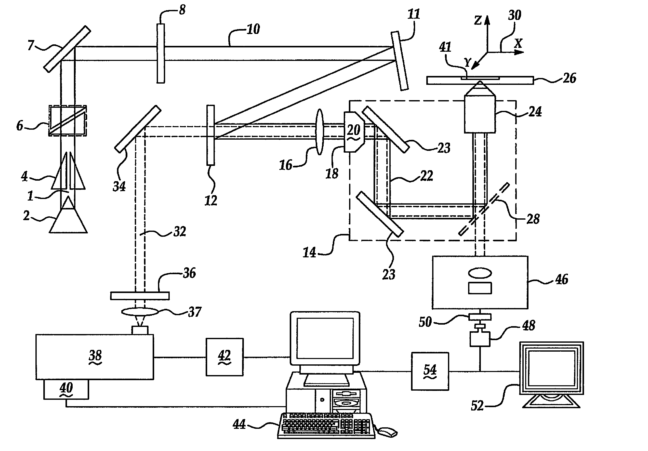
[0013]A particle identification system is provided that includes a digital database containing scattering spectra from a variety of different single particles that vary in spectral features. An algorithm applies a chemometrics protocol to an unknown particle spectrum to determine the relationship between the unknown particle spectrum and one or more standards spectra from the database. A computer is provided to run the algorithm and access the database. A user interface is coupled to the computer for manual data entry. A particle identification system is particularly well suited for endospore particles since the genealogy of a virulent endospore is critical in rapidly identifying the source of the outbreak.

Problems solved by technology

Particulate analyte characterization is currently limited to averaging techniques that sample a field of particles under comparatively mild sampling conditions, or single particle interrogation techniques that subject the particle to a harsh sampling condition in order to generate a sufficient signal relative to background noise.
Unfortunately, many forms of particulate, including organic polymers and biological cells cannot tolerate the spectroscopic techniques needed to characterize them directly.
Instead, one resorts to staining or shadowing techniques that are both labor intensive and destructive of the test subject.
The time required to characterize particulate also limits the use of such information in rapidly changing situations as diverse as a manufacturing, treating an infection, and managing a contamination event.
A difficulty that arises in biological detection is the relative paucity of molecules that are exclusively found in targets.
Additionally, the inability of a single technique to afford definitive characterization through the strain level of the microbe means that culturing the sample is often required thereby increasing the time-consumed in characterization.
However, un-enhanced Raman spectral features are considered to be relatively weak thus requiring relatively lengthy collection times. In many applications, high quality spectra may be achieved with shortened acquisition times and improved spectral features by exploiting various amplification techniques.
Currently, the mechanism of the SERS effect is not fully understood; however, a plausible explanation for at least a significant portion of the spectral amplification has been attributed to an increase in the electromagnetic field strength encountered by the analyte.
In spite of these features, SERS has gained only limited acceptance since early reports on this technique in the late 1970's.
This limited acceptance is in large part due to the lack of fabrication reproducibility in most SERS substrates.
Bacterial spore characterization is a subset of particulate characterization where the limitations of current techniques are profound.
These various Raman spectroscopies have been applied to the study of bulk endospore samples and have failed to afford information sufficient to distinguish between strains of the same endospore.

Method used


Image

  • Surface-enhanced-spectroscopic detection of optically trapped particulate
  • Surface-enhanced-spectroscopic detection of optically trapped particulate
  • Surface-enhanced-spectroscopic detection of optically trapped particulate

Examples


example 1

[0041]SERS-active substrates are prepared by a method similar to that suggested by Grabar, Freeman, Hommer and Natan (19) using freshly synthesized 60 nanometer gold colloids. Briefly, standard glass microscope slides are cleaned for 1-h in a bath consisting of 1:4 (v / v) 30% H2O2 to sulfuric acid bath at 60° C. Subsequent to cleaning, the microscope slides are profusely washed in methanol and stored in fresh CH3OH until needed. A self-assembled-monolayer (SAM) of 3-Aminopropyltriethoxy silane (APTES; Aldrich Chemical Company) is formed on the cleaned glass substrates by submerging the glass slides in a solution of the silane diluted 1:4 (v / v) in methanol. After 24-h, the APTES-derivatized glass is removed from the silane solution and washed repeatedly (˜15×) with methanol to remove any unbound silane from the surface. The APTES-derivatized glass is stored in fresh methanol until needed.

example 2

[0042]Gold colloids of 60 nanometer size freshly prepared by a previously published (26) method are coupled to the silanized substrate by immersing the washed glass in the aqueous colloidal suspension for 24-h at room temperature. Subsequent to the colloidal coupling the SERS substrates are washed with and stored in de-ionized water until needed for analysis. UV-visible absorbance measurements (not shown) of these substrates reveal a Surface Plasmon Resonance (SPR) optimally excited with 715 to 775 nanometer light. SERS-substrates fabricated through this method are near optimally excited using the NIR-RTDS.

example 3

[0043]Bacillus stearothermophilus (ATCC 10149 and ATCC 7953) are purchased from Raven Biological Laboratories, Inc. (Omaha, Nebr.) as 40% ethanol-deionized water suspensions. According to the manufacturer, these spores were prepared by incubation in soybean-casein digest broth at 55 to 60° C. for 7 days. Samples for measurement are prepared by diluting 100 microliters of the spore suspension in 40 milliliters of 18 MΩ deionized water to give a final concentration of 7.5×10⁴ CFU / ml (CFU=colony forming units).


PUM

PropertyMeasurementUnit
interrogation wavelengthaaaaaaaaaa
angleaaaaaaaaaa

Abstract

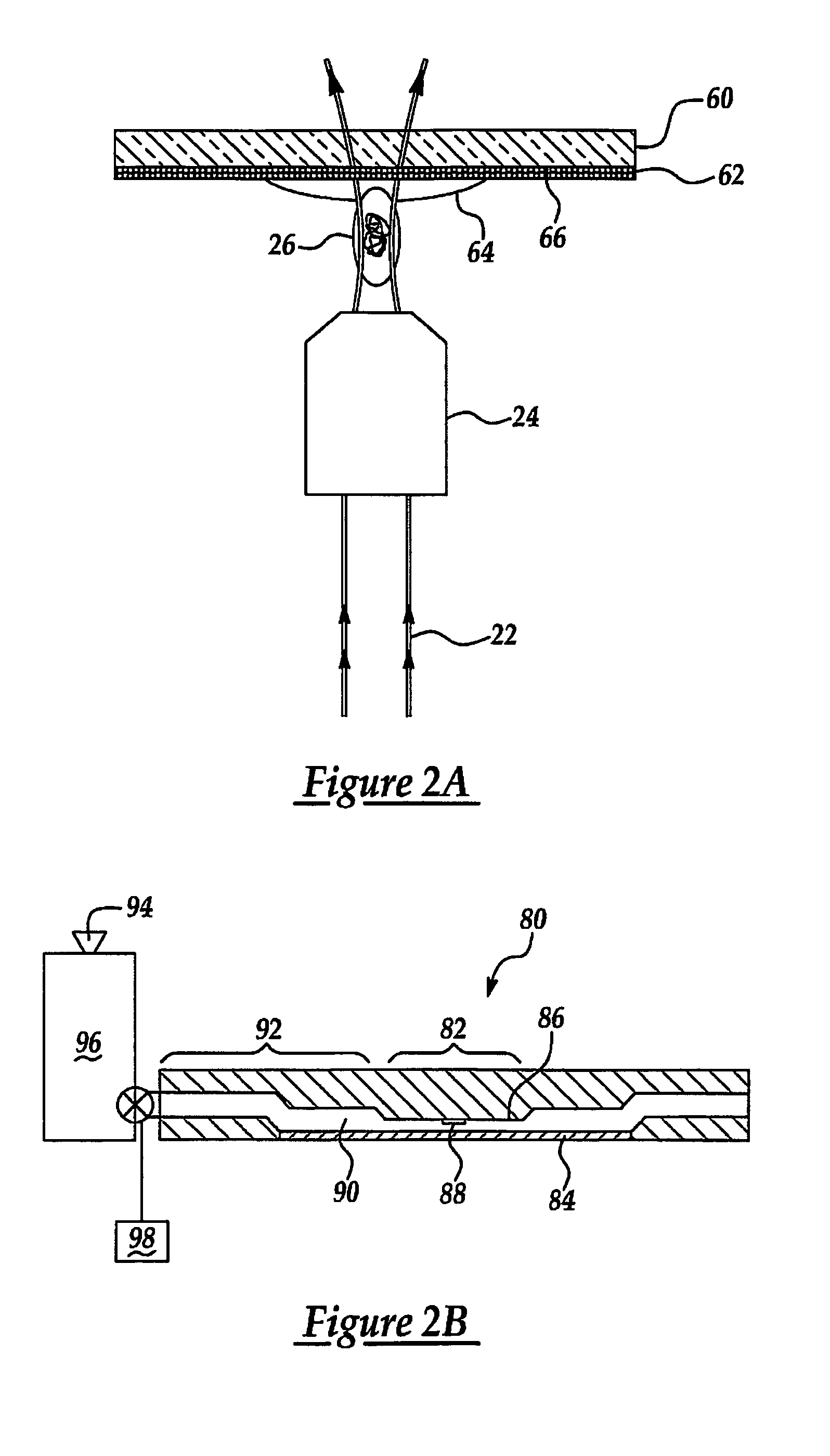
A method of particle detection includes optically trapping a single particle having an averaged linear dimension of between 0.10 microns and 50 microns in a focal region optical trap formed from a trap visible to near-infrared wavelength. The trap is proximal to a substrate having a discontinuous metal coating so as to exploit a surface plasmon resonance generated electric field caused by irradiation of the metal coating. The single particle is then exposed to an interrogation wavelength to yield scattering spectrum relating to the particle. A particle identification digital database containing scattering spectra from a variety of different single particles that vary in spectral features facilitates identification of unknown spectra. A microfluidic cell is detailed to facilitate the automated scanning of numerous particles from a sample.

Description

RELATED APPLICATIONS[0001]This application is a utility application claiming priority of U.S. provisional application 60 / 541,398 filed Feb. 3, 2004.GOVERNMENT INTEREST[0002]The invention described herein may be manufactured, used, and licensed by or for the United States Government.FIELD OF THE INVENTION[0003]This invention in general relates to a method and system for particulate optically trapped SERS characterization and in particular, to a method and system for non-destructive single particle characterization.BACKGROUND OF THE INVENTION[0004]Particulate analyte characterization is currently limited to averaging techniques that sample a field of particles under comparatively mild sampling conditions, or single particle interrogation techniques that subject the particle to a harsh sampling condition in order to generate a sufficient signal relative to background noise. Unfortunately, many forms of particulate, including organic polymers and biological cells cannot tolerate the spe...

Claims


Application Information

Patent Timeline
Patent Type & Authority Patents(United States)
IPC IPC(8): G01N21/55
CPCG01N21/658
Inventor ALEXANDER, TROY A.PELLEGRINO, PAUL M.GILLESPIE, JAMES B.
Owner ARMY US SEC THE
Why Eureka
Try Eureka