High affinity TCR proteins and methods

Inactive Publication Date: 2009-08-04
THE BOARD OF TRUSTEES OF THE UNIV OF ILLINOIS
View PDF137 Cites 48 Cited by
  • Summary
  • Abstract
  • Description
  • Claims
  • Application Information

AI Technical Summary

Benefits of technology

[0011]The high affinity TCR protein can be used in the laboratory as a tool for qualitative and quantitative measurements of a target ligand, in medical, veterinary or plant diagnostic setting or for tissue or plant material identification. Similarly, the high affinity TCRs of the present invention can be used as reagents for detecting and/or quantitating a target material or ligand. Also provided is a method of using high affinity TCRs to bind to a selected peptide/MHC ligand comprising: labeling said high affinity TCRs with a label that binds to the selected peptide/MHC ligand; contacting said labeled high affinity TCRs with cells containing MHC molecules. The high affinity protein of the present invention, where it specifically binds to a tumor cell antigen with high affinity and specificity can be used in diagnostic tests for the particular type of cancer or it can be used in an organism in imaging tests to locate and/or estimate size and number of tumors in an organism, preferably a mammal, and also preferably a human. Methods provided for using high affinity TCRs that bind to pMHCs for diagnostic tests comprise: labeling the high affinity TCR with a detectable label; contacting said high affinity TCR with cells containing the ligand to which the high affinity TCR has high affinity for; and detecting the label. In the method, the label may be chosen to bind to specific peptide/MHC ligands, whereby cells that express specific peptide/MHC ligands are targeted. Preferred methods for using high affinity TCRs as diagnostic probes for specific peptide/MHC molecules on surfaces of cells comprise: labeling high affinity TCRs with a detectable label that binds to specific peptide/MHC ligands; contacting said TCRs with cells; and detecting said label. The detectable label chosen for use depends on the particular use, and the choice of a suitable label is well within the ordinary skill of one in the relevant art. In general, the TCR proteins selected for high affinity binding to a ligand of interest can be used in methods in which antibodies specific for the ligand can be used, with procedural modifications made for the TCR vs. antibody protein, such modifications being known in the art.
[0012]The high affinity TCR, desirably a soluble single chain (sc) TCR, can be used to block autoimmune destruction of cells or tissues in autoimmune disease, where the site recognized by the cytotoxic lymphocytes on the surface of the target cell is the same as the site bound by the high affinity TCR. Preferred methods for blocking autoimmune destruction of cel

Problems solved by technology

One of the difficulties in exploring the basis of differences between Fab and TCR has been that the extensive sequence diversity in antibody and TCR variable (V) regions complicate

Method used


Image

  • High affinity TCR proteins and methods
  • High affinity TCR proteins and methods
  • High affinity TCR proteins and methods

Examples


example 1

Library Construction

[0088]The 2C scTCR used as the scaffold for directed evolution (T7) contained six mutations (βG17E, βG42E βL81S, αL43P, αW82R, and αI118N) that have been shown to increase the stability of the TCR but still allow pMHC binding [see, e.g., Shusta, E. V. et al. (1999) J. Mol. Biol. 292, 949-956].

[0089]Mutagenic PCR of the T7 scTCR VαCDR3 was performed using an AGA2-specific upstream primer (GGCAGCCCCATAAACACACAGTAT (SEQ ID NO:3)) and a degenerate downstream primer CTTTTGTGCCGGATCCAAATGTCAG(SNN)5GCTCACAGCACAGAAGTACACGGCCGA GTCGCTC (SEQ ID NO:4). Underlined bases indicate the positions of silent mutations introducing unique BamHI and EagI restriction sites. The purified PCR product was digested with NdeI and BamHI and ligated to NdeI-BamHI digested T7 / pCT302 [Boder and Wittrup (1997) supra; Kieke et al. (1999) supra; Shusta et al. (1999) supra]. The ligation mixture was transformed into DH10B electro-competent E. coli (Gibco BR1, Gaithersburg, Md.), and transformants ...

example 2

Cell Sorting

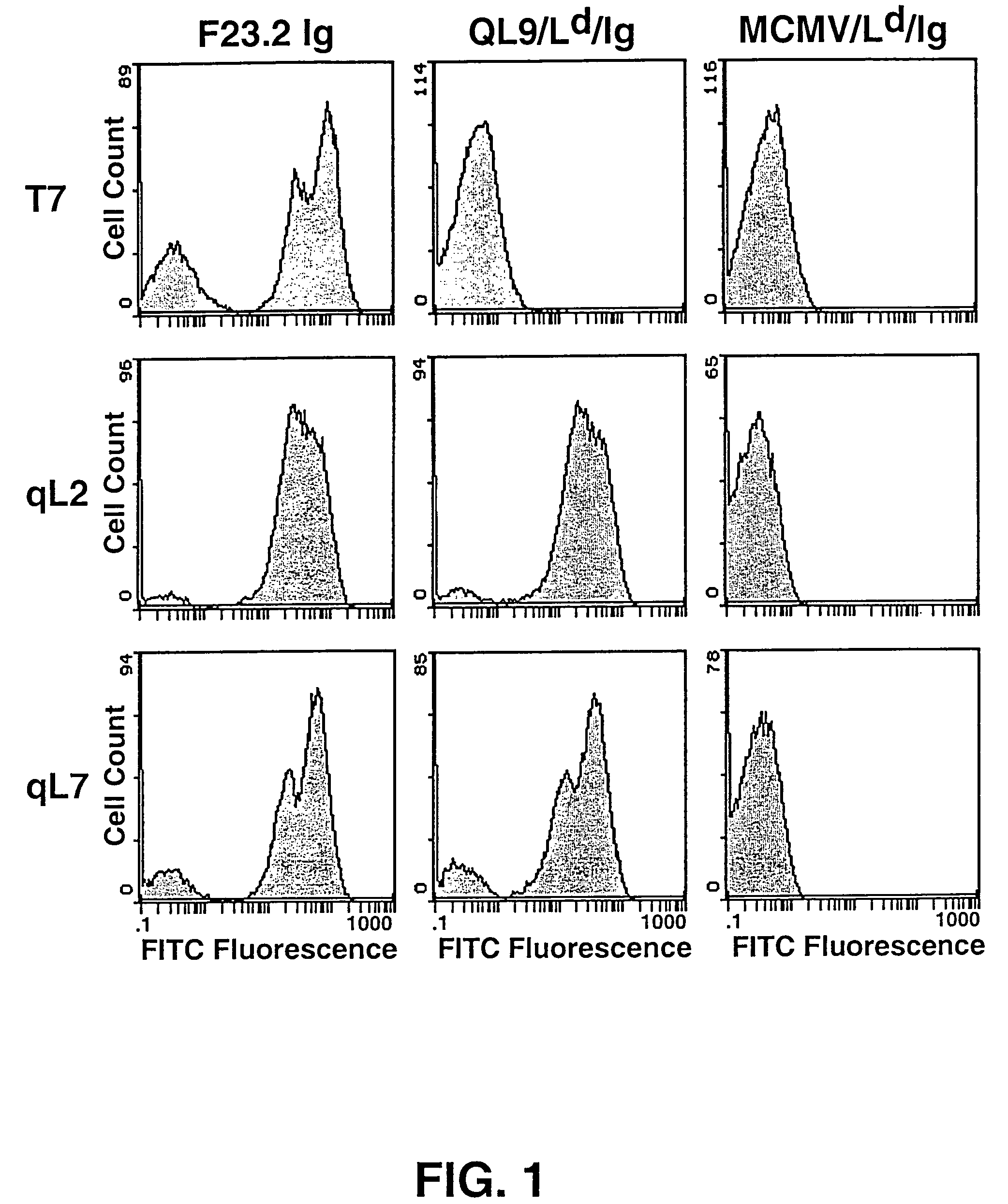
[0090]The yeast library [Shusta et al. (1999) Curr. Opin. Biotechnol. 10:117-122] was grown in SD-CAA (2% dextrose, 0.67% yeast nitrogen base, 1% casamino acids (Difco, Livonia, Mich.)) at 30° C. to an OD600=4.0. To induce surface scTCR expression, yeast were pelleted by centrifugation, resuspended to an OD600=1.0 in SG-CAA (2% galactose, 0.67% yeast nitrogen base, 1% casamino acids), and incubated at 20° C. for about 24 hr. In general, about 107 cells / tube were incubated on ice for 1 hr with 50 μl of QL9 / Ld / IgG dimers [Dal Porto et al. (1993) supra] diluted in phosphate buffered saline, pH 7.4 supplemented with 0.5 mg / ml BSA (PBS-BSA). After incubation, cells were washed and labeled for 30 min with FITC-conjugated goat anti-mouse IgG F(ab7)2 (Kirkegaard & Perry, Gaithersburg, Md.) in PBS-BSA. Yeast were then washed and resuspended in PBS-BSA immediately prior to sorting. Cells exhibiting the highest fluorescence were isolated by FACS sorting with a Coulter 753 bench. Af...

example 3

Soluble scTCR Production

[0091]The T7 and qL2 open reading frames were excised from pCT302 NheI-XhoI and ligated into NheI-XhoI digested pRSGALT, a yeast expression plasmid [Shusta et al. (1999) supra]. Ligated plasmids were transformed into DH10B electro-competent E. coli (Gibco BRL). Plasmid DNA was isolated from bacterial cultures and transformed into Saccharomyces cerevisiae BJ5464 (α ura3-52 trp1 leu2 1 his3 200 pep4: HIS3 prb1 1.6R can1 GAL) [Shusta et al. (1999) supra]. Yeast clones were grown in one liter SD-CAA / Trp (20 mg / L tryptophan) for 48 hr at 30° C. To induce scTCR secretion, cells were pelleted by centrifugation at 4000×g, resuspended in one liter SG-CAA / Trp supplemented with 1 mg / ml BSA, and incubated for 72 hr at 20° C. Culture supernatants were harvested by centrifugation at 4000×g, concentrated to about 50 ml, and dialyzed against PBS, pH 8.0. The 6His-tagged scTCRs were purified by native nickel affinity chromatography (Ni-NTA Superflow, Qiagen, Valencia, Calif.;...


PUM

PropertyMeasurementUnit
Fluorescenceaaaaaaaaaa
Chemiluminescenceaaaaaaaaaa
Affinityaaaaaaaaaa

Abstract

T cell receptors (TCRS) that have higher affinity for a ligand than wild type TCRs are provided. These high affinity TCRs are formed by mutagenizing a T cell receptor protein coding sequence to generate a variegated population of mutants of the T cell receptor protein coding sequence; transforming the T cell receptor mutant coding sequence into yeast cells; inducing expression of the T cell receptor mutant coding sequence on the surface of yeast cells; and selecting those cells expressing T cell receptor mutants that have higher affinity for the peptide/MHC ligand than the wild type T cell receptor protein. The high affinity TCRs can be used in place of an antibody or single chain antibody.

Description

CROSS REFERENCE TO RELATED APPLICATIONS[0001]This application is a divisional of U.S. application Ser. No. 09 / 731,242, filed Dec. 6, 2000 now U.S. Pat. No. 6,759,243, which is a continuation-in-part of U.S. application Ser. No. 09 / 009,388, filed Jan. 20, 1998 now U.S. Pat. No. 6,699,658, and wherein this application also claims the benefit of U.S. Provisional Application No. 60 / 169,179, filed Dec. 6, 1999.ACKNOWLEDGMENT OF FEDERAL RESEARCH SUPPORT[0002]This invention was made with Government support under Contract No. PHS-5-R01-GM55767-03 awarded by the National Institutes of Health (NIH). The Government has certain rights in this invention.BACKGROUND OF THE INVENTION[0003]The field of the present invention is molecular biology, in particular, as it is related to combinatorial libraries of immune cell receptors displayed on the cell surface of a recombinant host cell. More specifically, the present invention relates to a library of high affinity T cell receptor proteins displayed on...

Claims


Application Information

Patent Timeline
IPC IPC(8): G01N33/563G01N33/53C07K14/725C12N5/08C12N15/10C40B40/02G01N33/68
CPCC07K14/7051C12N15/1037C40B40/02G01N33/6842G01N33/6854G01N33/68G01N2333/39C12N5/0636C12N2510/00G01N33/56972G01N2333/7051
Inventor KRANZ, DAVID M.WITTRUP, K. DANEHOLLER, PHILLIP D.
Owner THE BOARD OF TRUSTEES OF THE UNIV OF ILLINOIS
Why Eureka
Try Eureka