Method and apparatus for low quantity detection of bioparticles in small sample volumes

a bioparticle and small sample volume technology, applied in the field of bioparticle detection, can solve the problems of difficult to see these particles with the naked eye, not always possible, and inability to use color change as an indicator

Inactive Publication Date: 2009-11-10
NANO SCI DIAGNOSTICS
View PDF38 Cites 19 Cited by
  • Summary
  • Abstract
  • Description
  • Claims
  • Application Information

AI Technical Summary

Problems solved by technology

Viewing these particles with the naked eye is difficult.
While it would be nice to have all color change indicating reagents for detection, it is not always possible.
Another problem arises when a nanoliter drop sample has only a few of the target particles.
In such cases, using color change as an indicator is impractical.
However, having only a few molecules limits the number of photons emitted from the sample drop.
In low cost diagnostic products, using photomultipliers to increase the photon counting efficiency or using complex optics and manual positioning are ruled out due to their increased cost.

Method used


Image

  • Method and apparatus for low quantity detection of bioparticles in small sample volumes
  • Method and apparatus for low quantity detection of bioparticles in small sample volumes
  • Method and apparatus for low quantity detection of bioparticles in small sample volumes

Examples


Embodiment Construction

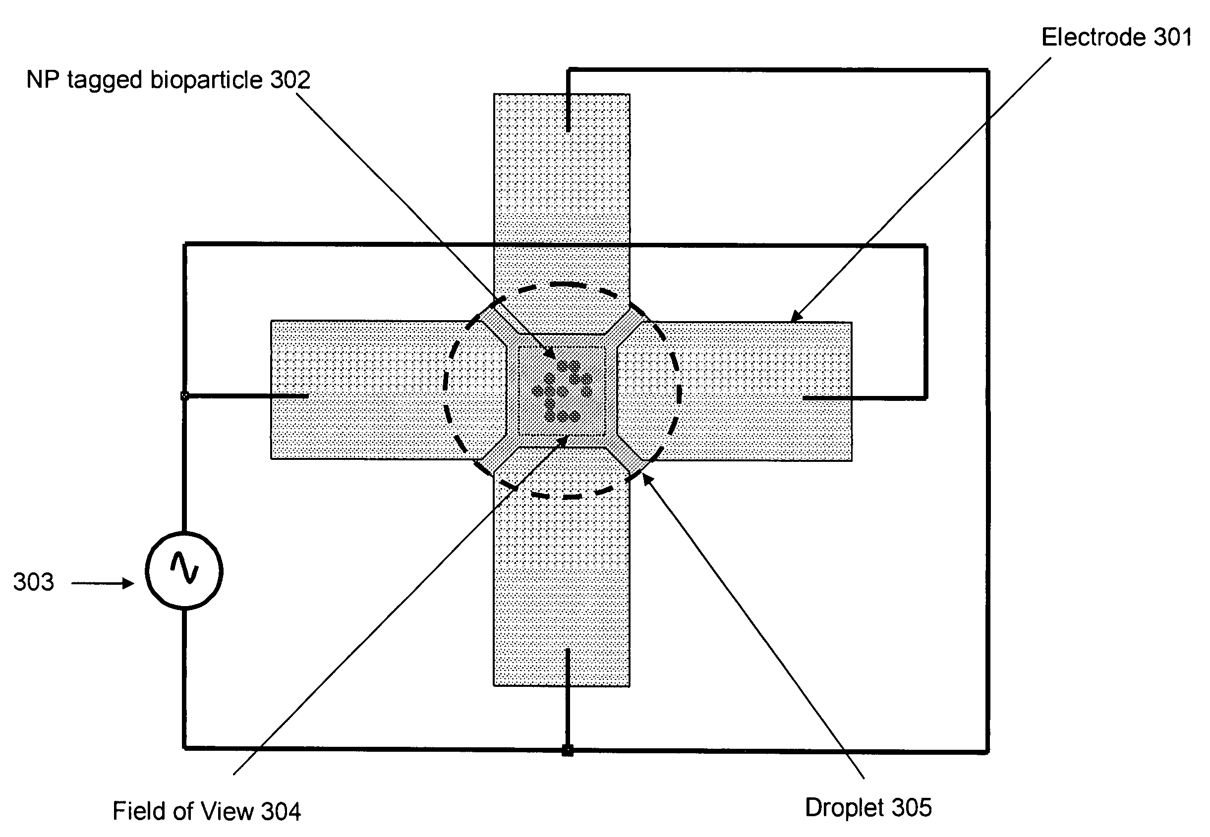
[0024]The threshold of detection of bioparticles can be improved using dielectrophoresis to move, mix and concentrate a few fluoro-tagged bioparticles into the field of view of the sensor that is small and does not have any focusing optics.

Dielectrophoresis

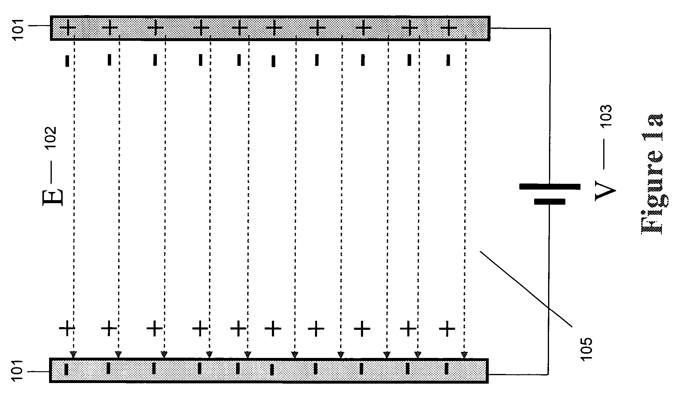
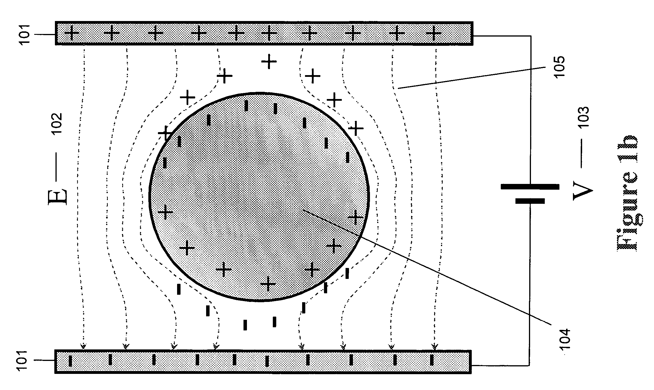
[0025]All particles in nature are made up of molecules. Molecules in turn have an electronic structure. In each particle (nanoparticle, virus, bacteria, cell, tagging antibody, antigen, DNA, RNA and protein) some molecules have loosely bound outer orbit electrons that are free to move about the confines of the particle. These are called free electrons. The degree of mobility of these electrons gives the particle a certain electrical conductivity. Metals have high conductivity, as these free electrons are very free and can be used to carry current. In semiconducting or insulating particles these free electrons allow an external electric field to convert the particle into a dipole. A dipole is a particle in which the free charge is ...


PUM

PropertyMeasurementUnit
fluorescenceaaaaaaaaaa
fluorescentaaaaaaaaaa
crossover frequencyaaaaaaaaaa

Abstract

A novel apparatus and method is described for detection of very small quantities (a few hundred molecules) of bioparticles in nanoliter / picoliter quantities of a sample. The apparatus involves a very small and low cost apparatus that contains a fluorometer. The detection process uses the fluorescence of nanoparticles. Dielectrophoresis is used to concentrate, mix and position the target particles with regard to the light sensor such that maximum detection efficiency is achieved. This allows low cost implementation of low cost point of care tests for disease (animal and plant), infection, food-borne bacteria detection, nucleotide sequencing and pathogen detection (bioterrorism) in real world applications.

Description

[0001]This application claims benefit of U.S. Provisional Application No. 60 / 632,791, filed Dec. 3, 2004.RELATED PATENT APPLICATION[0002]U.S. patent application Ser. No. 11 / 222,093 filed Sep. 8, 2005, claiming priority to U.S. patent application No. 60 / 614,688 filed Sep. 30, 2004 entitled “Method for Detection and Decontamination of Antigens by Nanoparticle-Raman Spectroscopy” having Sulatha Dwarakanath et al. as inventors and assigned to the Assignee of the present invention, which patent application is attached hereto as Attachment A.FIELD OF THE INVENTION[0003]The field of the invention relates generally to the detection of bioparticles using dielectrophoresis.BACKGROUND[0004]There is a great need for low cost detection of bioparticles in various applications like disease diagnostics in animals and plants, bioterrorism, food inspection, and scientific research. Samples in such applications are found in a liquid medium. These particles can range from a few nanometers (nm) in size ...

Claims


Application Information

Patent Timeline
Patent Type & Authority Patents(United States)
IPC IPC(8): G01N21/64
CPCB03C5/005B03C5/024B03C5/026G01N21/645G01N21/6458G01N21/6428G01N2021/6482
Inventor SATYANARAYANA, SRINAGESHDWARAKANATH, SULATHA
Owner NANO SCI DIAGNOSTICS
Why Eureka
Try Eureka