Apparatus for acquiring tomographic image formed by ultrasound-modulated fluorescence

a technology of fluorescence and tomography, applied in tomography, applications, catheters, etc., can solve the problem of at least approximately 1 mm resolution for acquiring fluorescence tomographic images of the region to be observed

Inactive Publication Date: 2009-11-17
FUJIFILM HLDG CORP +1
View PDF22 Cites 18 Cited by
  • Summary
  • Abstract
  • Description
  • Claims
  • Application Information

AI Technical Summary

Benefits of technology

[0011]The object of the present invention is to provide a fluorescence tomography apparatus which can obtain high-resolution fluorescence tomographic images of a region extending to a depth of several millimeters.
[0013]The fluorescence tomography apparatus according to the present invention can obtain a fluorescence tomographic image of a region which is to be observed and extends to a depth of several millimeters from a surface of the region, with high resolution (at most tens of micrometers). This is because when an ultrasonic wave is applied to the region to be observed, the ultrasonic wave causes an elastic wave (i.e., compression and rarefaction of tissue) in the region to be observed, and produces a distribution of the refractive index in the region. When fluorescence is emitted from the region which is to be observed and is in the above situation, the fluorescence is influenced by the compressed and rarefied state of the tissue, i.e., the fluorescence is modulated. It is possible to obtain a fluorescence (ultrasound-modulated-fluorescence) tomographic image by detecting and analyzing the ultrasound-modulated fluorescence.

Problems solved by technology

However, it is impossible to acquire a fluorescence tomographic image of a region which is to be observed and extends to a depth of 0.5 mm or greater from a surface of the region.
However, in this case, the resolution is at most approximately 1 mm, and insufficient.

Method used


Image

  • Apparatus for acquiring tomographic image formed by ultrasound-modulated fluorescence
  • Apparatus for acquiring tomographic image formed by ultrasound-modulated fluorescence
  • Apparatus for acquiring tomographic image formed by ultrasound-modulated fluorescence

Examples


embodiment

Outline of Embodiment

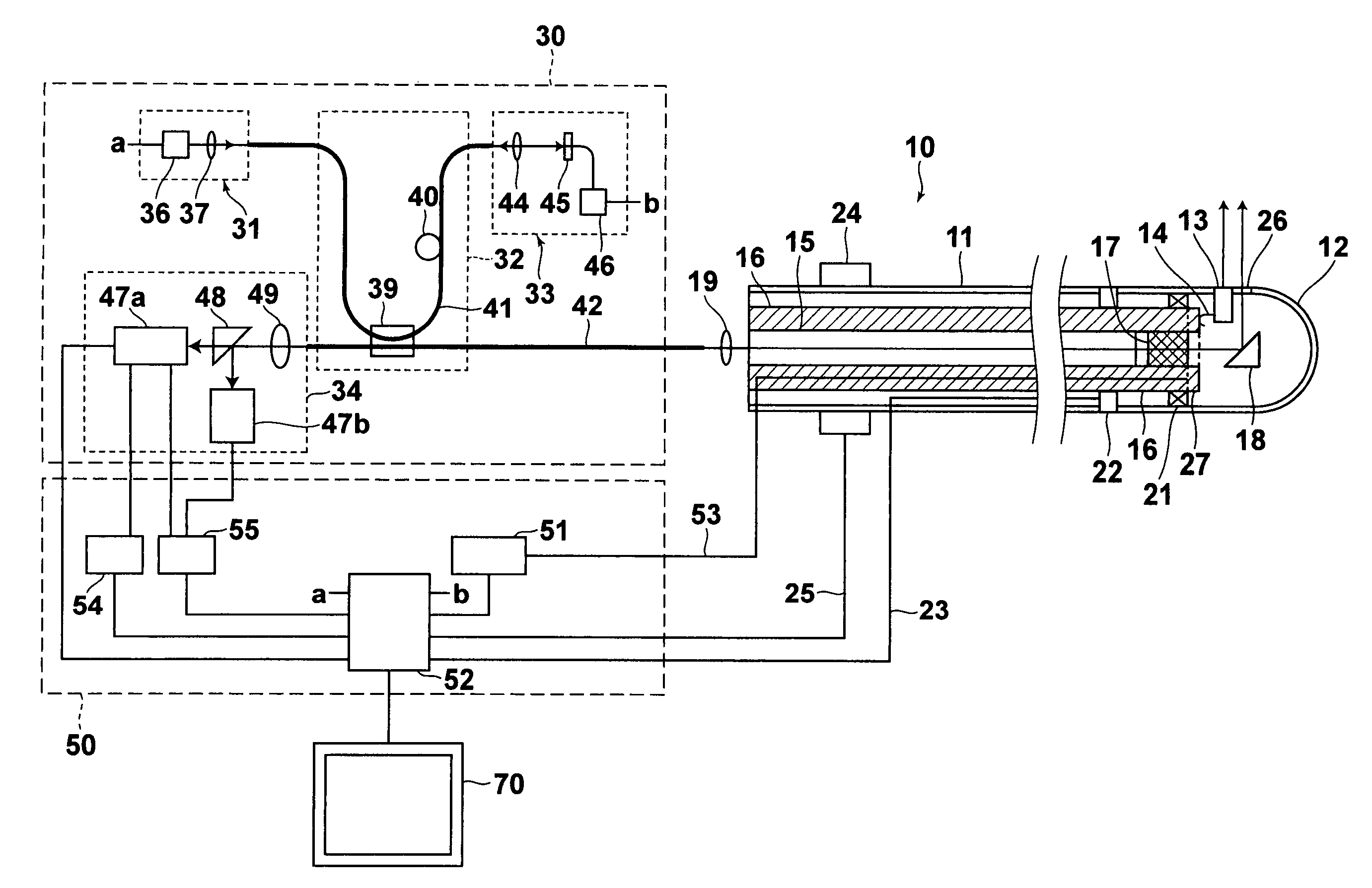
[0029]FIG. 1 is a diagram schematically illustrating the construction of the fluorescence tomography apparatus according to an embodiment of the present invention. The fluorescence tomography apparatus of FIG. 1 comprises a probe 10, an optical unit 30, a signal processing unit 50, and a monitor 70. The probe 10 can be inserted through a forceps channel of an endoscope, the optical unit 30 is connected to the probe 10, the signal processing unit 50 is connected to the probe 10 and the optical unit 30, and the monitor 70 is connected to the signal processing unit 50. In addition, the fluorescence tomography apparatus of FIG. 1 has a function of acquiring an optical tomographic image, a function of acquiring an ultrasonic tomographic image, a function of acquiring a fluorescence (ultrasound-modulated-fluorescence) tomographic image formed by ultrasound-modulated fluorescence, and a function of acquiring an ultrasound-modulated-light tomographic image. A (human) su...


PUM


Abstract

In a fluorescence tomography apparatus for obtaining fluorescence tomographic images of a region of an object to be observed: an application unit concurrently applies an ultrasonic wave and excitation light to the region so that ultrasound-modulated fluorescence is emitted from the region under the influence of the ultrasonic wave; and a first image acquisition unit acquires a fluorescence tomographic image of the region on the basis of the ultrasound-modulated fluorescence.

Description

BACKGROUND OF THE INVENTION[0001]1. Field of the Invention[0002]The present invention relates to a fluorescence tomography apparatus which applies excitation light to a region of an object to be observed, detects fluorescence emitted from the object, and acquires a fluorescence tomographic image of the region.[0003]2. Description of the Related Art[0004]In recent years, optical tomography apparatuses which acquire tomographic images by using light have been used in the field of medical diagnosis. The optical tomography apparatuses using confocal optical systems and the optical tomography apparatuses using time-resolved measurement systems are already in actual use.[0005]In addition, commercialization of the optical coherence tomography (OCT) apparatuses has been recently proceeding. In the OCT apparatuses, low-coherence light is emitted from a light source realized by a super luminescent diode (SLD) or the like, and split into measurement light (light applied to an object for measur...

Claims


Application Information

Patent Timeline
Patent Type & Authority Patents(United States)
IPC IPC(8): A61B8/14A61B6/00
CPCA61B5/0066A61B5/0071A61B5/0084A61B5/0073A61B8/12
Inventor TSUJITA, KAZUHIRO
Owner FUJIFILM HLDG CORP
Why Eureka
Try Eureka